|
Arcus Senilis
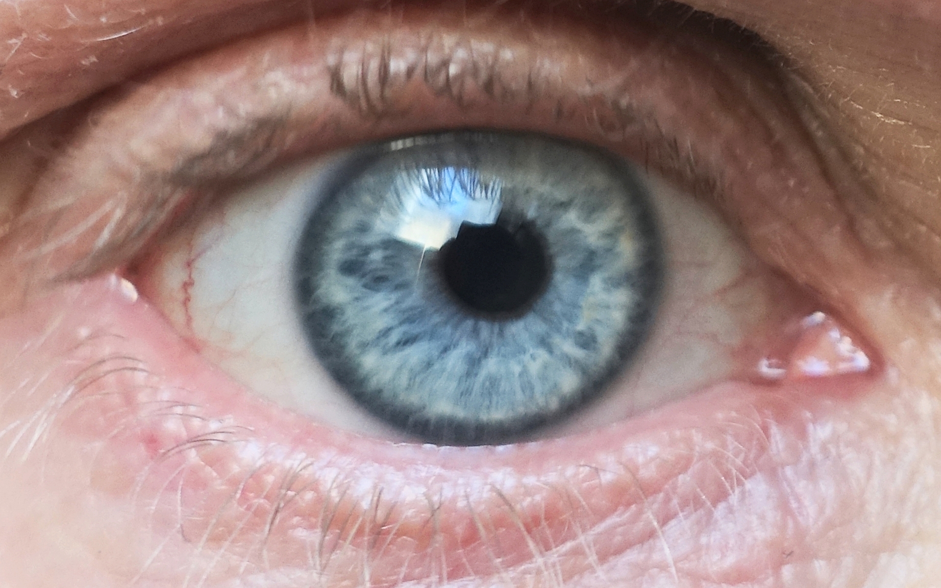
Arcus senilis (AS), also known as gerontoxon, arcus lipoides, arcus corneae, corneal arcus, arcus adiposus, or arcus cornealis, are rings in the peripheral cornea. It‘s usually caused by cholesterol deposits, so it may be a sign of high cholesterol. It is the most common peripheral corneal opacity, and is usually found in the elderly where it is considered a benign condition. When AS is found in patients less than 50 years old it is termed arcus juvenilis. The finding of arcus juvenilis in combination with hyperlipidemia in younger men represents an increased risk for cardiovascular disease. Pathophysiology AS is caused by leakage of lipoproteins from limbal capillaries into the corneal stroma. Deposits have been found to consist mostly of low-density lipoprotein (LDL). Deposition of lipids into the cornea begins at the superior and inferior aspects, and progresses to encircle the entire peripheral cornea. The interior border of AS has a diffuse appearance, while the exterior b ... [...More Info...] [...Related Items...] OR: [Wikipedia] [Google] [Baidu] |
Cornea
The cornea is the transparent front part of the eye that covers the iris, pupil, and anterior chamber. Along with the anterior chamber and lens, the cornea refracts light, accounting for approximately two-thirds of the eye's total optical power. In humans, the refractive power of the cornea is approximately 43 dioptres. The cornea can be reshaped by surgical procedures such as LASIK. While the cornea contributes most of the eye's focusing power, its focus is fixed. Accommodation (the refocusing of light to better view near objects) is accomplished by changing the geometry of the lens. Medical terms related to the cornea often start with the prefix "'' kerat-''" from the Greek word κέρας, ''horn''. Structure The cornea has unmyelinated nerve endings sensitive to touch, temperature and chemicals; a touch of the cornea causes an involuntary reflex to close the eyelid. Because transparency is of prime importance, the healthy cornea does not have or need blood ve ... [...More Info...] [...Related Items...] OR: [Wikipedia] [Google] [Baidu] |
Dystrophic Calcification
Dystrophic calcification (DC) is the calcification occurring in degenerated or necrotic tissue, as in hyalinized scars, degenerated foci in leiomyomas, and caseous nodules. This occurs as a reaction to tissue damage, including as a consequence of medical device implantation. Dystrophic calcification can occur even if the amount of calcium in the blood is not elevated (a systemic mineral imbalance would elevate calcium levels in the blood and all tissues) and cause metastatic calcification. Basophilic calcium salt deposits aggregate, first in the mitochondria, then progressively throughout the cell. These calcifications are an indication of previous microscopic cell injury, occurring in areas of cell necrosis when activated phosphatases bind calcium ions to phospholipids in the membrane. Calcification in dead tissue #Caseous necrosis in T.B. is most common site of dystrophic calcification. #Liquefactive necrosis in chronic abscesses may get calcified. #Fat necrosis following acut ... [...More Info...] [...Related Items...] OR: [Wikipedia] [Google] [Baidu] |
Limbal Ring
A limbal ring is a dark ring around the iris of the eye, where the sclera meets the cornea.Johnson and Johnson Vision Care Inc. Tinted contact lenses with combined limbal ring and iris patterns. US7246903B2. United States Patent and Trademark Office, July 24, 2007. http://patft.uspto.gov/netacgi/nph-Parser?Sect1=PTO1&Sect2=HITOFF&p=1&u=/netahtml/PTO/srchnum.html&r=1&f=G&l=50&d=PALL&s1=7246903.PN. It is a dark-colored manifestation of the corneal limbus resulting from optical properties of the region. The appearance and visibility of the limbal ring can be negatively affected by a variety of medical conditions concerning the peripheral cornea. It has been suggested that limbal ring thickness may correlate with health or youthfulness and may contribute to facial attractiveness. Some contact lenses are colored to simulate limbal rings. Youth, health, and attractiveness Both health and age are positively correlated with a prominent limbal ring. For instance, a darker limbal ring tends ... [...More Info...] [...Related Items...] OR: [Wikipedia] [Google] [Baidu] |
Atherosclerosis
Atherosclerosis is a pattern of the disease arteriosclerosis in which the wall of the artery develops abnormalities, called lesions. These lesions may lead to narrowing due to the buildup of atheromatous plaque. At onset there are usually no symptoms, but if they develop, symptoms generally begin around middle age. When severe, it can result in coronary artery disease, stroke, peripheral artery disease, or kidney problems, depending on which arteries are affected. The exact cause is not known and is proposed to be multifactorial. Risk factors include abnormal cholesterol levels, elevated levels of inflammatory markers, high blood pressure, diabetes, smoking, obesity, family history, genetic, and an unhealthy diet. Plaque is made up of fat, cholesterol, calcium, and other substances found in the blood. The narrowing of arteries limits the flow of oxygen-rich blood to parts of the body. Diagnosis is based upon a physical exam, electrocardiogram, and exercise stress test, amo ... [...More Info...] [...Related Items...] OR: [Wikipedia] [Google] [Baidu] |
Xanthoma
A xanthoma (pl. xanthomas or xanthomata) (condition: xanthomatosis) is a deposition of yellowish cholesterol-rich material that can appear anywhere in the body in various disease states. They are cutaneous manifestations of lipidosis in which lipids accumulate in large foam cells within the skin. They are associated with hyperlipidemias, both primary and secondary types. Tendon xanthomas are associated with type II hyperlipidemia, chronic biliary tract obstruction, primary biliary cirrhosis, sitosterolemia and the rare metabolic disease cerebrotendineous xanthomatosis. Palmar xanthomata and tuberoeruptive xanthomata (over knees and elbows) occur in type III hyperlipidemia. Etymology The term xanthoma stems from Greek ξανθός (xanthós) 'yellow', and -ωμα -oma, a suffix forming nouns indicating a mass or tumor. Types Xanthelasma A xanthelasma is a sharply demarcated yellowish collection of cholesterol underneath the skin, usually on or around the eyelids. Strictly, ... [...More Info...] [...Related Items...] OR: [Wikipedia] [Google] [Baidu] |
Coronary Artery Disease
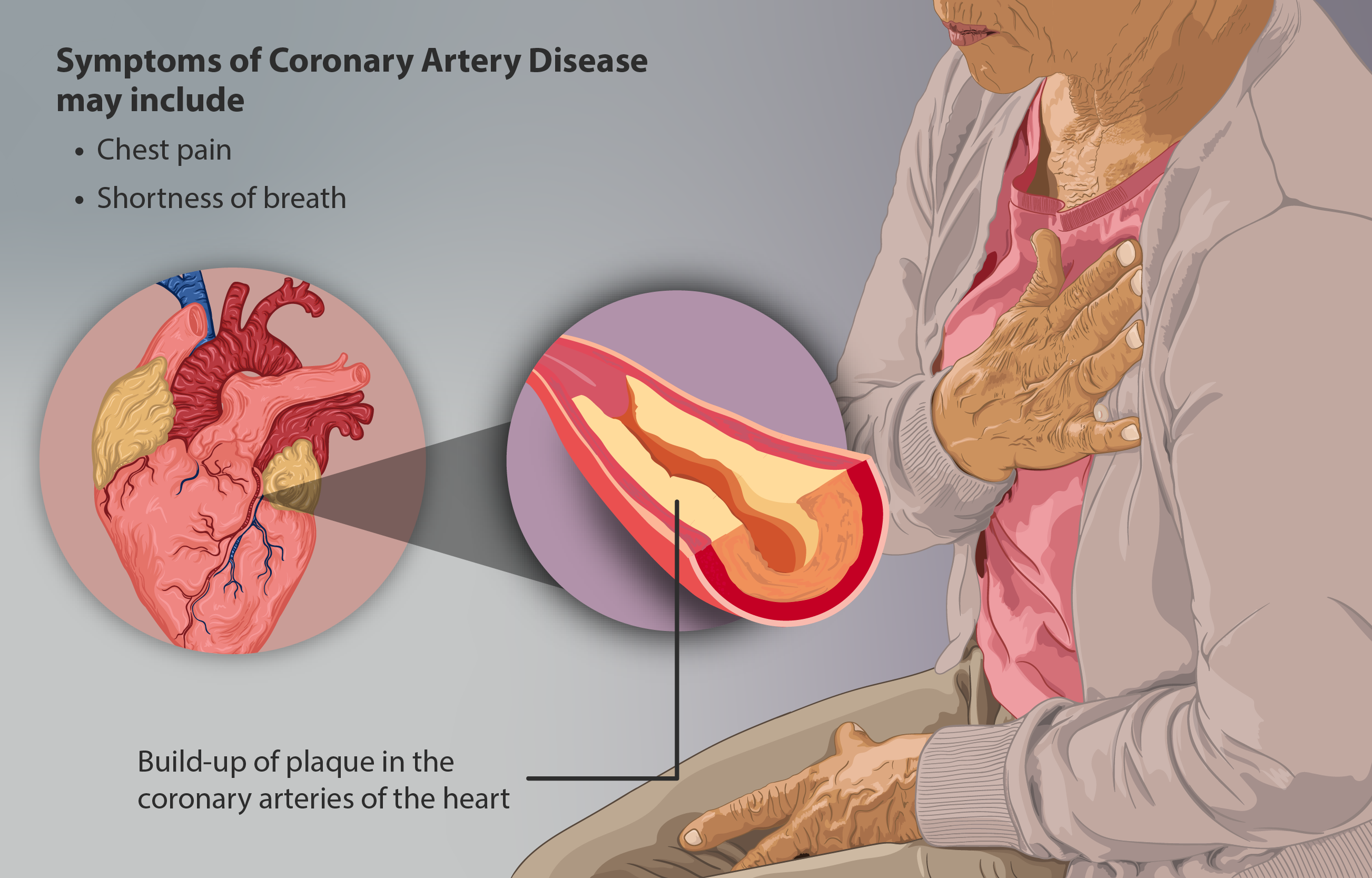
Coronary artery disease (CAD), also called coronary heart disease (CHD), ischemic heart disease (IHD), myocardial ischemia, or simply heart disease, involves the reduction of blood flow to the heart muscle due to build-up of atherosclerotic plaque in the arteries of the heart. It is the most common of the cardiovascular diseases. Types include stable angina, unstable angina, myocardial infarction, and sudden cardiac death. A common symptom is chest pain or discomfort which may travel into the shoulder, arm, back, neck, or jaw. Occasionally it may feel like heartburn. Usually symptoms occur with exercise or emotional stress, last less than a few minutes, and improve with rest. Shortness of breath may also occur and sometimes no symptoms are present. In many cases, the first sign is a heart attack. Other complications include heart failure or an abnormal heartbeat. Risk factors include high blood pressure, smoking, diabetes, lack of exercise, obesity, high blood choles ... [...More Info...] [...Related Items...] OR: [Wikipedia] [Google] [Baidu] |
Cardiovascular Disease
Cardiovascular disease (CVD) is a class of diseases that involve the heart or blood vessels. CVD includes coronary artery diseases (CAD) such as angina and myocardial infarction (commonly known as a heart attack). Other CVDs include stroke, heart failure, hypertensive heart disease, rheumatic heart disease, cardiomyopathy, abnormal heart rhythms, congenital heart disease, valvular heart disease, carditis, aortic aneurysms, peripheral artery disease, thromboembolic disease, and venous thrombosis. The underlying mechanisms vary depending on the disease. It is estimated that dietary risk factors are associated with 53% of CVD deaths. Coronary artery disease, stroke, and peripheral artery disease involve atherosclerosis. This may be caused by high blood pressure, smoking, diabetes mellitus, lack of exercise, obesity, high blood cholesterol, poor diet, excessive alcohol consumption, and poor sleep, among other things. High blood pressure is estimated to account for appro ... [...More Info...] [...Related Items...] OR: [Wikipedia] [Google] [Baidu] |
Relative Risk
The relative risk (RR) or risk ratio is the ratio of the probability of an outcome in an exposed group to the probability of an outcome in an unexposed group. Together with risk difference and odds ratio, relative risk measures the association between the exposure and the outcome. Statistical use and meaning Relative risk is used in the statistical analysis of the data of ecological, cohort, medical and intervention studies, to estimate the strength of the association between exposures (treatments or risk factors) and outcomes. Mathematically, it is the incidence rate of the outcome in the exposed group, I_e, divided by the rate of the unexposed group, I_u. As such, it is used to compare the risk of an adverse outcome when receiving a medical treatment versus no treatment (or placebo), or for environmental risk factors. For example, in a study examining the effect of the drug apixaban on the occurrence of thromboembolism, 8.8% of placebo-treated patients experienced the disease, ... [...More Info...] [...Related Items...] OR: [Wikipedia] [Google] [Baidu] |
Kayser–Fleischer Ring
Kayser–Fleischer rings (KF rings) are dark rings that appear to encircle the cornea of the eye. They are due to copper deposition in the Descemet's membrane as a result of particular liver diseases. They are named after German ophthalmologists Bernhard Kayser and Bruno Fleischer who first described them in 1902 and 1903. Initially thought to be due to the accumulation of silver, they were first demonstrated to contain copper in 1934. Presentation The rings, which consist of copper deposits where the cornea meets the sclera, in Descemet's membrane, first appear as a crescent at the top of the cornea. Eventually, a second crescent forms below, at the "six o'clock position", and ultimately completely encircles the cornea. Associations Kayser–Fleischer rings are a sign of Wilson's disease, which involves abnormal copper handling by the liver resulting in copper accumulation in the body and is characterised by abnormalities of the basal ganglia of the brain, liver cirrhosis, ... [...More Info...] [...Related Items...] OR: [Wikipedia] [Google] [Baidu] |
Limbal Ring
A limbal ring is a dark ring around the iris of the eye, where the sclera meets the cornea.Johnson and Johnson Vision Care Inc. Tinted contact lenses with combined limbal ring and iris patterns. US7246903B2. United States Patent and Trademark Office, July 24, 2007. http://patft.uspto.gov/netacgi/nph-Parser?Sect1=PTO1&Sect2=HITOFF&p=1&u=/netahtml/PTO/srchnum.html&r=1&f=G&l=50&d=PALL&s1=7246903.PN. It is a dark-colored manifestation of the corneal limbus resulting from optical properties of the region. The appearance and visibility of the limbal ring can be negatively affected by a variety of medical conditions concerning the peripheral cornea. It has been suggested that limbal ring thickness may correlate with health or youthfulness and may contribute to facial attractiveness. Some contact lenses are colored to simulate limbal rings. Youth, health, and attractiveness Both health and age are positively correlated with a prominent limbal ring. For instance, a darker limbal ring tends ... [...More Info...] [...Related Items...] OR: [Wikipedia] [Google] [Baidu] |
Schwalbe's Line
Schwalbe's line is the anatomical line found on the interior surface of the eye's cornea, and delineates the outer limit of the corneal endothelium layer. Specifically, it represents the termination of Descemet's membrane. In many cases it can be seen via gonioscopy Gonioscopy a routine ophthalmological procedure that measures the angle between the iris and the cornea (the iridocorneal angle), using a goniolens (also known as a gonioscope) together with a slit lamp or operating microscope. Its use is import .... Some evidence suggests that the corneal endothelium actually possesses stem cells that can produce endothelial cells, especially after injury, albeit on a limited scale. References Human eye anatomy {{Eye-stub ... [...More Info...] [...Related Items...] OR: [Wikipedia] [Google] [Baidu] |
Corneal Limbus
The corneal limbus (''Latin'': corneal border) is the border between the cornea and the sclera (the white of the eye). It contains stem cells in its palisades of Vogt. It may be affected by cancer or by aniridia (a developmental problem), among other issues. Structure The corneal limbus is the border between the cornea and the sclera. It is highly vascularised. Its stratified squamous epithelium is continuous with the epithelium covering the cornea. The corneal limbus contains radially-oriented fibrovascular ridges known as the palisades of Vogt that contain stem cells. The palisades of Vogt are more common in the superior and inferior quadrants around the eye. Clinical significance Cancer The corneal limbus is a common site for the occurrence of corneal epithelial neoplasm. Aniridia Aniridia, a developmental anomaly of the iris, disrupts the normal barrier of the cornea to the conjunctival epithelial cells at the limbus. Glaucoma The corneal limbus may be cut ... [...More Info...] [...Related Items...] OR: [Wikipedia] [Google] [Baidu] |


