|
Appendicular Arteries
The appendicular artery, also known as appendiceal artery, commonly arises from the terminal branch of the ileocolic artery, or less commonly from the posterior cecal artery or an ileal artery. It descends behind the termination of the ileum and enters the mesoappendix of the vermiform appendix The appendix (or vermiform appendix; also cecal r caecalappendix; vermix; or vermiform process) is a finger-like, blind-ended tube connected to the cecum, from which it develops in the embryo. The cecum is a pouch-like structure of the large .... It runs near the free margin of the mesoappendix and ends in branches which supply the appendix. External links * - "Branches of Superior Mesenteric Artery" References Arteries of the abdomen {{circulatory-stub ... [...More Info...] [...Related Items...] OR: [Wikipedia] [Google] [Baidu] |
Cecum
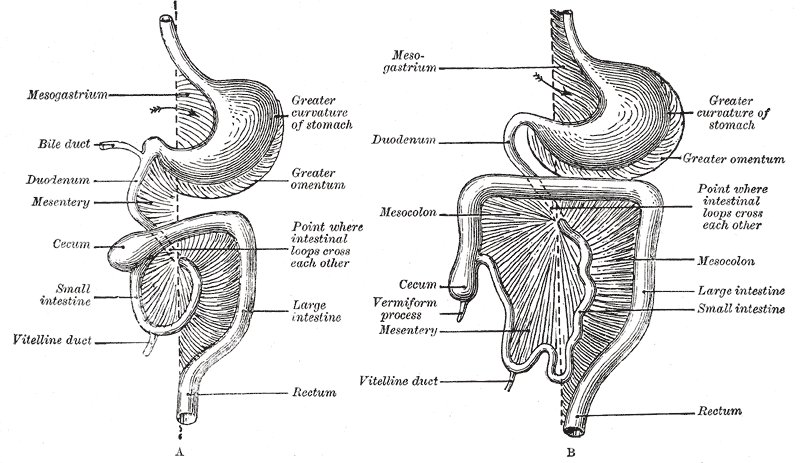
The cecum or caecum is a pouch within the peritoneum that is considered to be the beginning of the large intestine. It is typically located on the right side of the body (the same side of the body as the appendix, to which it is joined). The word cecum (, plural ceca ) stems from the Latin '' caecus'' meaning blind. It receives chyme from the ileum, and connects to the ascending colon of the large intestine. It is separated from the ileum by the ileocecal valve (ICV) or Bauhin's valve. It is also separated from the colon by the cecocolic junction. While the cecum is usually intraperitoneal, the ascending colon is retroperitoneal. In herbivores, the cecum stores food material where bacteria are able to break down the cellulose. In humans, the cecum is involved in absorption of salts and electrolytes and lubricates the solid waste that passes into the large intestine. Structure Development The cecum and appendix are formed by the enlargement of the postarterial segment of ... [...More Info...] [...Related Items...] OR: [Wikipedia] [Google] [Baidu] |
Vermiform Appendix
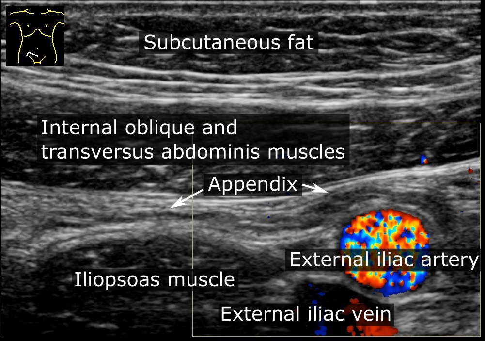
The appendix (or vermiform appendix; also cecal r caecalappendix; vermix; or vermiform process) is a finger-like, blind-ended tube connected to the cecum, from which it develops in the embryo. The cecum is a pouch-like structure of the large intestine, located at the junction of the small and the large intestines. The term " vermiform" comes from Latin and means "worm-shaped". The appendix was once considered a vestigial organ, but this view has changed since the early 2000s. Research suggests that the appendix may serve an important purpose. In particular, it may serve as a reservoir for beneficial gut bacteria. Structure The human appendix averages in length but can range from . The diameter of the appendix is , and more than is considered a thickened or inflamed appendix. The longest appendix ever removed was long. The appendix is usually located in the lower right quadrant of the abdomen, near the right hip bone. The base of the appendix is located beneath the ... [...More Info...] [...Related Items...] OR: [Wikipedia] [Google] [Baidu] |
Ileocolic Artery
The ileocolic artery is the lowest branch arising from the concavity of the superior mesenteric artery. It passes downward and to the right behind the peritoneum toward the right iliac fossa, where it divides into a superior and an inferior branch; the inferior gives rise to the appendicular artery and anastomoses with the end of the superior mesenteric artery, the superior with the right colic artery. It supplies the cecum, ileum, and appendix. Branches The inferior branch of the ileocolic runs toward the upper border of the ileocolic junction and supplies the following branches: * '' colic branch of ileocolic artery'', which passes upward on the ascending colon - from the posterior branch of the inferior branch of the ileocolic artery * ''ileocecal'' (some sources acknowledge this division while others do not) ** '' anterior cecal artery'' and '' posterior cecal artery'', which are distributed to the front and back of the cecum The cecum or caecum is a pouch within the ... [...More Info...] [...Related Items...] OR: [Wikipedia] [Google] [Baidu] |
Appendicular Vein
The appendicular vein is the vein which drains blood from the vermiform appendix. It is located in the mesoappendix and accompanies the appendicular artery. The appendicular vein drains into the ileocolic vein The ileocolic vein is a vein which drains the ileum, colon, and cecum The cecum or caecum is a pouch within the peritoneum that is considered to be the beginning of the large intestine. It is typically located on the right side of the body (t .... Veins of the torso {{circulatory-stub ... [...More Info...] [...Related Items...] OR: [Wikipedia] [Google] [Baidu] |
Vermiform Appendix
The appendix (or vermiform appendix; also cecal r caecalappendix; vermix; or vermiform process) is a finger-like, blind-ended tube connected to the cecum, from which it develops in the embryo. The cecum is a pouch-like structure of the large intestine, located at the junction of the small and the large intestines. The term " vermiform" comes from Latin and means "worm-shaped". The appendix was once considered a vestigial organ, but this view has changed since the early 2000s. Research suggests that the appendix may serve an important purpose. In particular, it may serve as a reservoir for beneficial gut bacteria. Structure The human appendix averages in length but can range from . The diameter of the appendix is , and more than is considered a thickened or inflamed appendix. The longest appendix ever removed was long. The appendix is usually located in the lower right quadrant of the abdomen, near the right hip bone. The base of the appendix is located beneath the ... [...More Info...] [...Related Items...] OR: [Wikipedia] [Google] [Baidu] |
Ileocolic Artery
The ileocolic artery is the lowest branch arising from the concavity of the superior mesenteric artery. It passes downward and to the right behind the peritoneum toward the right iliac fossa, where it divides into a superior and an inferior branch; the inferior gives rise to the appendicular artery and anastomoses with the end of the superior mesenteric artery, the superior with the right colic artery. It supplies the cecum, ileum, and appendix. Branches The inferior branch of the ileocolic runs toward the upper border of the ileocolic junction and supplies the following branches: * '' colic branch of ileocolic artery'', which passes upward on the ascending colon - from the posterior branch of the inferior branch of the ileocolic artery * ''ileocecal'' (some sources acknowledge this division while others do not) ** '' anterior cecal artery'' and '' posterior cecal artery'', which are distributed to the front and back of the cecum The cecum or caecum is a pouch within the ... [...More Info...] [...Related Items...] OR: [Wikipedia] [Google] [Baidu] |
Posterior Cecal Artery
The posterior cecal artery (or posterior caecal artery) is a branch of the ileocolic artery The ileocolic artery is the lowest branch arising from the concavity of the superior mesenteric artery. It passes downward and to the right behind the peritoneum toward the right iliac fossa, where it divides into a superior and an inferior branch .... External links * - "Intestines and Pancreas: Branches of Superior Mesenteric Artery" Arteries of the abdomen {{circulatory-stub ... [...More Info...] [...Related Items...] OR: [Wikipedia] [Google] [Baidu] |
Ileal Artery
The ileal arteries are branches of the superior mesenteric artery which supply blood to the ileum The ileum () is the final section of the small intestine in most higher vertebrates, including mammals, reptiles, and birds. In fish, the divisions of the small intestine are not as clear and the terms posterior intestine or distal intestine m .... Arteries of the abdomen {{circulatory-stub ... [...More Info...] [...Related Items...] OR: [Wikipedia] [Google] [Baidu] |
Ileum
The ileum () is the final section of the small intestine in most higher vertebrates, including mammals, reptiles, and birds. In fish, the divisions of the small intestine are not as clear and the terms posterior intestine or distal intestine may be used instead of ileum. Its main function is to absorb vitamin B12, bile salts, and whatever products of digestion that were not absorbed by the jejunum. The ileum follows the duodenum and jejunum and is separated from the cecum by the ileocecal valve (ICV). In humans, the ileum is about 2–4 m long, and the pH is usually between 7 and 8 (neutral or slightly basic). ''Ileum ''is derived from the Greek word ''eilein'', meaning "to twist up tightly". Structure The ileum is the third and final part of the small intestine. It follows the jejunum and ends at the ileocecal junction, where the terminal ileum communicates with the cecum of the large intestine through the ileocecal valve. The ileum, along with the jejunum, is susp ... [...More Info...] [...Related Items...] OR: [Wikipedia] [Google] [Baidu] |
Mesoappendix
The mesentery is an organ that attaches the intestines to the posterior abdominal wall in humans and is formed by the double fold of peritoneum. It helps in storing fat and allowing blood vessels, lymphatics, and nerves to supply the intestines, among other functions. The mesocolon was thought to be a fragmented structure, with all named parts—the ascending, transverse, descending, and sigmoid mesocolons, the mesoappendix, and the mesorectum—separately terminating their insertion into the posterior abdominal wall. However, in 2012, new microscopic and electron microscopic examinations showed the mesocolon to be a single structure derived from the duodenojejunal flexure and extending to the distal mesorectal layer. Thus, the mesentery is an internal organ. Structure The mesentery of the small intestine arises from the root of the mesentery (or mesenteric root) and is the part connected with the structures in front of the vertebral column. The root is narrow, about 15&n ... [...More Info...] [...Related Items...] OR: [Wikipedia] [Google] [Baidu] |
Micrograph Of Entry Point Of Appendicular Arteries
A micrograph or photomicrograph is a photograph or digital image taken through a microscope or similar device to show a magnified image of an object. This is opposed to a macrograph or photomacrograph, an image which is also taken on a microscope but is only slightly magnified, usually less than 10 times. Micrography is the practice or art of using microscopes to make photographs. A micrograph contains extensive details of microstructure. A wealth of information can be obtained from a simple micrograph like behavior of the material under different conditions, the phases found in the system, failure analysis, grain size estimation, elemental analysis and so on. Micrographs are widely used in all fields of microscopy. Types Photomicrograph A light micrograph or photomicrograph is a micrograph prepared using an optical microscope, a process referred to as ''photomicroscopy''. At a basic level, photomicroscopy may be performed simply by connecting a camera to a microscope, t ... [...More Info...] [...Related Items...] OR: [Wikipedia] [Google] [Baidu] |


