|
Anterior Interosseous Artery
The anterior interosseous artery (volar interosseous artery) is an artery in the forearm. It is a branch of the common interosseous artery. Course It passes down the forearm on the palmar surface of the interosseous membrane. It is accompanied by the palmar interosseous branch of the median nerve, and overlapped by the contiguous margins of the flexor digitorum profundus and flexor pollicis longus muscles, giving off in this situation muscular branches, and the nutrient arteries of the radius and ulna. At the upper border of the pronator quadratus muscle it pierces the interosseous membrane and reaches the back of the forearm, where it anastomoses with the dorsal interosseous artery. It then descends, in company with the terminal portion of the dorsal interosseous nerve, to the back of the wrist to join the dorsal carpal network. The anterior interosseous artery may give off a slender branch, the median artery, which accompanies the median nerve, and gives offsets to ... [...More Info...] [...Related Items...] OR: [Wikipedia] [Google] [Baidu] |
Ulnar Artery
The ulnar artery is the main blood vessel, with oxygenated blood, of the medial aspects of the forearm. It arises from the brachial artery and terminates in the superficial palmar arch, which joins with the superficial branch of the radial artery. It is palpable on the anterior and medial aspect of the wrist. Along its course, it is accompanied by a similarly named vein or veins, the ulnar vein or ulnar veins. The ulnar artery, the larger of the two terminal branches of the brachial, begins a little below the bend of the elbow in the cubital fossa, and, passing obliquely downward, reaches the ulnar side of the forearm at a point about midway between the elbow and the wrist. It then runs along the ulnar border to the wrist, crosses the transverse carpal ligament on the radial side of the pisiform bone, and immediately beyond this bone divides into two branches, which enter into the formation of the superficial and deep volar arches. Branches Forearm: Anterior ulnar recurr ... [...More Info...] [...Related Items...] OR: [Wikipedia] [Google] [Baidu] |
Anatomical Terms Of Location
Standard anatomical terms of location are used to unambiguously describe the anatomy of animals, including humans. The terms, typically derived from Latin or Greek roots, describe something in its standard anatomical position. This position provides a definition of what is at the front ("anterior"), behind ("posterior") and so on. As part of defining and describing terms, the body is described through the use of anatomical planes and anatomical axes. The meaning of terms that are used can change depending on whether an organism is bipedal or quadrupedal. Additionally, for some animals such as invertebrates, some terms may not have any meaning at all; for example, an animal that is radially symmetrical will have no anterior surface, but can still have a description that a part is close to the middle ("proximal") or further from the middle ("distal"). International organisations have determined vocabularies that are often used as standard vocabularies for subdisciplines of ana ... [...More Info...] [...Related Items...] OR: [Wikipedia] [Google] [Baidu] |
Anterior Compartment Of The Forearm
The anterior compartment of the forearm (or flexor compartment) contains the following muscles: The muscles are largely involved with extension and supination. The superficial muscles have their origin on the common flexor tendon. The ulnar nerve and artery are also contained within this compartment. The flexor digitorum superficialis lies in between the other four muscles of the superficial group and the three muscles of the deep group. This is why it is also classified as the intermediate group. See also * Compartment syndrome * Posterior compartment of the forearm References External links * Topographical Anatomy of the Upper Limb - Listed Alphabetically University of Arkansas Additional images Image:Gray421.png, Transverse section across distal ends of radius and ulna. Image:Gray422.png, Transverse section across the wrist In human anatomy, the wrist is variously defined as (1) the carpus or carpal bones, the complex of eight bones forming the proximal skele ... [...More Info...] [...Related Items...] OR: [Wikipedia] [Google] [Baidu] |
Median Artery
The median artery is an artery that is occasionally found in humans and other animals. The prevalence was around 10% in people born in the mid-1880s compared to 30% in those born in the late 20th century, and 35% of people born as of 2020; a significant increase in a fairly short period of time, when it comes to evolution. When the median artery prevalence reaches 50% or more, it should not be considered as a variant, but as a ‘normal’ human structure. "This increase could have resulted from mutations of genes involved in median artery development or health problems in mothers during pregnancy, or both. If this trend continues, a majority of people will have median artery of the forearm by 2100." When present, it is found in the forearm, between the radial artery and ulnar artery. It runs with the median nerve The median nerve is a nerve in humans and other animals in the upper limb. It is one of the five main nerves originating from the brachial plexus. The median nerve ... [...More Info...] [...Related Items...] OR: [Wikipedia] [Google] [Baidu] |
Dorsal Carpal Network
The dorsal carpal arch (dorsal carpal network, posterior carpal arch) is an anatomical term for the combination ( anastomosis) of dorsal carpal branch of the radial artery and the dorsal carpal branch of the ulnar artery near the back of the wrist In human anatomy, the wrist is variously defined as (1) the carpus or carpal bones, the complex of eight bones forming the proximal skeletal segment of the hand; "The wrist contains eight bones, roughly aligned in two rows, known as the carp .... It is made up of the dorsal carpal branches of both the ulnar and radial arteries. It also anastomoses with the anterior interosseous artery and the posterior interosseous artery. The arch gives off three dorsal metacarpal arteries. See also * Palmar carpal arch * Deep palmar arch * Superficial palmar arch References External links Arteries of the upper limb {{circulatory-stub ... [...More Info...] [...Related Items...] OR: [Wikipedia] [Google] [Baidu] |
Dorsal Interosseous Nerve
The posterior interosseous nerve (or dorsal interosseous nerve) is a nerve in the forearm. It is the continuation of the deep branch of the radial nerve, after this has crossed the supinator muscle. It is considerably diminished in size compared to the deep branch of the radial nerve. The nerve fibers originate from cervical segments C7 and C8 in the spinal column. Structure Course It descends along the interosseous membrane, anterior to the extensor pollicis longus muscle, to the back of the carpus, where it presents a gangliform enlargement from which filaments are distributed to the ligaments and articulations of the carpus. Supply The posterior interosseous nerve supplies all the muscles of the posterior compartment of the forearm, except anconeus muscle, brachioradialis muscle, and extensor carpi radialis longus muscle. In other words, it supplies the following muscles: * Extensor carpi radialis brevis muscle — deep branch of radial nerve * Extensor digitorum muscle ... [...More Info...] [...Related Items...] OR: [Wikipedia] [Google] [Baidu] |
Dorsal Interosseous Artery
The posterior interosseous artery (dorsal interosseous artery) is an artery of the forearm. It is a branch of the common interosseous artery, which is a branch of the ulnar artery. Structure The posterior interosseous artery passes backward between the oblique cord and the upper border of the interosseous membrane. It appears between the contiguous borders of supinator muscle and the abductor pollicis longus muscle, and runs down the back of the forearm between the superficial and deep layers of muscles, to both of which it distributes branches. Where it lies on abductor pollicis longus muscle and the extensor pollicis brevis muscle, it is accompanied by the dorsal interosseous nerve. At the lower part of the forearm it anastomoses with the termination of the volar interosseous artery, and with the dorsal carpal network. Branches Near its origin, it gives off the interosseous recurrent artery. This ascends to the interval between the lateral epicondyle and olecranon, on or ... [...More Info...] [...Related Items...] OR: [Wikipedia] [Google] [Baidu] |
Pronator Quadratus Muscle
Pronator quadratus is a square-shaped muscle on the distal forearm that acts to pronate (turn so the palm faces downwards) the hand. Structure Its fibres run perpendicular to the direction of the arm, running from the most distal quarter of the anterior ulna to the distal quarter of the radius. It has two heads: the superficial head originates from the anterior distal aspect of the diaphysis (shaft) of the ulna and inserts into the anterior distal diaphysis of the radius, as well as its anterior metaphysis. The deep head has the same origin, but inserts proximal to the ulnar notch. It is the only muscle that attaches only to the ulna at one end and the radius at the other end. Arterial blood comes via the anterior interosseous artery. Innervation Pronator quadratus muscle is innervated by the anterior interosseous nerve, a branch of the median nerve. Function When pronator quadratus contracts, it pulls the lateral side of the radius towards the ulna, thus pronating the hand. I ... [...More Info...] [...Related Items...] OR: [Wikipedia] [Google] [Baidu] |
Nutrient Artery
The nutrient artery (arteria nutricia, or medullary), usually accompanied by one or two veins, enters the bone through the nutrient foramen, runs obliquely through the cortex, sends branches upward and downward to the bone marrow, which ramify in the endosteum–the vascular membrane lining the medullary cavity–and give twigs to the adjoining canals. Nutrient arteries are the most apparent blood vessels of the bones. All bones possess larger or smaller foramina for the entrance of the nourishing blood-vessels; these are known as the nutrient foramina, and are particularly large in the shafts of the larger long bones, where they lead into a nutrient canal All bones possess larger or smaller foramina (openings) for the entrance of blood-vessels; these are known as the nutrient foramina, and are particularly large in the shafts of the larger long bones, where they lead into a nutrient canal, which ex ..., which extends into the medullary cavity (bone marrow cavity). References ... [...More Info...] [...Related Items...] OR: [Wikipedia] [Google] [Baidu] |
Median Nerve
The median nerve is a nerve in humans and other animals in the upper limb. It is one of the five main nerves originating from the brachial plexus. The median nerve originates from the lateral and medial cords of the brachial plexus, and has contributions from ventral roots of C5-C7 (lateral cord) and C8 and T1 (medial cord). The median nerve is the only nerve that passes through the carpal tunnel. Carpal tunnel syndrome is the disability that results from the median nerve being pressed in the carpal tunnel. Structure The median nerve arises from the branches from lateral and medial cords of the brachial plexus, courses through the anterior part of arm, forearm, and hand, and terminates by supplying the muscles of the hand. Arm After receiving inputs from both the lateral and medial cords of the brachial plexus, the median nerve enters the arm from the axilla at the inferior margin of the teres major muscle. It then passes vertically down and courses lateral to the brachial a ... [...More Info...] [...Related Items...] OR: [Wikipedia] [Google] [Baidu] |
Palmar Branch Of The Median Nerve
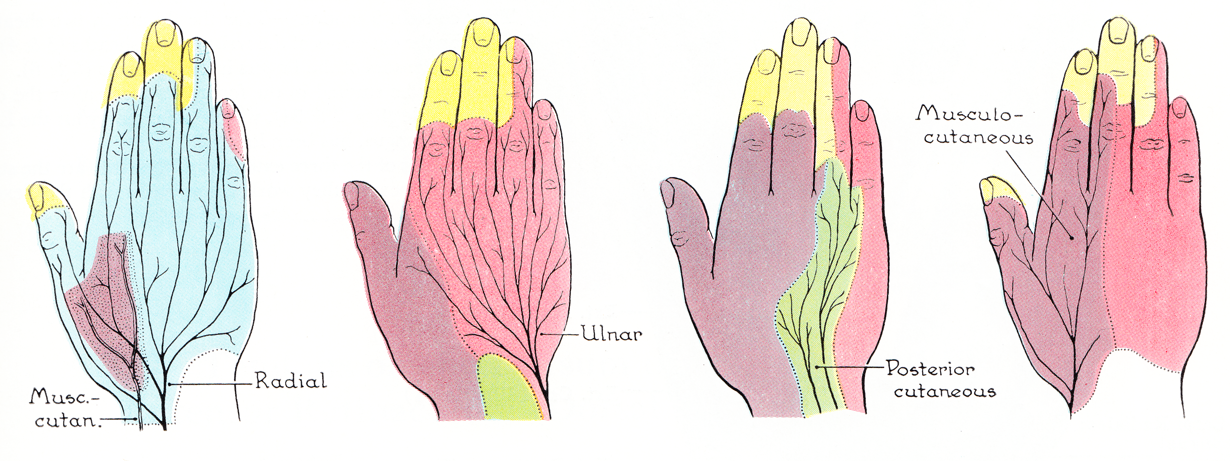
The palmar branch of the median nerve is a branch of the median nerve which arises at the distal part of the forearm. Branches It pierces the palmar carpal ligament, and divides into a lateral and a medial branch; * The ''lateral branch'' supplies the skin over the ball of the thumb, and communicates with the volar branch of the lateral antebrachial cutaneous nerve. * The ''medial branch'' supplies the skin of the Hand, palm and communicates with the palmar cutaneous branch of the ulnar. Clinical significance Unlike most of the median nerve innervation of the hand, the palmar branch travels superficial to the Flexor retinaculum of the hand. Therefore, this portion of the median nerve usually remains functioning during carpal tunnel syndrome. Additional images File:Gray812and814.PNG, Diagram of segmental distribution of the cutaneous nerves of the right upper extremity. References * http://nervesurgery.wustl.edu/NerveImages/Anatomy%20and%20Physiology/AP-Median-Nerve---I ... [...More Info...] [...Related Items...] OR: [Wikipedia] [Google] [Baidu] |
Interosseous Membrane
An interosseous membrane is a thick dense fibrous sheet of connective tissue that spans the space between two bones, forming a type of syndesmosis joint. Interosseous membranes in the human body: * Interosseous membrane of forearm * Interosseous membrane of leg The interosseous membrane of the leg (middle tibiofibular ligament) extends between the interosseous crests of the tibia and fibula, helps stabilize the Tib-Fib relationship and separates the muscles on the front from those on the back of the leg. ... Gallery File:5 ligaments of interosseous membrane of forearm.png, Five ligaments of interosseous membrane of forearm:* Central band (key portion to be reconstructed in case of injury)* Accessory band * Distal oblique bundle * Proximal oblique cord* Dorsal oblique accessory cord Notes External links * * {{Authority control Skeletal system ... [...More Info...] [...Related Items...] OR: [Wikipedia] [Google] [Baidu] |
