|
Vertebral
The vertebral column, also known as the backbone or spine, is part of the axial skeleton. The vertebral column is the defining characteristic of a vertebrate in which the notochord (a flexible rod of uniform composition) found in all chordates has been replaced by a segmented series of bone: vertebrae separated by intervertebral discs. Individual vertebrae are named according to their region and position, and can be used as anatomical landmarks in order to guide procedures such as lumbar punctures. The vertebral column houses the spinal canal, a cavity that encloses and protects the spinal cord. There are about 50,000 species of animals that have a vertebral column. The human vertebral column is one of the most-studied examples. Many different diseases in humans can affect the spine, with spina bifida and scoliosis being recognisable examples. The general structure of human vertebrae is fairly typical of that found in mammals, reptiles, and birds. The shape of the vertebral ... [...More Info...] [...Related Items...] OR: [Wikipedia] [Google] [Baidu] |
Vertebra
The spinal column, a defining synapomorphy shared by nearly all vertebrates, Hagfish are believed to have secondarily lost their spinal column is a moderately flexible series of vertebrae (singular vertebra), each constituting a characteristic irregular bone whose complex structure is composed primarily of bone, and secondarily of hyaline cartilage. They show variation in the proportion contributed by these two tissue types; such variations correlate on one hand with the cerebral/caudal rank (i.e., location within the backbone), and on the other with phylogenetic differences among the vertebrate taxa. The basic configuration of a vertebra varies, but the bone is its ''body'', with the central part of the body constituting the ''centrum''. The upper (closer to) and lower (further from), respectively, the cranium and its central nervous system surfaces of the vertebra body support attachment to the intervertebral discs. The posterior part of a vertebra forms a vertebral ar ... [...More Info...] [...Related Items...] OR: [Wikipedia] [Google] [Baidu] |
Transverse Process
The spinal column, a defining synapomorphy shared by nearly all vertebrates,Hagfish are believed to have secondarily lost their spinal column is a moderately flexible series of vertebrae (singular vertebra), each constituting a characteristic irregular bone whose complex structure is composed primarily of bone, and secondarily of hyaline cartilage. They show variation in the proportion contributed by these two tissue types; such variations correlate on one hand with the cerebral/caudal rank (i.e., location within the backbone), and on the other with phylogenetic differences among the vertebrate taxa. The basic configuration of a vertebra varies, but the bone is its ''body'', with the central part of the body constituting the ''centrum''. The upper (closer to) and lower (further from), respectively, the cranium and its central nervous system surfaces of the vertebra body support attachment to the intervertebral discs. The posterior part of a vertebra forms a vertebral arch ... [...More Info...] [...Related Items...] OR: [Wikipedia] [Google] [Baidu] |
Spinous Process
The spinal column, a defining synapomorphy shared by nearly all vertebrates, Hagfish are believed to have secondarily lost their spinal column is a moderately flexible series of vertebrae (singular vertebra), each constituting a characteristic irregular bone whose complex structure is composed primarily of bone, and secondarily of hyaline cartilage. They show variation in the proportion contributed by these two tissue types; such variations correlate on one hand with the cerebral/caudal rank (i.e., location within the backbone), and on the other with phylogenetic differences among the vertebrate taxa. The basic configuration of a vertebra varies, but the bone is its ''body'', with the central part of the body constituting the ''centrum''. The upper (closer to) and lower (further from), respectively, the cranium and its central nervous system surfaces of the vertebra body support attachment to the intervertebral discs. The posterior part of a vertebra forms a vertebral arch ... [...More Info...] [...Related Items...] OR: [Wikipedia] [Google] [Baidu] |
Intervertebral Disc
An intervertebral disc (or intervertebral fibrocartilage) lies between adjacent vertebrae in the vertebral column. Each disc forms a fibrocartilaginous joint (a symphysis), to allow slight movement of the vertebrae, to act as a ligament to hold the vertebrae together, and to function as a shock absorber for the spine. Structure Intervertebral discs consist of an outer fibrous ring, the anulus fibrosus disci intervertebralis, which surrounds an inner gel-like center, the nucleus pulposus. The ''anulus fibrosus'' consists of several layers (laminae) of fibrocartilage made up of both type I and type II collagen. Type I is concentrated toward the edge of the ring, where it provides greater strength. The stiff laminae can withstand compressive forces. The fibrous intervertebral disc contains the ''nucleus pulposus'' and this helps to distribute pressure evenly across the disc. This prevents the development of stress concentrations which could cause damage to the underlying vert ... [...More Info...] [...Related Items...] OR: [Wikipedia] [Google] [Baidu] |
Spinal Cord
The spinal cord is a long, thin, tubular structure made up of nervous tissue, which extends from the medulla oblongata in the brainstem to the lumbar region of the vertebral column (backbone). The backbone encloses the central canal of the spinal cord, which contains cerebrospinal fluid. The brain and spinal cord together make up the central nervous system (CNS). In humans, the spinal cord begins at the occipital bone, passing through the foramen magnum and then enters the spinal canal at the beginning of the cervical vertebrae. The spinal cord extends down to between the first and second lumbar vertebrae, where it ends. The enclosing bony vertebral column protects the relatively shorter spinal cord. It is around long in adult men and around long in adult women. The diameter of the spinal cord ranges from in the cervical and lumbar regions to in the thoracic area. The spinal cord functions primarily in the transmission of nerve signals from the motor cortex to the ... [...More Info...] [...Related Items...] OR: [Wikipedia] [Google] [Baidu] |
Lumbar Vertebrae
The lumbar vertebrae are, in human anatomy, the five vertebrae between the rib cage and the pelvis. They are the largest segments of the vertebral column and are characterized by the absence of the foramen transversarium within the transverse process (since it is only found in the cervical region) and by the absence of facets on the sides of the body (as found only in the thoracic region). They are designated L1 to L5, starting at the top. The lumbar vertebrae help support the weight of the body, and permit movement. Human anatomy General characteristics The adjacent figure depicts the general characteristics of the first through fourth lumbar vertebrae. The fifth vertebra contains certain peculiarities, which are detailed below. As with other vertebrae, each lumbar vertebra consists of a ''vertebral body'' and a ''vertebral arch''. The vertebral arch, consisting of a pair of ''pedicles'' and a pair of ''laminae'', encloses the ''vertebral foramen'' (opening) and sup ... [...More Info...] [...Related Items...] OR: [Wikipedia] [Google] [Baidu] |
Cervical Vertebrae
In tetrapods, cervical vertebrae (singular: vertebra) are the vertebrae of the neck, immediately below the skull. Truncal vertebrae (divided into thoracic and lumbar vertebrae in mammals) lie caudal (toward the tail) of cervical vertebrae. In sauropsid species, the cervical vertebrae bear cervical ribs. In lizards and saurischian dinosaurs, the cervical ribs are large; in birds, they are small and completely fused to the vertebrae. The vertebral transverse processes of mammals are homologous to the cervical ribs of other amniotes. Most mammals have seven cervical vertebrae, with the only three known exceptions being the manatee with six, the two-toed sloth with five or six, and the three-toed sloth with nine. In humans, cervical vertebrae are the smallest of the true vertebrae and can be readily distinguished from those of the thoracic or lumbar regions by the presence of a foramen (hole) in each transverse process, through which the vertebral artery, vertebral veins, and ... [...More Info...] [...Related Items...] OR: [Wikipedia] [Google] [Baidu] |
Thoracic Vertebrae
In vertebrates, thoracic vertebrae compose the middle segment of the vertebral column, between the cervical vertebrae and the lumbar vertebrae. In humans, there are twelve thoracic vertebrae and they are intermediate in size between the cervical and lumbar vertebrae; they increase in size going towards the lumbar vertebrae, with the lower ones being much larger than the upper. They are distinguished by the presence of facets on the sides of the bodies for articulation with the heads of the ribs, as well as facets on the transverse processes of all, except the eleventh and twelfth, for articulation with the tubercles of the ribs. By convention, the human thoracic vertebrae are numbered T1–T12, with the first one (T1) located closest to the skull and the others going down the spine toward the lumbar region. General characteristics These are the general characteristics of the second through eighth thoracic vertebrae. The first and ninth through twelfth vertebrae contain certa ... [...More Info...] [...Related Items...] OR: [Wikipedia] [Google] [Baidu] |
Sacrum
The sacrum (plural: ''sacra'' or ''sacrums''), in human anatomy, is a large, triangular bone at the base of the spine that forms by the fusing of the sacral vertebrae (S1S5) between ages 18 and 30. The sacrum situates at the upper, back part of the pelvic cavity, between the two wings of the pelvis. It forms joints with four other bones. The two projections at the sides of the sacrum are called the alae (wings), and articulate with the ilium at the L-shaped sacroiliac joints. The upper part of the sacrum connects with the last lumbar vertebra (L5), and its lower part with the coccyx (tailbone) via the sacral and coccygeal cornua. The sacrum has three different surfaces which are shaped to accommodate surrounding pelvic structures. Overall it is concave (curved upon itself). The base of the sacrum, the broadest and uppermost part, is tilted forward as the sacral promontory internally. The central part is curved outward toward the posterior, allowing greater room for the p ... [...More Info...] [...Related Items...] OR: [Wikipedia] [Google] [Baidu] |
Notochord
In anatomy, the notochord is a flexible rod which is similar in structure to the stiffer cartilage. If a species has a notochord at any stage of its life cycle (along with 4 other features), it is, by definition, a chordate. The notochord consists of inner, vacuolated cells covered by fibrous and elastic sheaths, lies along the anteroposterior axis (''front to back''), is usually closer to the dorsal than the ventral surface of the embryo, and is composed of cells derived from the mesoderm. The most commonly cited functions of the notochord are: as a midline tissue that provides directional signals to surrounding tissue during development, as a skeletal (structural) element, and as a vertebral precursor. In lancelets the notochord persists throughout life as the main structural support of the body. In tunicates the notochord is present only in the larval stage, being completely absent in the adult animal. In these invertebrate chordates, the notochord is not vacuolated. I ... [...More Info...] [...Related Items...] OR: [Wikipedia] [Google] [Baidu] |
Axis (anatomy)
In anatomy, the axis (from Latin ''axis'', "axle") or epistropheus is the second cervical vertebra (C2) of the spine, immediately inferior to the atlas, upon which the head rests. The axis' defining feature is its strong odontoid process (bony protrusion) known as the dens, which rises dorsally from the rest of the bone. Structure The body is deeper in front or in the back and is prolonged downward anteriorly to overlap the upper and front part of the third vertebra. It presents a median longitudinal ridge in front, separating two lateral depressions for the attachment of the longus colli muscles. Odontoid Process of Axis (Dens) The dens, also called the odontoid process or the peg, is the most pronounced projecting feature of the axis. The dens exhibits a slight constriction where it joins the main body of the vertebra. The condition where the dens is separated from the body of the axis is called ''os odontoideum'' and may cause nerve and circulation compression syndrome. ... [...More Info...] [...Related Items...] OR: [Wikipedia] [Google] [Baidu] |
Atlas (anatomy)
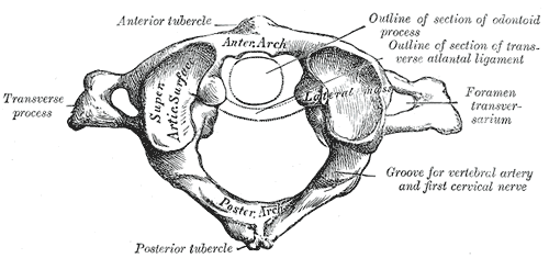
In anatomy, the atlas (C1) is the most superior (first) cervical vertebra of the spine and is located in the neck. It is named for Atlas of Greek mythology because, just as Atlas supported the globe, it supports the entire head. The atlas is the topmost vertebra and, with the axis (the vertebra below it), forms the joint connecting the skull and spine. The atlas and axis are specialized to allow a greater range of motion than normal vertebrae. They are responsible for the nodding and rotation movements of the head. The atlanto-occipital joint allows the head to nod up and down on the vertebral column. The dens acts as a pivot that allows the atlas and attached head to rotate on the axis, side to side. The atlas's chief peculiarity is that it has no body. It is ring-like and consists of an anterior and a posterior arch and two lateral masses. The atlas and axis are important neurologically because the brainstem extends down to the axis. Structure Anterior arch The anter ... [...More Info...] [...Related Items...] OR: [Wikipedia] [Google] [Baidu] |


