|
Ventricular Aneurysm
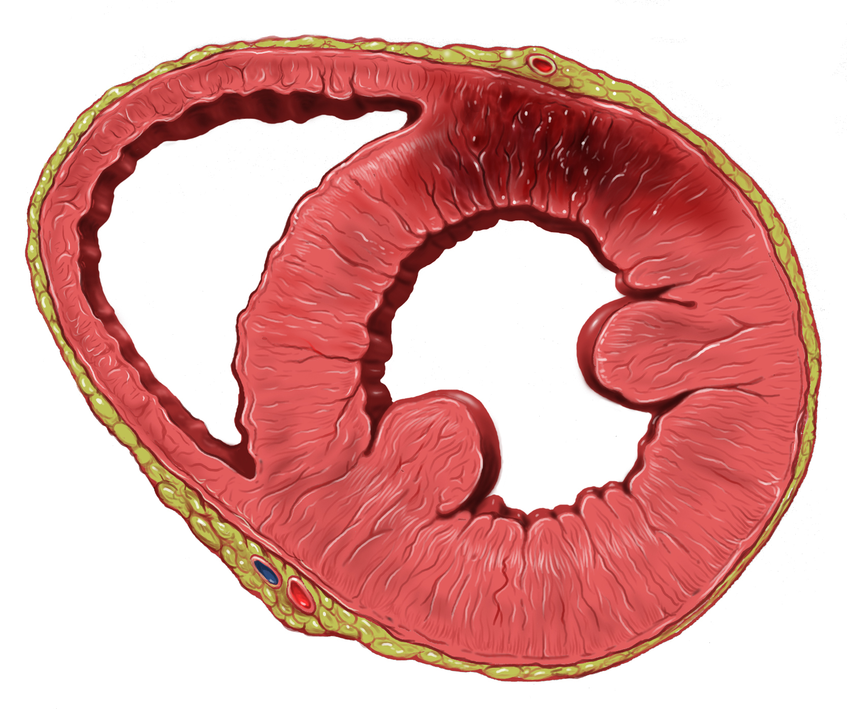
Ventricular aneurysms are one of the many complications that may occur after a heart attack. The word aneurysm refers to a bulge or 'pocketing' of the wall or lining of a vessel commonly occurring in the blood vessels at the base of the septum, or within the aorta. In the heart, they usually arise from a patch of weakened tissue in a ventricular wall, which swells into a bubble filled with blood. This, in turn, may block the passageways leading out of the heart, leading to severely constricted blood flow to the body. Ventricular aneurysms can be fatal. They are usually non-rupturing because they are lined by scar tissue. A left ventricular aneurysm can be associated with ST elevation. Signs and symptoms Ventricular aneurysms usually grow at a very slow pace, but can still pose problems. Usually, this type of aneurysm grows in the left ventricle. This bubble has the potential to block blood flow to the rest of the body, and thus limit the patient's stamina. In other cases, a s ... [...More Info...] [...Related Items...] OR: [Wikipedia] [Google] [Baidu] |
Myocardial Infarction
A myocardial infarction (MI), commonly known as a heart attack, occurs when Ischemia, blood flow decreases or stops in one of the coronary arteries of the heart, causing infarction (tissue death) to the heart muscle. The most common symptom is retrosternal Angina, chest pain or discomfort that classically radiates to the left shoulder, arm, or jaw. The pain may occasionally feel like heartburn. This is the dangerous type of acute coronary syndrome. Other symptoms may include shortness of breath, nausea, presyncope, feeling faint, a diaphoresis, cold sweat, Fatigue, feeling tired, and decreased level of consciousness. About 30% of people have atypical symptoms. Women more often present without chest pain and instead have neck pain, arm pain or feel tired. Among those over 75 years old, about 5% have had an MI with little or no history of symptoms. An MI may cause heart failure, an Cardiac arrhythmia, irregular heartbeat, cardiogenic shock or cardiac arrest. Most MIs occur d ... [...More Info...] [...Related Items...] OR: [Wikipedia] [Google] [Baidu] |
Myocardial Rupture
Myocardial rupture is a laceration of the ventricles or atria of the heart, of the interatrial or interventricular septum, or of the papillary muscles. It is most commonly seen as a serious sequela of an acute myocardial infarction (heart attack). It can also be caused by trauma. Signs and symptoms Symptoms of myocardial rupture are recurrent or persistent chest pain, syncope, and distension of jugular vein. Sudden death caused by a myocardial rupture is sometimes preceded by no symptoms. Causes The most common cause of myocardial rupture is a recent myocardial infarction, with the rupture typically occurring three to five days after infarction.Figueras J, Alcalde O, Barrabes JA, Serra V, Alguersuari J, Cortadellas J, et al. Changes in Hospital Mortality Rates in 425 Patients with Acute ST-Elevation Myocardial Infarction and Cardiac Rupure Over a 30-Year Period. Circulation. 2008;118:2783–9. Other causes of rupture include cardiac trauma, endocarditis (infection of the ... [...More Info...] [...Related Items...] OR: [Wikipedia] [Google] [Baidu] |
Coronary Artery Aneurysm
Coronary artery aneurysm is an abnormal dilatation of part of the coronary artery. This rare disorder occurs in about 0.3–4.9% of patients who undergo coronary angiography. Signs and symptoms The majority of individuals suffering from coronary artery aneurysms do not exhibit any symptoms; the development of complications or concurrent atherosclerotic coronary artery disease is what causes clinical manifestations to occur. The most common complications include coronary spasm, distal embolization, aneurysm rupture, local thrombosis, and compression of surrounding structures due to massive enlargement of coronary artery aneurysm. Causes Acquired causes include atherosclerosis in adults, Kawasaki disease in children and coronary catheterization. With the invention of drug eluting stents, there has been more cases implying stents lead to coronary aneurysms. The pathophysiology, although not completely understood, might be comparable to that of aneurysms of larger vessels. This ... [...More Info...] [...Related Items...] OR: [Wikipedia] [Google] [Baidu] |
Ventricular Reduction
Ventriculectomy, or ventricular reduction, is a type of operation in cardiac surgery to reduce enlargement of the heart from cardiomyopathy Cardiomyopathy is a group of primary diseases of the heart muscle. Early on there may be few or no symptoms. As the disease worsens, shortness of breath, feeling tired, and swelling of the legs may occur, due to the onset of heart failure. A ... or ischemic aneurysm formation. In these procedures, part of the ventricular wall is resected. A Batista procedure is a partial left ventriculectomy that is used to treat advanced heart failure. This procedure is not widely used because outcomes are often unsatisfactory. See also * Dor procedure References Cardiac surgery {{Surgery-stub ... [...More Info...] [...Related Items...] OR: [Wikipedia] [Google] [Baidu] |
Pericardiocentesis
Pericardiocentesis (PCC), also called pericardial tap, is a medical procedure where fluid is aspirated from the pericardium (the sac enveloping the heart). Anatomy and physiology The pericardium is a fibrous sac surrounding the heart composed of two layers: an inner visceral pericardium and an outer parietal pericardium. The area between these two layers is known as the pericardial space and normally contains 15 to 50 mL of serous fluid. This fluid protects the heart by serving as a shock absorber and provides lubrication to the heart during contraction. The elastic nature of the pericardium allows it to accommodate a small amount of extra fluid, roughly 80 to 120 mL, in the acute setting. However, once a critical volume is reached, even small amounts of extra fluid can rapidly increase pressure within the pericardium. This pressure can significantly hinder the ability of the heart to contract, leading to cardiac tamponade. If accumulation of fluid is slow and occurs over ... [...More Info...] [...Related Items...] OR: [Wikipedia] [Google] [Baidu] |
Trimesters
Pregnancy is the time during which one or more offspring gestates inside a woman's uterus. A multiple pregnancy involves more than one offspring, such as with twins. Conception usually occurs following vaginal intercourse, but can also occur through assisted reproductive technology procedures. A pregnancy may end in a live birth, a miscarriage, an induced abortion, or a stillbirth. Childbirth typically occurs around 40 weeks from the start of the last menstrual period (LMP), a span known as the ''gestational age''; this is just over nine months. Counting by ''fertilization age'', the length is about 38 weeks. Implantation occurs on average 8–9 days after fertilization. An ''embryo'' is the term for the developing offspring during the first seven weeks following implantation (i.e. ten weeks' gestational age), after which the term ''fetus'' is used until the birth of a ''baby''. Signs and symptoms of early pregnancy may include missed periods, tender b ... [...More Info...] [...Related Items...] OR: [Wikipedia] [Google] [Baidu] |
Aneurysm
An aneurysm is an outward :wikt:bulge, bulging, likened to a bubble or balloon, caused by a localized, abnormal, weak spot on a blood vessel wall. Aneurysms may be a result of a hereditary condition or an acquired disease. Aneurysms can also be a wikt:Special:Search/nidus, nidus (starting point) for clot formation (thrombosis) and Embolism, embolization. As an aneurysm increases in size, the risk of rupture increases, which could lead to uncontrolled bleeding. Although they may occur in any blood vessel, particularly lethal examples include aneurysms of the circle of Willis in the brain, aortic aneurysms affecting the thoracic aorta, and abdominal aortic aneurysms. Aneurysms can arise in the heart itself following a Myocardial infarction, heart attack, including both Ventricular aneurysm, ventricular and atrial septal aneurysms. There are congenital atrial septal defect, atrial septal aneurysms, a rare heart defect. Etymology The word is from Greek language, Greek: ἀνεύρ� ... [...More Info...] [...Related Items...] OR: [Wikipedia] [Google] [Baidu] |
Echocardiography
Echocardiography, also known as cardiac ultrasound, is the use of ultrasound to examine the heart. It is a type of medical imaging, using standard ultrasound or Doppler ultrasound. The visual image formed using this technique is called an echocardiogram, a cardiac echo, or simply an echo. Echocardiography is routinely used in the diagnosis, management, and follow-up of patients with any suspected or known heart diseases. It is one of the most widely used diagnostic imaging modalities in cardiology. It can provide a wealth of helpful information, including the size and shape of the heart (internal chamber size quantification), pumping capacity, location and extent of any tissue damage, and assessment of valves. An echocardiogram can also give physicians other estimates of heart function, such as a calculation of the cardiac output, ejection fraction, and diastolic function (how well the heart relaxes). Echocardiography is an important tool in assessing wall motion abnorma ... [...More Info...] [...Related Items...] OR: [Wikipedia] [Google] [Baidu] |
Congenital Heart Defect
A congenital heart defect (CHD), also known as a congenital heart anomaly, congenital cardiovascular malformation, and congenital heart disease, is a defect in the structure of the heart or great vessels that is present at birth. A congenital heart defect is classed as a cardiovascular disease. Signs and symptoms depend on the specific type of defect. Symptoms can vary from none to life-threatening. When present, symptoms are variable and may include rapid breathing, bluish skin (cyanosis), poor weight gain, and feeling tired. CHD does not cause chest pain. Most congenital heart defects are not associated with other diseases. A complication of CHD is heart failure. Congenital heart defects are the most common birth defect. In 2015, they were present in 48.9 million people globally. They affect between 4 and 75 per 1,000 live births, depending upon how they are diagnosed. In about 6 to 19 per 1,000 they cause a moderate to severe degree of problems. Congenital heart defects are t ... [...More Info...] [...Related Items...] OR: [Wikipedia] [Google] [Baidu] |
Cardiac
The heart is a muscular organ found in humans and other animals. This organ pumps blood through the blood vessels. The heart and blood vessels together make the circulatory system. The pumped blood carries oxygen and nutrients to the tissue, while carrying metabolic waste such as carbon dioxide to the lungs. In humans, the heart is approximately the size of a closed fist and is located between the lungs, in the middle compartment of the chest, called the mediastinum. In humans, the heart is divided into four chambers: upper left and right atria and lower left and right ventricles. Commonly, the right atrium and ventricle are referred together as the right heart and their left counterparts as the left heart. In a healthy heart, blood flows one way through the heart due to heart valves, which prevent backflow. The heart is enclosed in a protective sac, the pericardium, which also contains a small amount of fluid. The wall of the heart is made up of three layers: epicardi ... [...More Info...] [...Related Items...] OR: [Wikipedia] [Google] [Baidu] |
Coronary Artery Aneurysm
Coronary artery aneurysm is an abnormal dilatation of part of the coronary artery. This rare disorder occurs in about 0.3–4.9% of patients who undergo coronary angiography. Signs and symptoms The majority of individuals suffering from coronary artery aneurysms do not exhibit any symptoms; the development of complications or concurrent atherosclerotic coronary artery disease is what causes clinical manifestations to occur. The most common complications include coronary spasm, distal embolization, aneurysm rupture, local thrombosis, and compression of surrounding structures due to massive enlargement of coronary artery aneurysm. Causes Acquired causes include atherosclerosis in adults, Kawasaki disease in children and coronary catheterization. With the invention of drug eluting stents, there has been more cases implying stents lead to coronary aneurysms. The pathophysiology, although not completely understood, might be comparable to that of aneurysms of larger vessels. This ... [...More Info...] [...Related Items...] OR: [Wikipedia] [Google] [Baidu] |
Ventricle (heart)
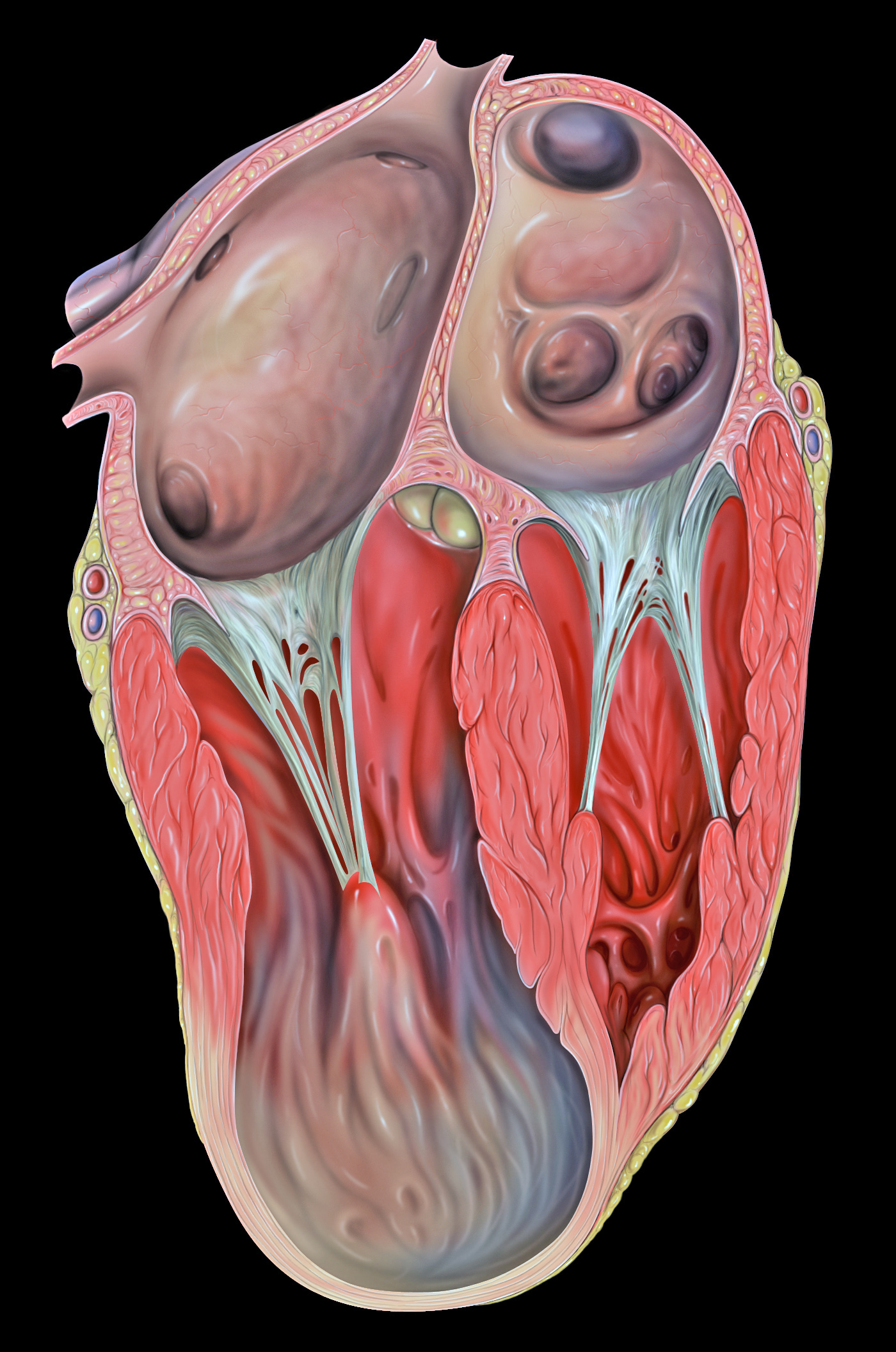
A ventricle is one of two large chambers located toward the bottom of the heart that collect and expel blood towards the peripheral beds within the body and lungs. The blood pumped by a ventricle is supplied by an atrium, an adjacent chamber in the upper heart that is smaller than a ventricle. Interventricular means between the ventricles (for example the interventricular septum), while intraventricular means within one ventricle (for example an intraventricular block). In a four-chambered heart, such as that in humans, there are two ventricles that operate in a double circulatory system: the right ventricle pumps blood into the pulmonary circulation to the lungs, and the left ventricle pumps blood into the systemic circulation through the aorta. Structure Ventricles have thicker walls than atria and generate higher blood pressures. The physiological load on the ventricles requiring pumping of blood throughout the body and lungs is much greater than the pressure generated by ... [...More Info...] [...Related Items...] OR: [Wikipedia] [Google] [Baidu] |


