|
Visual Phototransduction
Visual phototransduction is the sensory transduction process of the visual system by which light is detected by photoreceptor cells ( rods and cones) in the vertebrate retina. A photon is absorbed by a retinal chromophore (each bound to an opsin), which initiates a signal cascade through several intermediate cells, then through the retinal ganglion cells (RGCs) comprising the optic nerve. Overview Light enters the eye, passes through the optical media, then the inner neural layers of the retina before finally reaching the photoreceptor cells in the outer layer of the retina. The light may be absorbed by a chromophore bound to an opsin, which photoisomerizes the chromophore, initiating both the visual cycle, which "resets" the chromophore, and the phototransduction cascade, which transmits the visual signal to the brain. The cascade begins with graded polarisation (an analog signal) of the excited photoreceptor cell, as its membrane potential increases from a resting po ... [...More Info...] [...Related Items...] OR: [Wikipedia] [Google] [Baidu] |
Transduction (physiology)
In physiology, transduction is the translation of arriving stimulus into an action potential by a sensory receptor. It begins when stimulus changes the membrane potential of a sensory receptor. A sensory receptor converts the energy in a stimulus into an electrical signal. Receptors are broadly split into two main categories: exteroceptors, which receive external sensory stimuli, and interoceptors, which receive internal sensory stimuli. Sensory transduction The visual system In the visual system, sensory cells called Rod cell, rod and cone cells in the retina convert the physical energy of light signals into electrical impulses that travel to the brain. The light causes a conformational change in a protein called rhodopsin. This conformational change sets in motion a series of molecular events that result in a reduction of the electrochemical gradient of the photoreceptor. The decrease in the electrochemical gradient causes a reduction in the electrical signals going to the br ... [...More Info...] [...Related Items...] OR: [Wikipedia] [Google] [Baidu] |
Synaptic Terminal
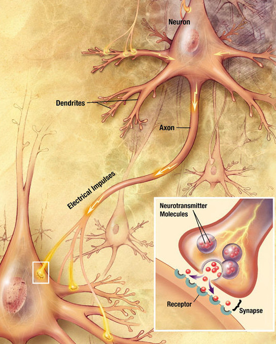
Chemical synapses are biological junctions through which neurons' signals can be sent to each other and to non-neuronal cells such as those in muscles or glands. Chemical synapses allow neurons to form circuits within the central nervous system. They are crucial to the biological computations that underlie perception and thought. They allow the nervous system to connect to and control other systems of the body. At a chemical synapse, one neuron releases neurotransmitter molecules into a small space (the synaptic cleft) that is adjacent to another neuron. The neurotransmitters are contained within small sacs called synaptic vesicles, and are released into the synaptic cleft by exocytosis. These molecules then bind to neurotransmitter receptors on the postsynaptic cell. Finally, the neurotransmitters are cleared from the synapse through one of several potential mechanisms including enzymatic degradation or re-uptake by specific transporters either on the presynaptic cell or ... [...More Info...] [...Related Items...] OR: [Wikipedia] [Google] [Baidu] |
Trichromatic
Trichromacy or trichromatism is the possession of three independent channels for conveying color information, derived from the three different types of cone cells in the eye. Organisms with trichromacy are called trichromats. The normal explanation of trichromacy is that the organism's retina contains three types of color receptors (called cone cells in vertebrates) with different absorption spectra. In actuality, the number of such receptor types may be greater than three, since different types may be active at different light intensities. In vertebrates with three types of cone cells, at low light intensities the rod cells may contribute to color vision. Humans and other animals that are trichromats Humans and some other mammals have evolved trichromacy based partly on pigments inherited from early vertebrates. In fish and birds, for example, four pigments are used for vision. These extra cone receptor visual pigments detect energy of other wavelengths, sometimes includin ... [...More Info...] [...Related Items...] OR: [Wikipedia] [Google] [Baidu] |
Wavelength
In physics and mathematics, wavelength or spatial period of a wave or periodic function is the distance over which the wave's shape repeats. In other words, it is the distance between consecutive corresponding points of the same ''phase (waves), phase'' on the wave, such as two adjacent crests, troughs, or zero crossings. Wavelength is a characteristic of both traveling waves and standing waves, as well as other spatial wave patterns. The multiplicative inverse, inverse of the wavelength is called the ''spatial frequency''. Wavelength is commonly designated by the Greek letter lambda (''λ''). For a modulated wave, ''wavelength'' may refer to the carrier wavelength of the signal. The term ''wavelength'' may also apply to the repeating envelope (mathematics), envelope of modulated waves or waves formed by Interference (wave propagation), interference of several sinusoids. Assuming a sinusoidal wave moving at a fixed phase velocity, wave speed, wavelength is inversely proportion ... [...More Info...] [...Related Items...] OR: [Wikipedia] [Google] [Baidu] |
Vitamin A
Vitamin A is a fat-soluble vitamin that is an essential nutrient. The term "vitamin A" encompasses a group of chemically related organic compounds that includes retinol, retinyl esters, and several provitamin (precursor) carotenoids, most notably Β-Carotene, β-carotene (''beta''-carotene). Vitamin A has multiple functions: growth during embryo development, maintaining the immune system, and healthy vision. For aiding vision specifically, it combines with the protein opsin to form rhodopsin, the light-absorbing molecule necessary for both low-light (scotopic vision) and color vision. Vitamin A occurs as two principal forms in foods: A) retinoids, found in Animal source foods, animal-sourced foods, either as retinol or bound to a fatty acid to become a retinyl ester, and B) the carotenoids Α-Carotene, α-carotene (''alpha''-carotene), β-carotene, Γ-Carotene, γ-carotene (''gamma''-carotene), and the xanthophyll beta-cryptoxanthin (all of which contain β-ionone rings) that ... [...More Info...] [...Related Items...] OR: [Wikipedia] [Google] [Baidu] |
Aldehyde
In organic chemistry, an aldehyde () (lat. ''al''cohol ''dehyd''rogenatum, dehydrogenated alcohol) is an organic compound containing a functional group with the structure . The functional group itself (without the "R" side chain) can be referred to as an aldehyde but can also be classified as a formyl group. Aldehydes are a common motif in many chemicals important in technology and biology. Structure and bonding Aldehyde molecules have a central carbon atom that is connected by a double bond to oxygen, a single bond to hydrogen and another single bond to a third substituent, which is carbon or, in the case of formaldehyde, hydrogen. The central carbon is often described as being sp2- hybridized. The aldehyde group is somewhat polar. The bond length is about 120–122 picometers. Physical properties and characterization Aldehydes have properties that are diverse and that depend on the remainder of the molecule. Smaller aldehydes such as formaldehyde and acetaldehyde are solubl ... [...More Info...] [...Related Items...] OR: [Wikipedia] [Google] [Baidu] |
11-cis Retinal
Retinal (also known as retinaldehyde) is a polyene chromophore. Retinal, bound to proteins called opsins, is the chemical basis of visual phototransduction, the light-detection stage of visual perception (vision). Some microorganisms use retinal to convert light into metabolic energy. One study suggests that approximately three billion years ago, most living organisms on Earth used retinal, rather than chlorophyll, to convert sunlight into energy. Because retinal absorbs mostly green light and transmits purple light, this gave rise to the Purple Earth hypothesis. Retinal itself is considered to be a form of vitamin A when eaten by an animal. There are many forms of vitamin A, all of which are converted to retinal, which cannot be made without them. The number of different molecules that can be converted to retinal varies from species to species. Retinal was originally called retinene, and was renamed after it was discovered to be vitamin A aldehyde. Vertebrate animals ingest ... [...More Info...] [...Related Items...] OR: [Wikipedia] [Google] [Baidu] |
Photosensitive Ganglion Cell
Intrinsically photosensitive retinal ganglion cells (ipRGCs), also called photosensitive retinal ganglion cells (pRGC), or melanopsin-containing retinal ganglion cells (mRGCs), are a type of neuron in the retina of the mammalian eye. The presence of an additional photoreceptor was first suspected in 1927 when mice lacking rod and cone cells still responded to changing light levels through pupil constriction; this suggested that rods and cones are not the only light-sensitive tissue. However, it was unclear whether this light sensitivity arose from an additional retinal photoreceptor or elsewhere in the body. Recent research has shown that these retinal ganglion cells, unlike other retinal ganglion cells, are intrinsically photosensitive due to the presence of melanopsin, a light-sensitive protein. Therefore, they constitute a third class of photoreceptors, in addition to rod and cone cells. Overview Compared to the rods and cones, the ipRGCs respond more sluggishly and ... [...More Info...] [...Related Items...] OR: [Wikipedia] [Google] [Baidu] |
Rod Cell
Rod cells are photoreceptor cells in the retina of the eye that can function in lower light better than the other type of visual photoreceptor, cone cells. Rods are usually found concentrated at the outer edges of the retina and are used in peripheral vision. On average, there are approximately 92 million rod cells (vs ~4.6 million cones) in the human retina. Rod cells are more sensitive than cone cells and are almost entirely responsible for night vision. However, rods have little role in color vision, which is the main reason why colors are much less apparent in dim light. Structure Rods are a little longer and leaner than cones but have the same basic structure. Opsin-containing disks lie at the end of the cell adjacent to the retinal pigment epithelium, which in turn is attached to the inside of the eye. The stacked-disc structure of the detector portion of the cell allows for very high efficiency. Rods are much more common than cones, with about 120 million rod cells ... [...More Info...] [...Related Items...] OR: [Wikipedia] [Google] [Baidu] |
Action Potential
An action potential (also known as a nerve impulse or "spike" when in a neuron) is a series of quick changes in voltage across a cell membrane. An action potential occurs when the membrane potential of a specific Cell (biology), cell rapidly rises and falls. This depolarization then causes adjacent locations to similarly depolarize. Action potentials occur in several types of Membrane potential#Cell excitability, excitable cells, which include animal cells like neurons and myocyte, muscle cells, as well as some plant cells. Certain endocrine cells such as pancreatic beta cells, and certain cells of the anterior pituitary gland are also excitable cells. In neurons, action potentials play a central role in cell–cell interaction, cell–cell communication by providing for—or with regard to saltatory conduction, assisting—the propagation of signals along the neuron's axon toward axon terminal, synaptic boutons situated at the ends of an axon; these signals can then connect wit ... [...More Info...] [...Related Items...] OR: [Wikipedia] [Google] [Baidu] |
Receptive Field
The receptive field, or sensory space, is a delimited medium where some physiological stimuli can evoke a sensory neuronal response in specific organisms. Complexity of the receptive field ranges from the unidimensional chemical structure of odorants to the multidimensional spacetime of human visual field, through the bidimensional skin surface, being a receptive field for touch perception. Receptive fields can positively or negatively alter the membrane potential with or without affecting the rate of action potentials. A sensory space can be dependent of an animal's location. For a particular sound wave traveling in an appropriate transmission medium, by means of sound localization, an auditory space would amount to a reference system that continuously shifts as the animal moves (taking into consideration the space inside the ears as well). Conversely, receptive fields can be largely independent of the animal's location, as in the case of place cells. A sensory space can also ... [...More Info...] [...Related Items...] OR: [Wikipedia] [Google] [Baidu] |
Amacrine Cell
In the anatomy of the eye, amacrine cells are interneurons in the retina. They are named , because of their short neuronal processes. Amacrine cells are inhibitory neurons which project their dendritic arbors onto the inner plexiform layer (IPL). They interact with retinal ganglion cells and bipolar cells. Structure Amacrine cells operate at the inner plexiform layer (IPL), the second synaptic retinal layer where bipolar cells and retinal ganglion cells form synapses. There are at least 33 different subtypes of amacrine cells based just on their dendrite morphology and stratification. Like horizontal cells, amacrine cells work laterally, but whereas horizontal cells are connected to the output of rod and cone cells, amacrine cells affect the output from bipolar cells, and are often more specialized. Each type of amacrine cell releases one or several neurotransmitters where it connects with other cells. They are often classified by the width of their field of connection, ... [...More Info...] [...Related Items...] OR: [Wikipedia] [Google] [Baidu] |


