|
Tip Link
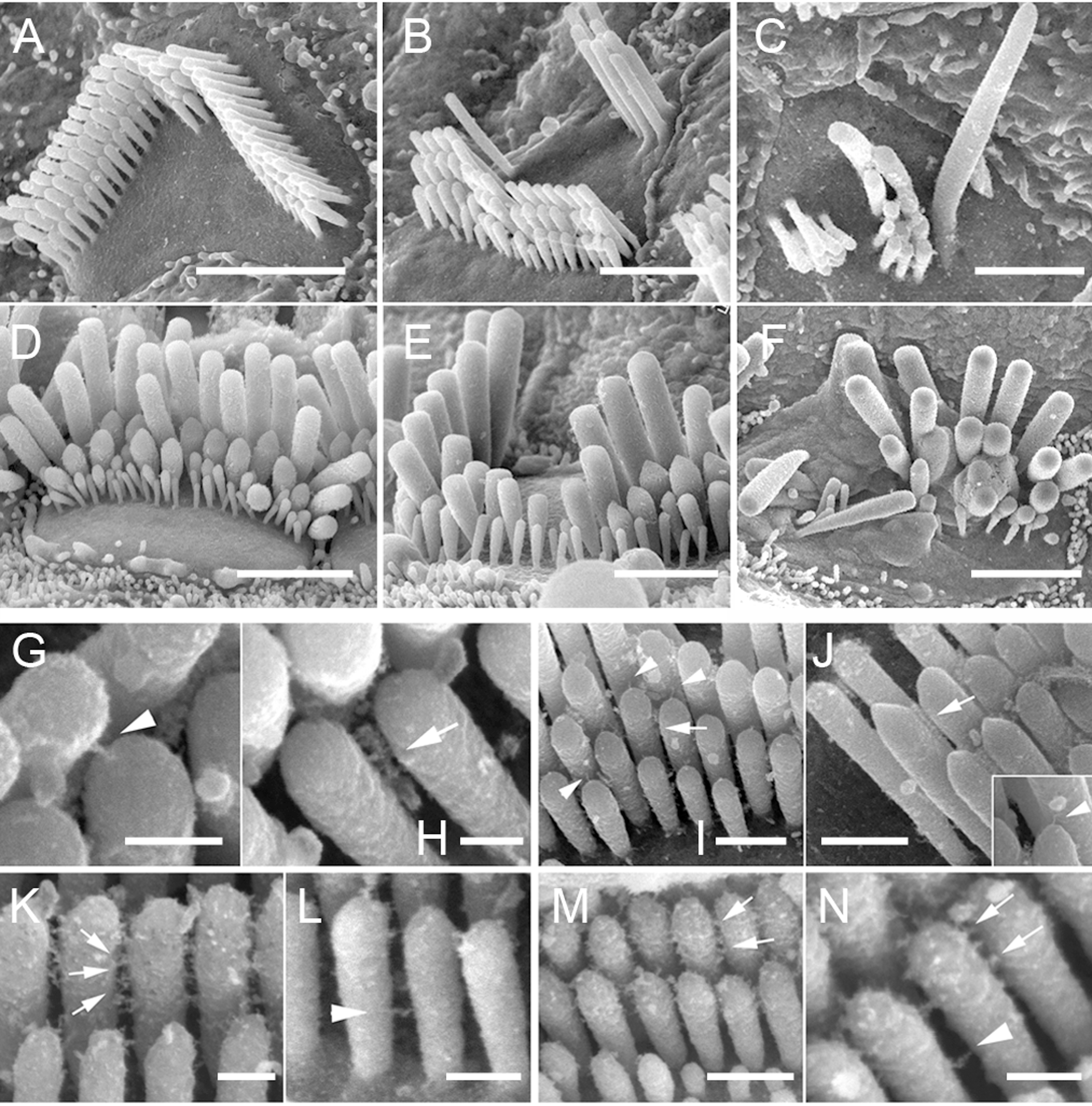
Tip links are extracellular filaments that connect stereocilia to each other or to the kinocilium in the hair cells of the inner ear.Pickles JO, Comis SD, Osborne MP. 1984.Cross-links between stereocilia in the guinea pig organ of Corti, and their possible relation to sensory transduction. Hearing Research 15:103-112. Mechanotransduction is thought to occur at the site of the tip links, which connect to spring-gated ion channels. These channels are cation-selective transduction channels that allow potassium and calcium ions to enter the hair cell from the endolymph that bathes its apical end. When the hair cells are deflected toward the kinocilium, depolarization occurs; when deflection is away from the kinocilium, hyperpolarization occurs. The tip link is made of two different cadherin molecules, protocadherin 15 and cadherin 23.Lewin GR, Moshourab R. 2004. Mechanosensation and pain. Journal of Neurobiology 61:30-44 It has been found that the tip links are relatively stiff, so ... [...More Info...] [...Related Items...] OR: [Wikipedia] [Google] [Baidu] |
The Tailchaser Mutation Does Not Affect Formation Of Interstereocilial Links
''The'' () is a grammatical article in English, denoting persons or things already mentioned, under discussion, implied or otherwise presumed familiar to listeners, readers, or speakers. It is the definite article in English. ''The'' is the most frequently used word in the English language; studies and analyses of texts have found it to account for seven percent of all printed English-language words. It is derived from gendered articles in Old English which combined in Middle English and now has a single form used with pronouns of any gender. The word can be used with both singular and plural nouns, and with a noun that starts with any letter. This is different from many other languages, which have different forms of the definite article for different genders or numbers. Pronunciation In most dialects, "the" is pronounced as (with the voiced dental fricative followed by a schwa) when followed by a consonant sound, and as (homophone of pronoun ''thee'') when followed by a ... [...More Info...] [...Related Items...] OR: [Wikipedia] [Google] [Baidu] |
Stereocilia
Stereocilia (or stereovilli or villi) are non-motile apical cell modifications. They are distinct from cilia and microvilli, but are closely related to microvilli. They form single "finger-like" projections that may be branched, with normal cell membrane characteristics. They contain actin. Stereocilia are found in the vas deferens, the epididymis, and the sensory cells of the inner ear. Structure Stereocilia are cylindrical and non-motile. They are much longer and thicker than microvilli, form single "finger-like" projections that may be branched, and have more of the characteristics of the cellular membrane proper. Like microvilli, they contain actin and lack an axoneme. This distinguishes them from cilia. They do not have a Basal body at their base since they do not contain microtubules. They may or may not be covered by a glycocalyx coating. They have no fixed arrangement, different to the structure present in kinocilium. Function Stereocilia are found in: *the vas ... [...More Info...] [...Related Items...] OR: [Wikipedia] [Google] [Baidu] |
Kinocilium
A kinocilium is a special type of cilium on the apex of hair cells located in the sensory epithelium of the vertebrate inner ear. Anatomy in humans Kinocilia are found on the apical surface of hair cells and are involved in both the morphogenesis of the hair bundle and mechanotransduction. Vibrations (either by movement or sound waves) cause displacement of the hair bundle, resulting in depolarization or hyperpolarization of the hair cell. The depolarization of the hair cells in both instances causes signal transduction via neurotransmitter release. Role in hair bundle morphogenesis Each hair cell has a single, microtubular kinocilium. Before morphogenesis of the hair bundle, the kinocilium is found in the center of the apical surface of the hair cell surrounded by 20-300 microvilli. During hair bundle morphogenesis, the kinocilium moves to the cell periphery dictating hair bundle orientation. As the kinocilium does not move, microvilli surrounding it begin to elongate and form ac ... [...More Info...] [...Related Items...] OR: [Wikipedia] [Google] [Baidu] |
Inner Ear
The inner ear (internal ear, auris interna) is the innermost part of the vertebrate ear. In vertebrates, the inner ear is mainly responsible for sound detection and balance. In mammals, it consists of the bony labyrinth, a hollow cavity in the temporal bone of the skull with a system of passages comprising two main functional parts: * The cochlea, dedicated to hearing; converting sound pressure patterns from the outer ear into electrochemical impulses which are passed on to the brain via the auditory nerve. * The vestibular system, dedicated to balance The inner ear is found in all vertebrates, with substantial variations in form and function. The inner ear is innervated by the eighth cranial nerve in all vertebrates. Structure The labyrinth can be divided by layer or by region. Bony and membranous labyrinths The bony labyrinth, or osseous labyrinth, is the network of passages with bony walls lined with periosteum. The three major parts of the bony labyrinth are the ve ... [...More Info...] [...Related Items...] OR: [Wikipedia] [Google] [Baidu] |
Mechanotransduction
In cellular biology, mechanotransduction ('' mechano'' + '' transduction'') is any of various mechanisms by which cells convert mechanical stimulus into electrochemical activity. This form of sensory transduction is responsible for a number of senses and physiological processes in the body, including proprioception, touch, balance, and hearing. The basic mechanism of mechanotransduction involves converting mechanical signals into electrical or chemical signals. In this process, a mechanically gated ion channel makes it possible for sound, pressure, or movement to cause a change in the excitability of specialized sensory cells and sensory neurons. The stimulation of a mechanoreceptor causes mechanically sensitive ion channels to open and produce a transduction current that changes the membrane potential of the cell. Typically the mechanical stimulus gets filtered in the conveying medium before reaching the site of mechanotransduction. Cellular responses to mechanotransduction ... [...More Info...] [...Related Items...] OR: [Wikipedia] [Google] [Baidu] |
Endolymph
Endolymph is the fluid contained in the membranous labyrinth of the inner ear. The major cation in endolymph is potassium, with the values of sodium and potassium concentration in the endolymph being 0.91 mM and 154 mM, respectively. It is also called ''Scarpa's fluid'', after Antonio Scarpa. Structure The inner ear has two parts: the bony labyrinth and the membranous labyrinth. The membranous labyrinth is contained within the bony labyrinth, and within the membranous labyrinth is a fluid called endolymph. Between the outer wall of the membranous labyrinth and the wall of the bony labyrinth is the location of perilymph. Composition Perilymph and endolymph have unique ionic compositions suited to their functions in regulating electrochemical impulses of hair cells. The electric potential of endolymph is ~80-90 mV more positive than perilymph due to a higher concentration of K compared to Na. The main component of this unique extracellular fluid is potassium, which i ... [...More Info...] [...Related Items...] OR: [Wikipedia] [Google] [Baidu] |
Cadherin
Cadherins (named for "calcium-dependent adhesion") are a type of cell adhesion molecule (CAM) that is important in the formation of adherens junctions to allow cells to adhere to each other . Cadherins are a class of type-1 transmembrane proteins, and they are dependent on calcium (Ca2+) ions to function, hence their name. Cell-cell adhesion is mediated by extracellular cadherin domains, whereas the intracellular cytoplasmic tail associates with numerous adaptors and signaling proteins, collectively referred to as the cadherin adhesome. The cadherin family is essential in maintaining the cell-cell contact and regulating cytoskeletal complexes. The cadherin superfamily includes cadherins, protocadherins, desmogleins, desmocollins, and more. In structure, they share ''cadherin repeats'', which are the extracellular Ca2+-binding domains. There are multiple classes of cadherin molecules, each designated with a prefix (in general, noting the types of tissue with which it is associated ... [...More Info...] [...Related Items...] OR: [Wikipedia] [Google] [Baidu] |
Protocadherin 15
Protocadherin-15 is a protein that in humans is encoded by the ''PCDH15'' gene. Function This gene is a member of the cadherin superfamily. Family members encode integral membrane proteins that mediate calcium-dependent cell-cell adhesion. The protein product of this gene consists of a signal peptide, 11 extracellular calcium-binding domains, a transmembrane domain and a unique cytoplasmic domain. It plays an essential role in maintenance of normal retinal and cochlear function. It is thought to interact with CDH23 to form tip-link filaments. Clinical significance Mutations in this gene have been associated with hearing loss, which is consistent with its location at the Usher syndrome Usher syndrome, also known as Hallgren syndrome, Usher–Hallgren syndrome, retinitis pigmentosa–dysacusis syndrome or dystrophia retinae dysacusis syndrome, is a rare genetic disorder caused by a mutation in any one of at least 11 genes result ... type 1F (USH1F) critical region on ch ... [...More Info...] [...Related Items...] OR: [Wikipedia] [Google] [Baidu] |
CDH23
Cadherin-23 is a protein that in humans is encoded by the ''CDH23'' gene. Function This gene is a member of the cadherin superfamily, genes encoding calcium dependent cell-cell adhesion glycoproteins. The protein encoded by this gene is a large, single-pass transmembrane protein composed of an extracellular domain containing 27 repeats that show significant homology to the cadherin ectodomain. Expressed in the neurosensory epithelium, the protein is thought to be involved in stereocilia organization and hair bundle formation. Specifically, it is thought to interact with protocadherin 15 to form tip-link filaments. Clinical significance The gene is located in a region containing the human deafness loci DFNB12 and USH1D. Usher syndrome 1D and nonsyndromic autosomal recessive deafness DFNB12 are caused by allelic mutations of this novel cadherin-like gene. The gene is associated with kidney function decline. Interactions CDH23 has been shown to interact Advocates for ... [...More Info...] [...Related Items...] OR: [Wikipedia] [Google] [Baidu] |
Actin
Actin is a family of globular multi-functional proteins that form microfilaments in the cytoskeleton, and the thin filaments in muscle fibrils. It is found in essentially all eukaryotic cells, where it may be present at a concentration of over 100 μM; its mass is roughly 42 kDa, with a diameter of 4 to 7 nm. An actin protein is the monomeric subunit of two types of filaments in cells: microfilaments, one of the three major components of the cytoskeleton, and thin filaments, part of the contractile apparatus in muscle cells. It can be present as either a free monomer called G-actin (globular) or as part of a linear polymer microfilament called F-actin (filamentous), both of which are essential for such important cellular functions as the mobility and contraction of cells during cell division. Actin participates in many important cellular processes, including muscle contraction, cell motility, cell division and cytokinesis, vesicle and organelle movement ... [...More Info...] [...Related Items...] OR: [Wikipedia] [Google] [Baidu] |
Neuronal Encoding Of Sound
The neural encoding of sound is the representation of auditory sensation and perception in the nervous system. This article explores the basic physiological principles of sound perception, and traces hearing mechanisms from sound as pressure waves in air to the transduction of these waves into electrical impulses (action potentials) along auditory nerve fibers, and further processing in the brain. Introduction The complexities of contemporary neuroscience are continually redefined. Thus what is known of the auditory system has been continually changing. This article is structured in a format that starts with a small exploration of what sound is, followed by the general anatomy of the ear, which in turn will finally give way to explaining the encoding mechanism of the engineering marvel that is the ear. This article traces the route that sound waves first take from generation at an unknown source to their integration and perception by the auditory cortex. Basic physics of sou ... [...More Info...] [...Related Items...] OR: [Wikipedia] [Google] [Baidu] |
Vestibular System
The vestibular system, in vertebrates, is a sensory system that creates the sense of balance and spatial orientation for the purpose of coordinating movement with balance. Together with the cochlea, a part of the auditory system, it constitutes the labyrinth of the inner ear in most mammals. As movements consist of rotations and translations, the vestibular system comprises two components: the semicircular canals, which indicate rotational movements; and the otoliths, which indicate linear accelerations. The vestibular system sends signals primarily to the neural structures that control eye movement; these provide the anatomical basis of the vestibulo-ocular reflex, which is required for clear vision. Signals are also sent to the muscles that keep an animal upright and in general control posture; these provide the anatomical means required to enable an animal to maintain its desired position in space. The brain uses information from the vestibular system in the head a ... [...More Info...] [...Related Items...] OR: [Wikipedia] [Google] [Baidu] |

.png)


