|
Syncytin-1
Syncytin-1 also known as enverin is a protein found in humans and other primates that is encoded by the ERVW-1 gene ( endogenous retrovirus group W envelope member 1). Syncytin-1 is a cell-cell fusion protein whose function is best characterized in placental development. The placenta in turn aids in embryo attachment to the uterus and establishment of a nutrient supply. The gene encoding this protein is an endogenous retroviral element that is the remnant of an ancient retroviral infection integrated into the primate germ line. In the case of syncytin-1 (which is found in humans, apes, and Old World but not New World monkeys), this integration likely occurred more than 25 million years ago. Syncytin-1 is one of two known syncytin proteins expressed in catarrhini primates (the other being syncytin-2) and one of many viral genomes incorporated on multiple occasions over evolutionary time in diverse mammalian species. ERVW-1 is located within ERVWE1, a full length provirus on ... [...More Info...] [...Related Items...] OR: [Wikipedia] [Google] [Baidu] |
Membrane Fusion Protein
Membrane fusion proteins (not to be confused with chimeric or fusion proteins) are proteins that cause fusion of biological membranes. Membrane fusion is critical for many biological processes, especially in eukaryotic development and viral entry. Fusion proteins can originate from genes encoded by infectious enveloped viruses, ancient retroviruses integrated into the host genome, or solely by the host genome. Post-transcriptional modifications made to the fusion proteins by the host, namely addition and modification of glycans and acetyl groups, can drastically affect fusogenicity (the ability to fuse). Fusion in eukaryotes Eukaryotic genomes contain several gene families, of host and viral origin, which encode products involved in driving membrane fusion. While adult somatic cells do not typically undergo membrane fusion under normal conditions, gametes and embryonic cells follow developmental pathways to non-spontaneously drive membrane fusion, such as in placental for ... [...More Info...] [...Related Items...] OR: [Wikipedia] [Google] [Baidu] |
Protein
Proteins are large biomolecules and macromolecules that comprise one or more long chains of amino acid residues. Proteins perform a vast array of functions within organisms, including catalysing metabolic reactions, DNA replication, responding to stimuli, providing structure to cells and organisms, and transporting molecules from one location to another. Proteins differ from one another primarily in their sequence of amino acids, which is dictated by the nucleotide sequence of their genes, and which usually results in protein folding into a specific 3D structure that determines its activity. A linear chain of amino acid residues is called a polypeptide. A protein contains at least one long polypeptide. Short polypeptides, containing less than 20–30 residues, are rarely considered to be proteins and are commonly called peptides. The individual amino acid residues are bonded together by peptide bonds and adjacent amino acid residues. The sequence of amino acid residue ... [...More Info...] [...Related Items...] OR: [Wikipedia] [Google] [Baidu] |
Pol (HIV)
Pol (DNA polymerase) refers to a gene in retroviruses, or the protein produced by that gene. Products of pol include: Reverse transcriptase Common to all retroviruses, this enzyme transcribes the viral RNA into double-stranded DNA. Integrase This enzyme integrates the DNA produced by reverse transcriptase into the host's genome. Protease A protease is any enzyme Enzymes () are proteins that act as biological catalysts by accelerating chemical reactions. The molecules upon which enzymes may act are called substrates, and the enzyme converts the substrates into different molecules known as products. A ... that cuts proteins into segments. HIV's ''gag'' and ''pol'' genes do not produce their proteins in their final form, but as larger combination proteins; the specific protease used by HIV cleaves these into separate functional units. Protease inhibitor drugs block this step. See also * Gag/pol translational readthrough site External links * * {{Viral proteins ... [...More Info...] [...Related Items...] OR: [Wikipedia] [Google] [Baidu] |
SLC1A4
Neutral amino acid transporter A is a protein that in humans is encoded by the ''SLC1A4'' gene. Function The transporter is responsible for transport of L-serine, L-alanine, L-cysteine, and L-threonine. Pathology Mutations of the gene cause a disease called spastic tetraplegia, thin corpus callosum, and progressive microcephaly ( SPATCCM). This disorder is inherited in an autosomal recessive fashion. Interactions In melanocytic cells SLC1A4 gene expression may be regulated by MITF. See also * Glutamate transporter Glutamate transporters are a family of neurotransmitter transporter proteins that move glutamate – the principal excitatory neurotransmitter – across a membrane. The family of glutamate transporters is composed of two primary subclasses: the ex ... * Solute carrier family References Further reading * * * * * * * * * * * * * * Solute carrier family {{membrane-protein-stub ... [...More Info...] [...Related Items...] OR: [Wikipedia] [Google] [Baidu] |
SLC1A5
Neutral amino acid transporter B(0) is a protein that in humans is encoded by the ''SLC1A5'' gene. See also * Glutamate transporter Glutamate transporters are a family of neurotransmitter transporter proteins that move glutamate – the principal excitatory neurotransmitter – across a membrane. The family of glutamate transporters is composed of two primary subclasses: the ex ... * Solute carrier family References Further reading * * * * * * * * * * * * * * * * Solute carrier family {{membrane-protein-stub ... [...More Info...] [...Related Items...] OR: [Wikipedia] [Google] [Baidu] |
Senescence
Senescence () or biological aging is the gradual deterioration of functional characteristics in living organisms. The word ''senescence'' can refer to either cellular senescence or to senescence of the whole organism. Organismal senescence involves an increase in death rates and/or a decrease in fecundity with increasing age, at least in the latter part of an organism's life cycle. Senescence is the inevitable fate of almost all multicellular organisms with germ-soma separation, but it can be delayed. The discovery, in 1934, that calorie restriction can extend lifespan by 50% in rats, and the existence of species having negligible senescence and potentially immortal organisms such as '' Hydra'', have motivated research into delaying senescence and thus age-related diseases. Rare human mutations can cause accelerated aging diseases. Environmental factors may affect aging – for example, overexposure to ultraviolet radiation accelerates skin aging. Different parts of the body ... [...More Info...] [...Related Items...] OR: [Wikipedia] [Google] [Baidu] |
Placental Barrier
The placenta is a temporary embryonic and later fetal organ that begins developing from the blastocyst shortly after implantation. It plays critical roles in facilitating nutrient, gas and waste exchange between the physically separate maternal and fetal circulations, and is an important endocrine organ, producing hormones that regulate both maternal and fetal physiology during pregnancy. The placenta connects to the fetus via the umbilical cord, and on the opposite aspect to the maternal uterus in a species-dependent manner. In humans, a thin layer of maternal decidual (endometrial) tissue comes away with the placenta when it is expelled from the uterus following birth (sometimes incorrectly referred to as the 'maternal part' of the placenta). Placentas are a defining characteristic of placental mammals, but are also found in marsupials and some non-mammals with varying levels of development. Mammalian placentas probably first evolved about 150 million to 200 million years ag ... [...More Info...] [...Related Items...] OR: [Wikipedia] [Google] [Baidu] |
Basal Membrane
The cell membrane (also known as the plasma membrane (PM) or cytoplasmic membrane, and historically referred to as the plasmalemma) is a biological membrane that separates and protects the interior of all cells from the outside environment (the extracellular space). The cell membrane consists of a lipid bilayer, made up of two layers of phospholipids with cholesterols (a lipid component) interspersed between them, maintaining appropriate membrane fluidity at various temperatures. The membrane also contains membrane proteins, including integral proteins that span the membrane and serve as membrane transporters, and peripheral proteins that loosely attach to the outer (peripheral) side of the cell membrane, acting as enzymes to facilitate interaction with the cell's environment. Glycolipids embedded in the outer lipid layer serve a similar purpose. The cell membrane controls the movement of substances in and out of cells and organelles, being selectively permeable to ions an ... [...More Info...] [...Related Items...] OR: [Wikipedia] [Google] [Baidu] |
Syncytium
A syncytium (; plural syncytia; from Greek: σύν ''syn'' "together" and κύτος ''kytos'' "box, i.e. cell") or symplasm is a multinucleate cell which can result from multiple cell fusions of uninuclear cells (i.e., cells with a single nucleus), in contrast to a coenocyte, which can result from multiple nuclear divisions without accompanying cytokinesis. The muscle cell that makes up animal skeletal muscle is a classic example of a syncytium cell. The term may also refer to cells interconnected by specialized membranes with gap junctions, as seen in the heart muscle cells and certain smooth muscle cells, which are synchronized electrically in an action potential. The field of embryogenesis uses the word ''syncytium'' to refer to the coenocytic blastoderm embryos of invertebrates, such as ''Drosophila melanogaster''. Physiological examples Protists In protists, syncytia can be found in some rhizarians (e.g., chlorarachniophytes, plasmodiophorids, haplosporidians) and acellula ... [...More Info...] [...Related Items...] OR: [Wikipedia] [Google] [Baidu] |
Syncytiotrophoblast
Syncytiotrophoblast (from the Greek 'syn'- "together"; 'cytio'- "of cells"; 'tropho'- "nutrition"; 'blast'- "bud") is the epithelial covering of the highly vascular embryonic placental villi, which invades the wall of the uterus to establish nutrient circulation between the embryo and the mother. It is a multi-nucleate, terminally differentiated syncytium, extending to 13cm. Function It is the outer layer of the trophoblasts and actively invades the uterine wall, during implantation, rupturing maternal capillaries and thus establishing an interface between maternal blood and embryonic extracellular fluid, facilitating passive exchange of material between the mother and the embryo. The syncytial property is important since the mother's immune system includes white blood cells that are able to migrate into tissues by "squeezing" in between cells. If they were to reach the fetal side of the placenta many foreign proteins would be recognised, triggering an immune reaction. Howeve ... [...More Info...] [...Related Items...] OR: [Wikipedia] [Google] [Baidu] |
Cytotrophoblast
"Cytotrophoblast" is the name given to both the inner layer of the trophoblast (also called layer of Langhans) or the cells that live there. It is interior to the syncytiotrophoblast and external to the wall of the blastocyst in a developing embryo. The cytotrophoblast is considered to be the trophoblastic stem cell because the layer surrounding the blastocyst remains while daughter cells differentiate and proliferate to function in multiple roles. There are two lineages that cytotrophoblastic cells may differentiate through: fusion and invasive. The fusion lineage yields syncytiotrophoblast and the invasive lineage yields interstitial cytotrophoblast cells. Cytotrophoblastic cells play an important role in the implantation of an embryo in the uterus. Fusion lineage The formation of all syncytiotrophoblast is from the fusion of two or more cytotrophoblasts via this fusion pathway. This pathway is important because the syncytiotrophoblast plays an important role in fetal-maternal ... [...More Info...] [...Related Items...] OR: [Wikipedia] [Google] [Baidu] |
Placenta
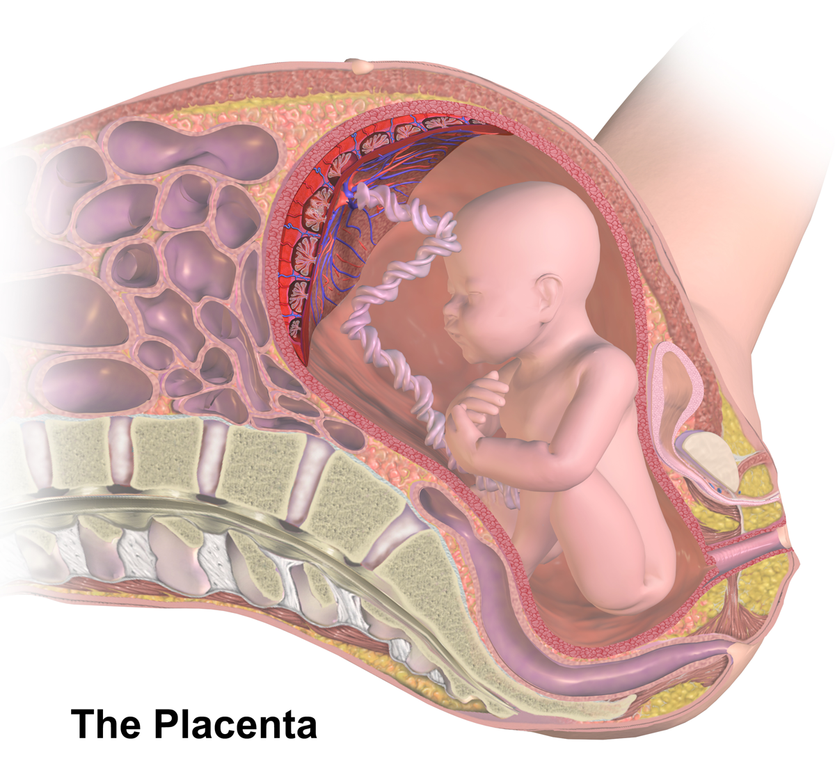
The placenta is a temporary embryonic and later fetal organ that begins developing from the blastocyst shortly after implantation. It plays critical roles in facilitating nutrient, gas and waste exchange between the physically separate maternal and fetal circulations, and is an important endocrine organ, producing hormones that regulate both maternal and fetal physiology during pregnancy. The placenta connects to the fetus via the umbilical cord, and on the opposite aspect to the maternal uterus in a species-dependent manner. In humans, a thin layer of maternal decidual (endometrial) tissue comes away with the placenta when it is expelled from the uterus following birth (sometimes incorrectly referred to as the 'maternal part' of the placenta). Placentas are a defining characteristic of placental mammals, but are also found in marsupials and some non-mammals with varying levels of development. Mammalian placentas probably first evolved about 150 million to 200 million years ... [...More Info...] [...Related Items...] OR: [Wikipedia] [Google] [Baidu] |


