|
Suprahyoid Muscles
The suprahyoid muscles are four muscles located above the hyoid bone in the neck. They are the digastric, stylohyoid, geniohyoid, and mylohyoid muscles. They are all pharyngeal muscles, with the exception of the geniohyoid muscle. The digastric is uniquely named for its two bellies. Its posterior belly rises from the mastoid process of the cranium and slopes downward and forward. The anterior belly arises from the digastric fossa on the inner surface of the mandibular body, which slopes downward and backward. The two bellies connect at the intermediate tendon. The intermediate tendon passes through a connective tissue loop attached to the hyoid bone. The mylohyoid muscles are thin, flat muscles that form a sling inferior to the tongue supporting the floor of the mouth. The geniohyoids are short, narrow muscles that contact each other in the midline. The stylohyoids are long, thin muscles that are nearly parallel with the posterior belly of the digastric muscle. Function These fo ... [...More Info...] [...Related Items...] OR: [Wikipedia] [Google] [Baidu] |
Hyoid Bone
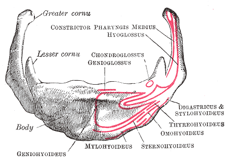
The hyoid bone (lingual bone or tongue-bone) () is a horseshoe-shaped bone situated in the anterior midline of the neck between the chin and the thyroid cartilage. At rest, it lies between the base of the mandible and the third cervical vertebra. Unlike other bones, the hyoid is only distantly articulated to other bones by muscles or ligaments. It is the only bone in the human body that is not connected to any other bones nearby. The hyoid is anchored by muscles from the anterior, posterior and inferior directions, and aids in tongue movement and swallowing. The hyoid bone provides attachment to the muscles of the floor of the mouth and the tongue above, the larynx below, and the epiglottis and pharynx behind. Its name is derived . Structure The hyoid bone is classed as an irregular bone and consists of a central part called the body, and two pairs of horns, the greater and lesser horns. Body The body of the hyoid bone is the central part of the hyoid bone. *At the ... [...More Info...] [...Related Items...] OR: [Wikipedia] [Google] [Baidu] |
Swallowing
Swallowing, sometimes called deglutition in scientific contexts, is the process in the human or animal body that allows for a substance to pass from the mouth, to the pharynx, and into the esophagus, while shutting the epiglottis. Swallowing is an important part of eating and drinking. If the process fails and the material (such as food, drink, or medicine) goes through the trachea, then choking or pulmonary aspiration can occur. In the human body the automatic temporary closing of the epiglottis is controlled by the swallowing reflex. The portion of food, drink, or other material that will move through the neck in one swallow is called a bolus. In colloquial English, the term "swallowing" is also used to describe the action of taking in a large mouthful of food without any biting, where the word gulping is more adequate. In humans Swallowing comes so easily to most people that the process rarely prompts much thought. However, from the viewpoints of physiology, of spe ... [...More Info...] [...Related Items...] OR: [Wikipedia] [Google] [Baidu] |
Mylohyoid Line
The mylohyoid line is a bony ridge on the internal surface of the mandible. It runs posterosuperiorly. It is the site of origin of the mylohyoid muscle, the superior pharyngeal constrictor muscle, and the pterygomandibular raphe. Structure The mylohyoid line is a bony ridge on the internal surface of the body of the mandible. The mylohyoid line extends posterosuperiorly. The mylohyoid line continues as the mylohyoid groove on the internal surface of the ramus. The mylohyoid muscle originates from the anterior (front) part of the mylohyoid line. Rarely, the mylohyoid muscle may originate partially from other surfaces of the mandible. The posterior (back) part of this line, near the alveolar margin, gives attachment to a small part of the superior pharyngeal constrictor muscle, and to the pterygomandibular raphe. Function The mylohyoid line is the site of attachment of many muscles, including the mylohyoid muscle, and the superior pharyngeal constrictor muscle. It is also ... [...More Info...] [...Related Items...] OR: [Wikipedia] [Google] [Baidu] |
Temporal Styloid Process
The temporal styloid process is a slender bony process of the temporal bone extending downward and forward from the undersurface of the temporal bone just below the ear. The styloid process gives attachments to several muscles, and ligaments. Structure The styloid process is a slender and pointed bony process of the temporal bone projecting anteroinferiorly from the inferior surface of the temporal bone just below the ear. Its length normally ranges from just under 3 cm to just over 4 cm. It is usually nearly straight, but may be curved in some individuals. Its ''proximal'' (''tympanohyal'') ''part'' is ensheathed by the tympanic part of the temporal bone ''(vaginal process), whereas'' its ''distal (stylohyal)'' ''part'' gives attachment to several structures. Attachments The styloid process gives attachments to several muscles, and ligaments. It serves as an anchor point for several muscles associated with the tongue and larynx. * stylohyoid ligament * stylomandibu ... [...More Info...] [...Related Items...] OR: [Wikipedia] [Google] [Baidu] |
Stylohyoid Muscle
The stylohyoid muscle is a slender muscle, lying anterior and superior of the posterior belly of the digastric muscle. It is one of the suprahyoid muscles. It shares this muscle's innervation by the facial nerve, and functions to draw the hyoid bone backwards and elevate the tongue. Its origin is the styloid process of the temporal bone. It inserts on the body of the hyoid. Structure The stylohyoid muscle originates from the posterior and lateral surface of the styloid process of the temporal bone, near the base. Passing inferior and anterior, it inserts into the body of the hyoid bone, at its junction with the greater cornu, and just superior to the omohyoid muscle. It belongs to the group of suprahyoid muscles. It is perforated, near its insertion, by the intermediate tendon of the digastric muscle. The stylohyoid muscle has vascular supply from the lingual artery, a branch of the external carotid artery. Nerve supply A branch of the facial nerve (CN VII) innervates the sty ... [...More Info...] [...Related Items...] OR: [Wikipedia] [Google] [Baidu] |
Cervical Spinal Nerve 1
The cervical spinal nerve 1 (C1) is a spinal nerve of the cervical segment. from spinalcordinjuryzone.com. Published February 23, 2004 Archived Dec 23, 2011. Retrieved June 12, 2018. C1 carries predominantly motor fibres, but also a small meningeal branch that supplies sensation to parts of the dura around the foramen magnum (via dorsal rami). It originates from the spinal column from above the cervical vertebra 1 (C1). The dorsal root and ganglion of the first cervical nerve ... [...More Info...] [...Related Items...] OR: [Wikipedia] [Google] [Baidu] |
Symphysis Menti
In human anatomy, the facial skeleton of the skull the external surface of the mandible is marked in the median line by a faint ridge, indicating the mandibular symphysis (Latin: ''symphysis menti'') or line of junction where the two lateral halves of the mandible typically fuse at an early period of life (1-2 years). It is not a true symphysis as there is no cartilage between the two sides of the mandible. This ridge divides below and encloses a triangular eminence, the mental protuberance, the base of which is depressed in the center but raised on either side to form the mental tubercle. The lowest (most inferior) end of the mandibular symphysis — the point of the chin — is called the "menton". It serves as the origin for the geniohyoid and the genioglossus muscles. Other animals Solitary mammalian carnivores that rely on a powerful canine bite to subdue their prey have a strong mandibular symphysis, while pack hunters delivering shallow bites have a weaker one. When filter ... [...More Info...] [...Related Items...] OR: [Wikipedia] [Google] [Baidu] |
Mental Spine
A mental spine is a small projection of bone on the posterior aspect of the mandible in the midline. There are usually four mental spines: two superior and two inferior. Collectively they are also known as the ''genial tubercle'',"Genial tubercle." The American Heritage Stedman's Medical Dictionary. Houghton Mifflin Company, 2002. Accessed: 22 Oct. 2007. ''genial apophysis'' and the Latin name ''spinae mentalis''. The inferior mental spines are the points of origin of the geniohyoid muscle,"Genial tubercle." Mosby's Dental Dictionary. Elsevier, Inc., 2004. Accessed: 22 Oct. 2007. one of the suprahyoid muscles, and the superior mental spines are the origin of the genioglossus muscle, one of the muscles of the tongue. Mental spines are important landmarks in clinical practice. Structure Mental spines are found on the posterior aspect of the mandible (lower jaw bone) in the midline. They usually surround spinous mental foramina in the midline. Variation Mental spines may be foun ... [...More Info...] [...Related Items...] OR: [Wikipedia] [Google] [Baidu] |
Geniohyoid Muscle
The geniohyoid muscle is a narrow muscle situated superior to the medial border of the mylohyoid muscle. It is named for its passage from the chin ("genio-" is a standard prefix for "chin") to the hyoid bone. Structure It arises from the inferior mental spine, on the back of the mandibular symphysis, and runs backward and slightly downward, to be inserted into the anterior surface of the body of the hyoid bone. It lies in contact with its fellow of the opposite side. It thus belongs to the suprahyoid muscles. The muscle is supplied by branches of the lingual artery. Innervation The geniohyoid muscle is innervated by fibres from the first cervical spinal nerve travelling alongside the hypoglossal nerve. Although the first three cervical nerves give rise to the ansa cervicalis, the geniohyoid muscle is said to be innervated by the first cervical nerve, as some of its efferent fibers do not contribute to ansa cervicalis. Variations It may be blended with the one on opposite side ... [...More Info...] [...Related Items...] OR: [Wikipedia] [Google] [Baidu] |
Mastoid Notch
The mastoid part of the temporal bone is the posterior (back) part of the temporal bone, one of the bones of the skull. Its rough surface gives attachment to various muscles (via tendons) and it has openings for blood vessels. From its borders, the mastoid part articulates with two other bones. Etymology The word "mastoid" is derived from the Greek word for "breast", a reference to the shape of this bone. Surfaces Outer surface Its outer surface is rough and gives attachment to the occipitalis and posterior auricular muscles. It is perforated by numerous foramina (holes); for example, the mastoid foramen is situated near the posterior border and transmits a vein to the transverse sinus and a small branch of the occipital artery to the dura mater. The position and size of this foramen are very variable; it is not always present; sometimes it is situated in the occipital bone, or in the suture between the temporal and the occipital. Mastoid process The mastoid process is ... [...More Info...] [...Related Items...] OR: [Wikipedia] [Google] [Baidu] |
Digastric Muscle
The digastric muscle (also digastricus) (named ''digastric'' as it has two 'bellies') is a small muscle located under the jaw. The term "digastric muscle" refers to this specific muscle. However, other muscles that have two separate muscle bellies include the suspensory muscle of duodenum, omohyoid, occipitofrontalis. It lies below the body of the mandible, and extends, in a curved form, from the mastoid notch to the mandibular symphysis. It belongs to the suprahyoid muscles group. A broad aponeurotic layer is given off from the tendon of the digastric muscle on either side, to be attached to the body and greater cornu of the hyoid bone; this is termed the suprahyoid aponeurosis. Structure The digastricus (digastric muscle) consists of two muscular bellies united by an intermediate rounded tendon. The two bellies of the digastric muscle have different embryological origins, and are supplied by different cranial nerves. Each person has a right and left digastric muscle ... [...More Info...] [...Related Items...] OR: [Wikipedia] [Google] [Baidu] |
Stylohyoid
The stylohyoid muscle is a slender muscle, lying anterior and superior of the posterior belly of the digastric muscle. It is one of the suprahyoid muscles. It shares this muscle's innervation by the facial nerve, and functions to draw the hyoid bone backwards and elevate the tongue. Its origin is the styloid process of the temporal bone. It inserts on the body of the hyoid. Structure The stylohyoid muscle originates from the posterior and lateral surface of the styloid process of the temporal bone, near the base. Passing inferior and anterior, it inserts into the body of the hyoid bone, at its junction with the greater cornu, and just superior to the omohyoid muscle. It belongs to the group of suprahyoid muscles. It is perforated, near its insertion, by the intermediate tendon of the digastric muscle. The stylohyoid muscle has vascular supply from the lingual artery, a branch of the external carotid artery. Nerve supply A branch of the facial nerve (CN VII) innervates the sty ... [...More Info...] [...Related Items...] OR: [Wikipedia] [Google] [Baidu] |


