|
Seborrheic Keratosis
A seborrheic keratosis is a non-cancerous (benign) skin tumour that originates from cells, namely keratinocytes, in the outer layer of the skin called the epidermis. Like liver spots, seborrheic keratoses are seen more often as people age. The tumours (also called lesions) appear in various colours, from light tan to black. They are round or oval, feel flat or slightly elevated, like the scab from a healing wound, and range in size from very small to more than across. They are often associated with other skin conditions, including basal cell carcinoma. Rarely seborrheic keratosis and basal cell carcinoma occur at the same location. At clinical examination the differential diagnosis include a wart and melanoma. Because only the top layers of the epidermis are involved, seborrheic keratoses are often described as having a "pasted on" appearance. Some dermatologists refer to seborrheic keratoses as "seborrheic warts", because they resemble warts, but strictly speaking the term "warts ... [...More Info...] [...Related Items...] OR: [Wikipedia] [Google] [Baidu] |
Leser–Trélat Sign
The Leser–Trélat sign is the explosive onset of multiple seborrheic keratoses (many pigmented skin lesions), often with an inflammation, inflammatory base. This can be a sign of internal malignancy as part of a paraneoplastic syndrome. In addition to the development of new lesions, preexisting ones frequently increase in size and become symptomatic. Associations Although most associated neoplasms are gastrointestinal adenocarcinomas (Gastric adenocarcinoma, stomach, Liver cancer, liver, Colorectal adenocarcinoma, colorectal and Pancreatic adenocarcinoma, pancreas), malignancies of the Breast cancer, breast, Lung cancer, lung, and Bladder cancer, urinary tract, as well as Lymphoma, lymphoid tissue, have been associated with this impressive rash. It is likely that various cytokines and other growth factors produced by the neoplasm are responsible for the abrupt appearance of the seborrheic keratoses. In some cases, paraneoplastic acanthosis nigricans (35% of patients), florid cut ... [...More Info...] [...Related Items...] OR: [Wikipedia] [Google] [Baidu] |
Lentigo Maligna
Lentigo maligna is where melanocyte cells have become malignant and grow continuously along the stratum basale of the skin, but have not invaded below the epidermis. Lentigo maligna is not the same as lentigo maligna melanoma, as detailed below. It typically progresses very slowly and can remain in a non-invasive form for years. It is normally found in the elderly (peak incidence in the 9th decade), on skin areas with high levels of sun exposure like the face and forearms. Incidence of evolution to lentigo maligna melanoma is low, about 2.2% to 5% in elderly patients. It is also known as "Hutchinson's melanotic freckle". This is named for Jonathan Hutchinson. The word lentiginous comes from the latin for freckle. Relation to melanoma Lentigo maligna is a histopathological variant of melanoma ''in situ''. Lentigo maligna is sometimes classified as a very early melanoma, and sometimes as a precursor to melanoma. When malignant melanocytes from a lentigo maligna have invaded be ... [...More Info...] [...Related Items...] OR: [Wikipedia] [Google] [Baidu] |
Papule
A papule is a small, well-defined bump in the skin. It may have a rounded, pointed or flat top, and may have a dip. It can appear with a stalk, be thread-like or look warty. It can be soft or firm and its surface may be rough or smooth. Some have crusts or scales. A papule can be flesh colored, yellow, white, brown, red, blue or purplish. There may be just one or many, and they may occur irregularly in different parts of the body or appear in clusters. It does not contain fluid but may progress to a pustule or vesicle. A papule is smaller than a nodule; it can be as tiny as a pinhead and is typically less than 1 cm in width, according to some sources, and 0.5 cm according to others. When merged together, it appears as a plaque. Its color might indicate its cause, such as white in milia, red in eczema, yellowish in xanthoma and black in melanoma. They may open when scratched and become infected and crusty. Definition A papule is a small, well-defined bump in the ... [...More Info...] [...Related Items...] OR: [Wikipedia] [Google] [Baidu] |
Melanocyte
Melanocytes are melanin-producing neural crest-derived cells located in the bottom layer (the stratum basale) of the skin's epidermis, the middle layer of the eye (the uvea), the inner ear, vaginal epithelium, meninges, bones, and heart. Melanin is a dark pigment primarily responsible for skin color. Once synthesized, melanin is contained in special organelles called melanosomes which can be transported to nearby keratinocytes to induce pigmentation. Thus darker skin tones have more melanosomes present than lighter skin tones. Functionally, melanin serves as protection against UV radiation. Melanocytes also have a role in the immune system. Function Through a process called melanogenesis, melanocytes produce melanin, which is a pigment found in the skin, eyes, hair, nasal cavity, and inner ear. This melanogenesis leads to a long-lasting pigmentation, which is in contrast to the pigmentation that originates from oxidation of already-existing melanin. There a ... [...More Info...] [...Related Items...] OR: [Wikipedia] [Google] [Baidu] |
Keratinocyte
Keratinocytes are the primary type of cell found in the epidermis, the outermost layer of the skin. In humans, they constitute 90% of epidermal skin cells. Basal cells in the basal layer (''stratum basale'') of the skin are sometimes referred to as basal keratinocytes. Keratinocytes form a barrier against environmental damage by heat, UV radiation, water loss, pathogenic bacteria, fungi, parasites, and viruses. A number of structural proteins, enzymes, lipids, and antimicrobial peptides contribute to maintain the important barrier function of the skin. Keratinocytes differentiate from epidermal stem cells in the lower part of the epidermis and migrate towards the surface, finally becoming corneocytes and eventually be shed off, which happens every 40 to 56 days in humans. Function The primary function of keratinocytes is the formation of a barrier against environmental damage by heat, UV radiation, water loss, pathogenic bacteria, fungi, parasites, and viruses. Pathogens ... [...More Info...] [...Related Items...] OR: [Wikipedia] [Google] [Baidu] |
Distal Tibia
The tibia (; ), also known as the shinbone or shankbone, is the larger, stronger, and anterior (frontal) of the two bones in the leg below the knee in vertebrates (the other being the fibula, behind and to the outside of the tibia); it connects the knee with the ankle. The tibia is found on the medial side of the leg next to the fibula and closer to the median plane. The tibia is connected to the fibula by the interosseous membrane of leg, forming a type of fibrous joint called a syndesmosis with very little movement. The tibia is named for the flute ''tibia''. It is the second largest bone in the human body, after the femur. The leg bones are the strongest long bones as they support the rest of the body. Structure In human anatomy, the tibia is the second largest bone next to the femur. As in other vertebrates the tibia is one of two bones in the lower leg, the other being the fibula, and is a component of the knee and ankle joints. The ossification or formation of the bone st ... [...More Info...] [...Related Items...] OR: [Wikipedia] [Google] [Baidu] |
Histologically
Histology, also known as microscopic anatomy or microanatomy, is the branch of biology which studies the microscopic anatomy of biological tissues. Histology is the microscopic counterpart to gross anatomy, which looks at larger structures visible without a microscope. Although one may divide microscopic anatomy into ''organology'', the study of organs, ''histology'', the study of tissues, and ''cytology'', the study of cells, modern usage places all of these topics under the field of histology. In medicine, histopathology is the branch of histology that includes the microscopic identification and study of diseased tissue. In the field of paleontology, the term paleohistology refers to the histology of fossil organisms. Biological tissues Animal tissue classification There are four basic types of animal tissues: muscle tissue, nervous tissue, connective tissue, and epithelial tissue. All animal tissues are considered to be subtypes of these four principal tissue types (for ... [...More Info...] [...Related Items...] OR: [Wikipedia] [Google] [Baidu] |
Collagen
Collagen () is the main structural protein in the extracellular matrix found in the body's various connective tissues. As the main component of connective tissue, it is the most abundant protein in mammals, making up from 25% to 35% of the whole-body protein content. Collagen consists of amino acids bound together to form a triple helix of elongated fibril known as a collagen helix. It is mostly found in connective tissue such as cartilage, bones, tendons, ligaments, and skin. Depending upon the degree of mineralization, collagen tissues may be rigid (bone) or compliant (tendon) or have a gradient from rigid to compliant (cartilage). Collagen is also abundant in corneas, blood vessels, the gut, intervertebral discs, and the dentin in teeth. In muscle tissue, it serves as a major component of the endomysium. Collagen constitutes one to two percent of muscle tissue and accounts for 6% of the weight of the skeletal muscle tissue. The fibroblast is the most common ... [...More Info...] [...Related Items...] OR: [Wikipedia] [Google] [Baidu] |
Epidermis (skin)
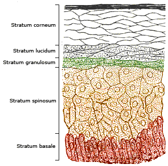
The epidermis is the outermost of the three layers that comprise the skin, the inner layers being the dermis and hypodermis. The epidermis layer provides a barrier to infection from environmental pathogens and regulates the amount of water released from the body into the atmosphere through transepidermal water loss. The epidermis is composed of multiple layers of flattened cells that overlie a base layer ( stratum basale) composed of columnar cells arranged perpendicularly. The layers of cells develop from stem cells in the basal layer. The human epidermis is a familiar example of epithelium, particularly a stratified squamous epithelium. The word epidermis is derived through Latin , itself and . Something related to or part of the epidermis is termed epidermal. Structure Cellular components The epidermis primarily consists of keratinocytes ( proliferating basal and differentiated suprabasal), which comprise 90% of its cells, but also contains melanocytes, Langerha ... [...More Info...] [...Related Items...] OR: [Wikipedia] [Google] [Baidu] |
Skin Biopsy
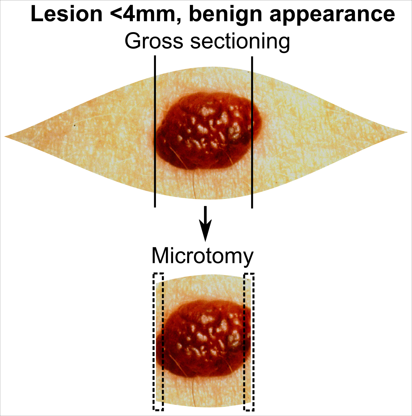
Skin biopsy is a biopsy technique in which a skin lesion is removed to be sent to a pathologist to render a microscopic diagnosis. It is usually done under local anesthetic in a physician's office, and results are often available in 4 to 10 days. It is commonly performed by dermatologists. Skin biopsies are also done by family physicians, internists, surgeons, and other specialties. However, performed incorrectly, and without appropriate clinical information, a pathologist's interpretation of a skin biopsy can be severely limited, and therefore doctors and patients may forgo traditional biopsy techniques and instead choose Mohs surgery. There are four main types of skin biopsies: shave biopsy, punch biopsy, excisional biopsy, and incisional biopsy. The choice of the different skin biopsies is dependent on the suspected diagnosis of the skin lesion. Like most biopsies, patient consent and anesthesia (usually lidocaine injected into the skin) are prerequisites. Types Shave bio ... [...More Info...] [...Related Items...] OR: [Wikipedia] [Google] [Baidu] |
Gold Standard (test)
In medicine and statistics, a gold standard test is usually the diagnostic test or benchmark that is the best available under reasonable conditions. In other words, a gold standard is the most accurate test possible without restrictions. Both meanings are different because for example, in medicine, dealing with conditions that would require an autopsy to have a perfect diagnosis, the gold standard test would be the best one that keeps the patient alive instead of the autopsy. In medicine "Gold standard" can refer to the criteria by which scientific evidence is evaluated. For example, in resuscitation research, the "gold standard" test of a medication or procedure is whether or not it leads to an increase in the number of neurologically intact survivors that walk out of the hospital.''ACLS: Principles and Practice''. p. 62. Dallas: American Heart Association, 2003. . Other types of medical research might regard a significant decrease in 30-day mortality as the gold standard. The ... [...More Info...] [...Related Items...] OR: [Wikipedia] [Google] [Baidu] |
Squamous Cell Carcinoma
Squamous-cell carcinomas (SCCs), also known as epidermoid carcinomas, comprise a number of different types of cancer that begin in squamous cells. These cells form on the surface of the skin, on the lining of hollow organs in the body, and on the lining of the respiratory and digestive tracts. Common types include: * Squamous-cell skin cancer: A type of skin cancer * Squamous-cell carcinoma of the lung: A type of lung cancer * Squamous-cell thyroid carcinoma: A type of thyroid cancer * Esophageal squamous-cell carcinoma: A type of esophageal cancer * Squamous-cell carcinoma of the vagina: A type of vaginal cancer Despite sharing the name "squamous-cell carcinoma", the SCCs of different body sites can show differences in their presented symptoms, natural history, prognosis, and response to treatment. By body location Human papillomavirus infection has been associated with SCCs of the oropharynx, lung, fingers, and anogenital region. Head and neck cancer About 90% of ... [...More Info...] [...Related Items...] OR: [Wikipedia] [Google] [Baidu] |
.jpg)


