|
Synaptic Pruning
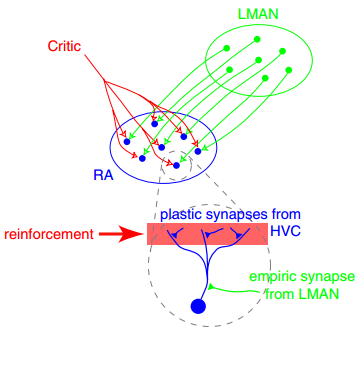
Synaptic pruning is the process of synapse elimination or weakening. Though it occurs throughout the lifespan of a mammal, the most active period of synaptic pruning in the development of the nervous system occurs between early childhood and the onset of puberty in many mammals, including humans. Pruning starts near the time of birth and continues into the late-20s. During elimination of a synapse, the axon withdraws or dies off, and the dendrite decays and die off. Synaptic pruning was traditionally considered to be complete by the time of sexual maturation, but magnetic resonance imaging studies have discounted this idea. The infant brain will increase in size by a factor of up to 5 by adulthood. Two factors contribute to this growth: the growth of synaptic connections between neurons and the myelination of nerve fibers. The total number of neurons, however, remains approximately the same, containing approximately 86 (± 8) billion neurons. After adolescence, the volume of the ... [...More Info...] [...Related Items...] OR: [Wikipedia] [Google] [Baidu] |
Visual Cortex
The visual cortex of the brain is the area of the cerebral cortex that processes visual information. It is located in the occipital lobe. Sensory input originating from the eyes travels through the lateral geniculate nucleus in the thalamus and then reaches the visual cortex. The area of the visual cortex that receives the sensory input from the lateral geniculate nucleus is the primary visual cortex, also known as visual area 1 ( V1), Brodmann area 17, or the striate cortex. The extrastriate areas consist of visual areas 2, 3, 4, and 5 (also known as V2, V3, V4, and V5, or Brodmann area 18 and all Brodmann area 19). Both hemispheres of the brain include a visual cortex; the visual cortex in the left hemisphere receives signals from the right visual field, and the visual cortex in the right hemisphere receives signals from the left visual field. Introduction The primary visual cortex (V1) is located in and around the calcarine fissure in the occipital lobe. Each h ... [...More Info...] [...Related Items...] OR: [Wikipedia] [Google] [Baidu] |
Fractionator
Fractionation is a separation process in which a certain quantity of a mixture (of gasses, solids, liquids, enzymes, or isotopes, or a suspension) is divided during a phase transition, into a number of smaller quantities (fractions) in which the composition varies according to a gradient. Fractions are collected based on differences in a specific property of the individual components. A common trait in fractionations is the need to find an optimum between the amount of fractions collected and the desired purity in each fraction. Fractionation makes it possible to isolate more than two components in a mixture in a single run. This property sets it apart from other separation techniques. Fractionation is widely employed in many branches of science and technology. Mixtures of liquids and gasses are separated by fractional distillation by difference in boiling point. Fractionation of components also takes place in column chromatography by a difference in affinity between stationar ... [...More Info...] [...Related Items...] OR: [Wikipedia] [Google] [Baidu] |
Stereology
Stereology is the three-dimensional interpretation of two-dimensional cross sections of materials or tissues. It provides practical techniques for extracting quantitative information about a three-dimensional material from measurements made on two-dimensional planar sections of the material. Stereology is a method that utilizes random, systematic sampling to provide unbiased and quantitative data. It is an important and efficient tool in many applications of microscopy (such as petrography, materials science, and biosciences including histology, bone and neuroanatomy). Stereology is a developing science with many important innovations being developed mainly in Europe. New innovations such as the proportionator continue to make important improvements in the efficiency of stereological procedures. In addition to two-dimensional plane sections, stereology also applies to three-dimensional slabs (e.g. 3D microscope images), one-dimensional probes (e.g. needle biopsy), projected image ... [...More Info...] [...Related Items...] OR: [Wikipedia] [Google] [Baidu] |
Oxford University
The University of Oxford is a collegiate research university in Oxford, England. There is evidence of teaching as early as 1096, making it the oldest university in the English-speaking world and the second-oldest continuously operating university globally. It expanded rapidly from 1167, when Henry II prohibited English students from attending the University of Paris. When disputes erupted between students and the Oxford townspeople, some Oxford academics fled northeast to Cambridge, where they established the University of Cambridge in 1209. The two English ancient universities share many common features and are jointly referred to as ''Oxbridge''. The University of Oxford comprises 43 constituent colleges, consisting of 36 semi-autonomous colleges, four permanent private halls and three societies (colleges that are departments of the university, without their own royal charter). and a range of academic departments that are organised into four divisions. Each college ... [...More Info...] [...Related Items...] OR: [Wikipedia] [Google] [Baidu] |
Medial Dorsal Nucleus
The medial dorsal nucleus (or mediodorsal nucleus of thalamus, dorsomedial nucleus, dorsal medial nucleus, or medial nucleus group) is a large nucleus in the thalamus. It is separated from the other thalamic nuclei by the internal medullary lamina. The medial dorsal nucleus is interconnected with the prefrontal cortex, therefore involved in prefrontal functions. Damage to the interconnected tract or the nucleus itself will result in similar damage to the prefrontal cortex. It is also believed to play a role in memory. Structure The medial dorsal nucleus relays inputs from the amygdala and olfactory cortex and projects to the prefrontal cortex and the limbic system, and in turn relays them to the prefrontal association cortex. As a result, it plays a crucial role in attention, planning, organization, abstract thinking, multi-tasking, and active memory. The connections of the medial dorsal nucleus have even been used to delineate the prefrontal cortex of the Göttingen minip ... [...More Info...] [...Related Items...] OR: [Wikipedia] [Google] [Baidu] |
Glial Cell
Glia, also called glial cells (gliocytes) or neuroglia, are non-neuronal cells in the central nervous system (the brain and the spinal cord) and in the peripheral nervous system that do not produce electrical impulses. The neuroglia make up more than one half the volume of neural tissue in the human body. They maintain homeostasis, form myelin, and provide support and protection for neurons. In the central nervous system, glial cells include oligodendrocytes (that produce myelin), astrocytes, ependymal cells and microglia, and in the peripheral nervous system they include Schwann cells (that produce myelin), and satellite cells. Function They have four main functions: * to surround neurons and hold them in place * to supply nutrients and oxygen to neurons * to insulate one neuron from another * to destroy pathogens and remove dead neurons. They also play a role in neurotransmission and synaptic connections, and in physiological processes such as breathing. While glia w ... [...More Info...] [...Related Items...] OR: [Wikipedia] [Google] [Baidu] |
Central Nervous System
The central nervous system (CNS) is the part of the nervous system consisting primarily of the brain, spinal cord and retina. The CNS is so named because the brain integrates the received information and coordinates and influences the activity of all parts of the bodies of bilateria, bilaterally symmetric and triploblastic animals—that is, all multicellular animals except sponges and Coelenterata, diploblasts. It is a structure composed of nervous tissue positioned along the Anatomical_terms_of_location#Rostral,_cranial,_and_caudal, rostral (nose end) to caudal (tail end) axis of the body and may have an enlarged section at the rostral end which is a brain. Only arthropods, cephalopods and vertebrates have a true brain, though precursor structures exist in onychophorans, gastropods and lancelets. The rest of this article exclusively discusses the vertebrate central nervous system, which is radically distinct from all other animals. Overview In vertebrates, the brain and spinal ... [...More Info...] [...Related Items...] OR: [Wikipedia] [Google] [Baidu] |
Climbing Fiber
Climbing fibers are the name given to a series of neuronal projections from the inferior olivary nucleus located in the medulla oblongata. These axons pass through the pons and enter the cerebellum via the inferior cerebellar peduncle where they form synapses with the deep cerebellar nuclei and Purkinje cells. Each climbing fiber will form synapses with 1-10 Purkinje cells. Early in development, Purkinje cells are innervated by multiple climbing fibers, but as the cerebellum matures, these inputs gradually become eliminated resulting in a single climbing fiber input per Purkinje cell. These fibers provide very powerful, excitatory input to the cerebellum which results in the generation of complex spike excitatory postsynaptic potential (EPSP) in Purkinje cells. In this way climbing fibers (CFs) perform a central role in motor behaviors. The climbing fibers carry information from various sources such as the spinal cord, vestibular system, red nucleus, superior collicul ... [...More Info...] [...Related Items...] OR: [Wikipedia] [Google] [Baidu] |
Peripheral Nervous System
The peripheral nervous system (PNS) is one of two components that make up the nervous system of Bilateria, bilateral animals, with the other part being the central nervous system (CNS). The PNS consists of nerves and ganglia, which lie outside the brain and the spinal cord. The main function of the PNS is to connect the CNS to the Limb (anatomy), limbs and Organ (anatomy), organs, essentially serving as a relay between the brain and spinal cord and the rest of the body. Unlike the CNS, the PNS is not protected by the vertebral column and skull, or by the blood–brain barrier, which leaves it exposed to toxins. The peripheral nervous system can be divided into a somatic nervous system, somatic division and an autonomic nervous system, autonomic division. Each of these can further be differentiated into a sensory and a motor sector. In the somatic nervous system, the cranial nerves are part of the PNS with the exceptions of the olfactory nerve and epithelia and the optic nerve (c ... [...More Info...] [...Related Items...] OR: [Wikipedia] [Google] [Baidu] |
Neuromuscular Junction
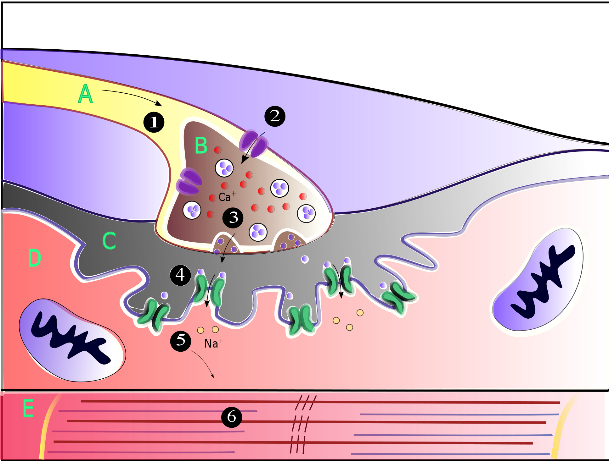
A neuromuscular junction (or myoneural junction) is a chemical synapse between a motor neuron and a muscle fiber. It allows the motor neuron to transmit a signal to the muscle fiber, causing muscle contraction. Muscles require innervation to function—and even just to maintain muscle tone, avoiding atrophy. In the neuromuscular system, nerves from the central nervous system and the peripheral nervous system are linked and work together with muscles. Synaptic transmission at the neuromuscular junction begins when an action potential reaches the presynaptic terminal of a motor neuron, which activates voltage-gated calcium channels to allow calcium ions to enter the neuron. Calcium ions bind to sensor proteins (synaptotagmins) on synaptic vesicles, triggering vesicle fusion with the cell membrane and subsequent neurotransmitter release from the motor neuron into the synaptic cleft. In vertebrates, motor neurons release acetylcholine (ACh), a small molecule neurotransmitter, which ... [...More Info...] [...Related Items...] OR: [Wikipedia] [Google] [Baidu] |
Synaptic Plasticity
In neuroscience, synaptic plasticity is the ability of synapses to Chemical synapse#Synaptic strength, strengthen or weaken over time, in response to increases or decreases in their activity. Since memory, memories are postulated to be represented by vastly interconnected neural circuits in the brain, synaptic plasticity is one of the important neurochemical foundations of learning and memory (''see Hebbian theory''). Plastic change often results from the alteration of the number of neurotransmitter receptors located on a synapse. There are several underlying mechanisms that cooperate to achieve synaptic plasticity, including changes in the quantity of neurotransmitters released into a synapse and changes in how effectively cells respond to those neurotransmitters. Synaptic plasticity in both Excitatory synapse, excitatory and Inhibitory synapse, inhibitory synapses has been found to be dependent upon postsynaptic calcium release. Historical discoveries In 1973, Terje Lømo and ... [...More Info...] [...Related Items...] OR: [Wikipedia] [Google] [Baidu] |


