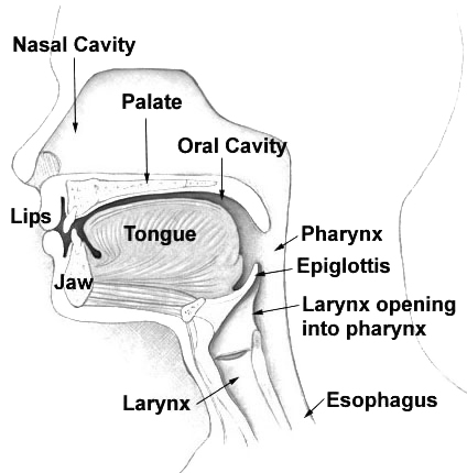
|
Soft Palate
The soft palate (also known as the velum, palatal velum, or muscular palate) is, in mammals, the soft biological tissue, tissue constituting the back of the roof of the mouth. The soft palate is part of the palate of the mouth; the other part is the hard palate. The soft palate is distinguished from the hard palate at the front of the mouth in that it does not contain bone. Structure Muscles The five muscles of the soft palate play important roles in swallowing and breathing. The muscles are: # Tensor veli palatini, which is involved in swallowing # Palatoglossus, involved in swallowing # Palatopharyngeus, involved in breathing # Levator veli palatini, involved in swallowing # Musculus uvulae, which moves the palatine uvula, uvula These muscles are innervated by the pharyngeal plexus of vagus nerve, pharyngeal plexus via the vagus nerve, with the exception of the tensor veli palatini. The tensor veli palatini is innervated by the mandibular division of the trigeminal nerve (V ... [...More Info...] [...Related Items...] OR: [Wikipedia] [Google] [Baidu] |
Lesser Palatine Arteries
The lesser palatine arteries are Artery, arteries of the head. It is a branch of the descending palatine artery. They supply the Palatine tonsil, palatine tonsils and the soft palate. Structure The lesser palatine arteries are branches of the descending palatine artery. They go through the lesser palatine foramina. They Anastomosis, anastomose with the ascending pharyngeal artery. Function The lesser palatine arteries give off tonsillary branches to supply the Palatine tonsil, palatine tonsils. They also gives off mucosal branches that usually supply the soft palate, and potentially the hard palate. See also * Lesser palatine nerve References Arteries of the head and neck {{circulatory-stub ... [...More Info...] [...Related Items...] OR: [Wikipedia] [Google] [Baidu] |
Breathing
Breathing (spiration or ventilation) is the rhythmical process of moving air into ( inhalation) and out of ( exhalation) the lungs to facilitate gas exchange with the internal environment, mostly to flush out carbon dioxide and bring in oxygen. All aerobic creatures need oxygen for cellular respiration, which extracts energy from the reaction of oxygen with molecules derived from food and produces carbon dioxide as a waste product. Breathing, or external respiration, brings air into the lungs where gas exchange takes place in the alveoli through diffusion. The body's circulatory system transports these gases to and from the cells, where cellular respiration takes place. The breathing of all vertebrates with lungs consists of repetitive cycles of inhalation and exhalation through a highly branched system of tubes or airways which lead from the nose to the alveoli. The number of respiratory cycles per minute is the breathing or respiratory rate, and is one of the fou ... [...More Info...] [...Related Items...] OR: [Wikipedia] [Google] [Baidu] |
Tongue
The tongue is a Muscle, muscular organ (anatomy), organ in the mouth of a typical tetrapod. It manipulates food for chewing and swallowing as part of the digestive system, digestive process, and is the primary organ of taste. The tongue's upper surface (dorsum) is covered by taste buds housed in numerous lingual papillae. It is sensitive and kept moist by saliva and is richly supplied with nerves and blood vessels. The tongue also serves as a natural means of cleaning the teeth. A major function of the tongue is to enable speech in humans and animal communication, vocalization in other animals. The human tongue is divided into two parts, an oral cavity, oral part at the front and a pharynx, pharyngeal part at the back. The left and right sides are also separated along most of its length by a vertical section of connective tissue, fibrous tissue (the lingual septum) that results in a groove, the median sulcus, on the tongue's surface. There are two groups of glossal muscles. The f ... [...More Info...] [...Related Items...] OR: [Wikipedia] [Google] [Baidu] |
Pharyngeal Reflex
The pharyngeal reflex or gag reflex is a reflex muscular contraction of the back of the throat, evoked by touching the roof of the mouth, back of the tongue, area around the tonsils, uvula, and back of the throat. It, along with other aerodigestive reflexes such as reflexive pharyngeal swallowing, prevents objects in the oral cavity from entering the throat except as part of normal swallowing and helps prevent choking, and is a form of coughing. The pharyngeal reflex is different from the laryngeal spasm, which is a reflex muscular contraction of the vocal cords. Reflex arc In a reflex arc, a series of physiological steps occur very rapidly to produce a reflex. Generally, a sensory receptor receives an environmental stimulus, in this case from objects reaching nerves in the back of the throat, and sends a message via an afferent nerve to the central nervous system (CNS). The CNS receives this message and sends an appropriate response via an efferent nerve (also known as a mot ... [...More Info...] [...Related Items...] OR: [Wikipedia] [Google] [Baidu] |
Human
Humans (''Homo sapiens'') or modern humans are the most common and widespread species of primate, and the last surviving species of the genus ''Homo''. They are Hominidae, great apes characterized by their Prehistory of nakedness and clothing#Evolution of hairlessness, hairlessness, bipedality, bipedalism, and high Human intelligence, intelligence. Humans have large Human brain, brains, enabling more advanced cognitive skills that facilitate successful adaptation to varied environments, development of sophisticated tools, and formation of complex social structures and civilizations. Humans are Sociality, highly social, with individual humans tending to belong to a Level of analysis, multi-layered network of distinct social groups — from families and peer groups to corporations and State (polity), political states. As such, social interactions between humans have established a wide variety of Value theory, values, norm (sociology), social norms, languages, and traditions (co ... [...More Info...] [...Related Items...] OR: [Wikipedia] [Google] [Baidu] |
Nasal Cavity
The nasal cavity is a large, air-filled space above and behind the nose in the middle of the face. The nasal septum divides the cavity into two cavities, also known as fossae. Each cavity is the continuation of one of the two nostrils. The nasal cavity is the uppermost part of the respiratory system and provides the nasal passage for inhaled air from the nostrils to the nasopharynx and rest of the respiratory tract. The paranasal sinuses surround and drain into the nasal cavity. Structure The term "nasal cavity" can refer to each of the two cavities of the nose, or to the two sides combined. The lateral wall of each nasal cavity mainly consists of the maxilla. However, there is a deficiency that is compensated for by the perpendicular plate of the palatine bone, the medial pterygoid plate, the labyrinth of ethmoid and the inferior concha. The paranasal sinuses are connected to the nasal cavity through small orifices called ostia. Most of these ostia communicat ... [...More Info...] [...Related Items...] OR: [Wikipedia] [Google] [Baidu] |
Mucous Membrane
A mucous membrane or mucosa is a membrane that lines various cavities in the body of an organism and covers the surface of internal organs. It consists of one or more layers of epithelial cells overlying a layer of loose connective tissue. It is mostly of endodermal origin and is continuous with the skin at body openings such as the eyes, eyelids, ears, inside the nose, inside the mouth, lips, the genital areas, the urethral opening and the anus. Some mucous membranes secrete mucus, a thick protective fluid. The function of the membrane is to stop pathogens and dirt from entering the body and to prevent bodily tissues from becoming dehydrated. Structure The mucosa is composed of one or more layers of epithelial cells that secrete mucus, and an underlying lamina propria of loose connective tissue. The type of cells and type of mucus secreted vary from organ to organ and each can differ along a given tract. Mucous membranes line the digestive, respiratory and rep ... [...More Info...] [...Related Items...] OR: [Wikipedia] [Google] [Baidu] |
Muscle
Muscle is a soft tissue, one of the four basic types of animal tissue. There are three types of muscle tissue in vertebrates: skeletal muscle, cardiac muscle, and smooth muscle. Muscle tissue gives skeletal muscles the ability to muscle contraction, contract. Muscle tissue contains special Muscle contraction, contractile proteins called actin and myosin which interact to cause movement. Among many other muscle proteins, present are two regulatory proteins, troponin and tropomyosin. Muscle is formed during embryonic development, in a process known as myogenesis. Skeletal muscle tissue is striated consisting of elongated, multinucleate muscle cells called muscle fibers, and is responsible for movements of the body. Other tissues in skeletal muscle include tendons and perimysium. Smooth and cardiac muscle contract involuntarily, without conscious intervention. These muscle types may be activated both through the interaction of the central nervous system as well as by innervation ... [...More Info...] [...Related Items...] OR: [Wikipedia] [Google] [Baidu] |
Mandibular Division Of The Trigeminal Nerve
In neuroanatomy, the mandibular nerve (V) is the largest of the three divisions of the trigeminal nerve, the fifth cranial nerve (CN V). Unlike the other divisions of the trigeminal nerve ( ophthalmic nerve, maxillary nerve) which contain only afferent fibers, the mandibular nerve contains both afferent and efferent fibers. These nerve fibers innervate structures of the lower jaw and face, such as the tongue, lower lip, and chin. The mandibular nerve also innervates the muscles of mastication. Structure Course The large sensory root of mandibular nerve emerges from the lateral part of the trigeminal ganglion and exits the cranial cavity through the foramen ovale. The motor root (Latin: ''radix motoria'' s. ''portio minor''), the small motor root of the trigeminal nerve, passes under the trigeminal ganglion and through the foramen ovale to unite with the sensory root just outside the skull. The mandibular nerve immediately passes between tensor veli palatini, which is med ... [...More Info...] [...Related Items...] OR: [Wikipedia] [Google] [Baidu] |
Tensor Veli Palatini
The tensor veli palatini muscle (tensor palati or tensor muscle of the velum palatinum) is a thin, triangular muscle of the head that tenses the soft palate and opens the Eustachian tube to equalise pressure in the middle ear. Structure The tensor veli palatini muscle is thin and triangular in shape. Origin It arises from the scaphoid fossa of the pterygoid process of the sphenoid anteriorly, the (medial aspect of the) spine of sphenoid bone posteriorly, and - between the aforementioned anterior and posterior attachments - from the anterolateral aspect of the membranous wall of the pharyngotympanic tube. At the muscle's origin, some of its muscle fibres may be continuous with those of the tensor tympani muscle. Insertion Inferiorly, the muscle converges to form a tendon of attachment. This tendon winds medially around the pterygoid hamulus (with a small bursa interposed between the two) to insert into the palatine aponeurosis and into the bony surface posterior to ... [...More Info...] [...Related Items...] OR: [Wikipedia] [Google] [Baidu] |
Vagus Nerve
The vagus nerve, also known as the tenth cranial nerve (CN X), plays a crucial role in the autonomic nervous system, which is responsible for regulating involuntary functions within the human body. This nerve carries both sensory and motor fibers and serves as a major pathway that connects the brain to various organs, including the heart, lungs, and digestive tract. As a key part of the parasympathetic nervous system, the vagus nerve helps regulate essential involuntary functions like heart rate, breathing, and digestion. By controlling these processes, the vagus nerve contributes to the body's "rest and digest" response, helping to calm the body after stress, lower heart rate, improve digestion, and maintain homeostasis. The vagus nerve consists of two branches: the right and left vagus nerves. In the neck, the right vagus nerve contains approximately 105,000 fibers, while the left vagus nerve has about 87,000 fibers, according to one source. However, other sources report sl ... [...More Info...] [...Related Items...] OR: [Wikipedia] [Google] [Baidu] |
Pharyngeal Plexus Of Vagus Nerve
The pharyngeal plexus is a nerve plexus located upon the outer surface of the pharynx. It contains a motor component (derived from the vagus nerve (cranial nerve X)), a sensory component (derived from the glossopharyngeal nerve (cranial nerve IX)), and sympathetic component (derived from the superior cervical ganglion). The plexus provides motor innervation to most muscles of the soft palate (all but the tensor veli palatini muscle) and most muscles of the pharynx (all but the stylopharyngeus muscle). The larynx meanwhile receives motor innervation from the vagus nerve (CN X) via its external branch of the superior laryngeal nerve and its recurrent laryngeal nerve, and ''not'' through the pharyngeal plexus. Anatomy The pharyngeal plexus occurs upon the outer surface of the pharynx - especially superficial to the middle pharyngeal constrictor muscle. Afferents It has the following components: * Motor – pharyngeal branch of vagus nerve (CN X) which arises from the ... [...More Info...] [...Related Items...] OR: [Wikipedia] [Google] [Baidu] |


