|
Skull Bossing
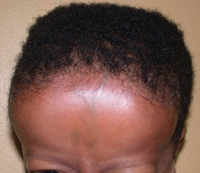
Skull bossing is a descriptive term in medical physical examination indicating a protuberance of the skull, most often in the frontal bones of the forehead ("frontal bossing"). Although prominence of the skull bones may be normal, skull bossing may be associated with certain medical conditions, including nutritional, metabolic, hormonal, and hematologic disorders. Frontal bossing Frontal bossing is the development of an unusually pronounced forehead which may also be associated with a heavier than normal brow ridge. It is caused by enlargement of the frontal bone, often in conjunction with abnormal enlargement of other facial bones, skull, mandible, and bones of the hands and feet. Frontal bossing may be seen in a few rare medical syndromes such as acromegaly – a chronic medical disorder in which the anterior pituitary gland produces excess growth hormone (GH). Frontal bossing may also occur in diseases resulting in chronic anemia, where there is increased hematopoiesis and enl ... [...More Info...] [...Related Items...] OR: [Wikipedia] [Google] [Baidu] [Amazon] |
Frontal Bone
In the human skull, the frontal bone or sincipital bone is an unpaired bone which consists of two portions.'' Gray's Anatomy'' (1918) These are the vertically oriented squamous part, and the horizontally oriented orbital part, making up the bony part of the forehead, part of the bony orbital cavity holding the eye, and part of the bony part of the nose respectively. The name comes from the Latin word ''frons'' (meaning "forehead"). Structure The frontal bone is made up of two main parts. These are the squamous part, and the orbital part. The squamous part marks the vertical, flat, and also the biggest part, and the main region of the forehead. The orbital part is the horizontal and second biggest region of the frontal bone. It enters into the formation of the roofs of the orbital and nasal cavities. Sometimes a third part is included as the nasal part of the frontal bone, and sometimes this is included with the squamous part. The nasal part is between the brow ridges, ... [...More Info...] [...Related Items...] OR: [Wikipedia] [Google] [Baidu] [Amazon] |
Crouzon Syndrome
Crouzon syndrome is an autosomal dominant genetic disorder known as a branchial arch syndrome. Specifically, this syndrome affects the first branchial (or pharyngeal) arch, which is the precursor of the maxilla and mandible. Because the branchial arches are important developmental features in a growing embryo, disturbances in their development create lasting and widespread effects. The syndrome is caused by a mutation in a gene on chromosome 10 that controls the body's production of fibroblast growth factor receptor 2 (''FGFR2''). Crouzon syndrome is named for Octave Crouzon, a French physician who first described this disorder. First called "craniofacial dysostosis" ("craniofacial" refers to the skull and face, and " dysostosis" refers to malformation of bone), the disorder was characterized by a number of clinical features which can be described by the rudimentary meanings of its former name. The developing fetus's skull and facial bones fuse early or are unable to expand. Thu ... [...More Info...] [...Related Items...] OR: [Wikipedia] [Google] [Baidu] [Amazon] |
Beta-thalassemia
Beta-thalassemia (β-thalassemia) is an inherited blood disorder, a form of thalassemia resulting in variable outcomes ranging from clinically asymptomatic to severe anemia individuals. It is caused by reduced or absent synthesis of the beta chains of hemoglobin, the molecule that carries oxygen in the blood. Symptoms depend on the extent to which hemoglobin is deficient, and include anemia, pallor, tiredness, enlargement of the spleen, jaundice, and gallstones. In severe cases death ensues. Beta thalassemia occurs due to a mutation of the HBB gene leading to deficient production of the hemoglobin subunit beta-globin; the severity of the disease depends on the nature of the mutation, and whether or not the mutation is homozygous. The body's inability to construct beta-globin leads to reduced or zero production of adult hemoglobin thus causing anemia. The other component of hemoglobin, alpha-globin, accumulates in excess leading to ineffective production of red blood cells, ... [...More Info...] [...Related Items...] OR: [Wikipedia] [Google] [Baidu] [Amazon] |
Trimethadione
Trimethadione (Tridione) is an oxazolidinedione anticonvulsant. It is most commonly used to treat epileptic conditions that are resistant to other treatments. It is primarily effective in treating absence seizures, but can also be used in refractory temporal lobe epilepsy. It is usually administered 3 or 4 times daily, with the total daily dose ranging from 900 mg to 2.4 g. Treatment is most effective when the concentration of its active metabolite, dimethadione, is above 700 μg/mL. Severe adverse reactions are possible, including Steven Johnson syndrome, nephrotoxicity, hepatitis, aplastic anemia, neutropenia, or agranulocytosis. More common adverse effects include drowsiness, hemeralopia, and hiccups. Fetal trimethadione syndrome If administered during pregnancy, fetal trimethadione syndrome may result causing facial dysmorphism (short upturned nose, slanted eyebrows), cardiac defects, intrauterine growth restriction Intrauterine growth restriction (IUGR), or fetal grow ... [...More Info...] [...Related Items...] OR: [Wikipedia] [Google] [Baidu] [Amazon] |
Thanatophoric Dysplasia
Thanatophoric dysplasia is a severe skeleton, skeletal disorder characterized by a disproportionately small Rib cage, ribcage, extremely short limbs and folds of extra skin on the arms and legs. Symptoms and signs Infants with this condition have disproportionately short arms and legs with extra folds of skin. Other signs of the disorder include a narrow chest, small ribs, underdeveloped lungs, and an enlarged head with a large forehead and prominent, wide-spaced eyes. Thanatophoric dysplasia is a lethal skeletal dysplasia divided into two subtypes. Type I is characterized by extreme rhizomelia, bowed long bones, narrow thorax, a relatively large head, normal trunk length and absent Kleeblattschaedel, cloverleaf skull. The spine shows platyspondyly, the cranium has a short base, and, frequently, the foramen magnum is decreased in size. The forehead is prominent, and hypertelorism and a saddle nose may be present. Hands and feet are normal, but fingers are short. Type II is charact ... [...More Info...] [...Related Items...] OR: [Wikipedia] [Google] [Baidu] [Amazon] |
Pfeiffer Syndrome
Pfeiffer syndrome is a rare genetic disorder, characterized by the premature fusion of certain bones of the human skull, skull (craniosynostosis), which affects the shape of the head and face. The syndrome includes abnormalities of the hands and feet, such as wide and deviated thumbs and big toes. Pfeiffer syndrome is caused by mutations in the fibroblast growth factor receptors ''FGFR1'' and ''FGFR2''. The syndrome is grouped into three types: type 1 (classic Pfeiffer syndrome) is milder and caused by mutations in either gene; types 2 and 3 are more severe, often leading to death in infancy, caused by mutations in ''FGFR2''. There is no cure for the syndrome. Treatment is supportive and often involves surgery in the earliest years of life to correct skull deformities and respiratory function. Most persons with Pfeiffer syndrome type 1 have a normal intelligence and life span; types 2 and 3 typically cause neurodevelopmental disorders and early death. Later in life, surgery c ... [...More Info...] [...Related Items...] OR: [Wikipedia] [Google] [Baidu] [Amazon] |
Hurler Syndrome
Hurler syndrome, also known as mucopolysaccharidosis Type IH (MPS-IH), Hurler's disease, and formerly gargoylism, is a genetic disorder that results in the buildup of large sugar molecules called glycosaminoglycans (GAGs) in lysosomes. The inability to break down these molecules results in a wide variety of symptoms caused by damage to several different organ systems, including but not limited to the nervous system, skeletal system, eyes, and heart. The underlying mechanism is a deficiency of alpha-L iduronidase, an enzyme responsible for breaking down GAGs. Without this enzyme, a buildup of dermatan sulfate and heparan sulfate occurs in the body. Symptoms appear during childhood, and early death usually occurs. Other, less severe forms of MPS Type I include Hurler–Scheie syndrome (MPS-IHS) and Scheie syndrome (MPS-IS). Hurler syndrome is classified as a lysosomal storage disease. It is clinically related to Hunter syndrome (MPS II); however, Hunter syndrome is X-linked, whi ... [...More Info...] [...Related Items...] OR: [Wikipedia] [Google] [Baidu] [Amazon] |
Fragile X Syndrome
Fragile X syndrome (FXS) is a genetic neurodevelopmental disorder. The average IQ in males with FXS is under 55, while affected females tend to be in the borderline to normal range, typically around 70–85. Physical features may include a long and narrow face, large ears, flexible fingers, and large testicles. About a third of those affected have features of autism such as problems with social interactions and delayed speech. Hyperactivity is common, and seizures occur in about 10%. Males are usually more affected than females. This disorder and finding of fragile X syndrome has an X-linked dominant inheritance. It is typically caused by an expansion of the CGG triplet repeat within the '' FMR1'' (fragile X messenger ribonucleoprotein 1) gene on the X chromosome. This results in silencing ( methylation) of this part of the gene and a deficiency of the resultant protein (FMRP), which is required for the normal development of connections between neurons. Diagnosis req ... [...More Info...] [...Related Items...] OR: [Wikipedia] [Google] [Baidu] [Amazon] |

