|
Sign Language In The Brain
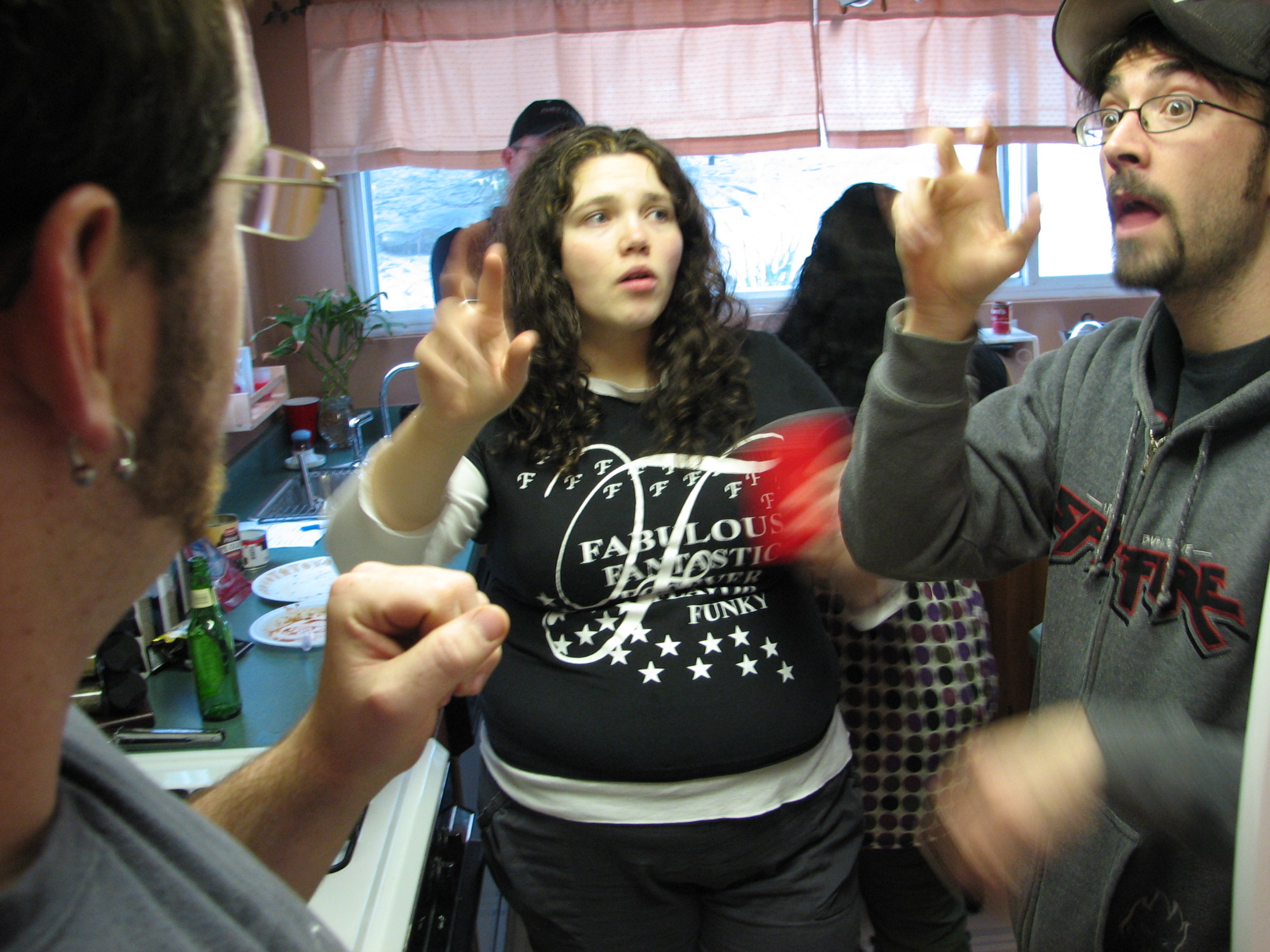
Sign language refers to any natural language which uses visual gestures produced by the hands and body language to express meaning. The brain's left side is the dominant side utilized for producing and understanding sign language, just as it is for speech. In 1861, Paul Broca studied patients with the ability to understand spoken languages but the inability to produce them. The damaged area was named Broca's area, and located in the left hemisphere’s inferior frontal gyrus (Brodmann areas 44, 45). Soon after, in 1874, Carl Wernicke studied patients with the reverse deficits: patients could produce spoken language, but could not comprehend it. The damaged area was named Wernicke's area, and is located in the left hemisphere’s posterior superior temporal gyrus (Brodmann area 22). Signers with damage in Broca's area have problems producing signs. Those with damage in the Wernicke's area (left hemisphere) in the temporal lobe of the brain have problems comprehending signed languages. ... [...More Info...] [...Related Items...] OR: [Wikipedia] [Google] [Baidu] |
Sign Language
Sign languages (also known as signed languages) are languages that use the visual-manual modality to convey meaning, instead of spoken words. Sign languages are expressed through manual articulation in combination with #Non-manual elements, non-manual markers. Sign languages are full-fledged natural languages with their own grammar and lexicon. Sign languages are not universal and are usually not mutual intelligibility, mutually intelligible, although there are similarities among different sign languages. Linguists consider both spoken and signed communication to be types of natural language, meaning that both emerged through an abstract, protracted aging process and evolved over time without meticulous planning. This is supported by the fact that there is substantial overlap between the neural substrates of sign and spoken language processing, despite the obvious differences in modality. Sign language should not be confused with body language, a type of non verbal communicati ... [...More Info...] [...Related Items...] OR: [Wikipedia] [Google] [Baidu] |
Auditory Cortex
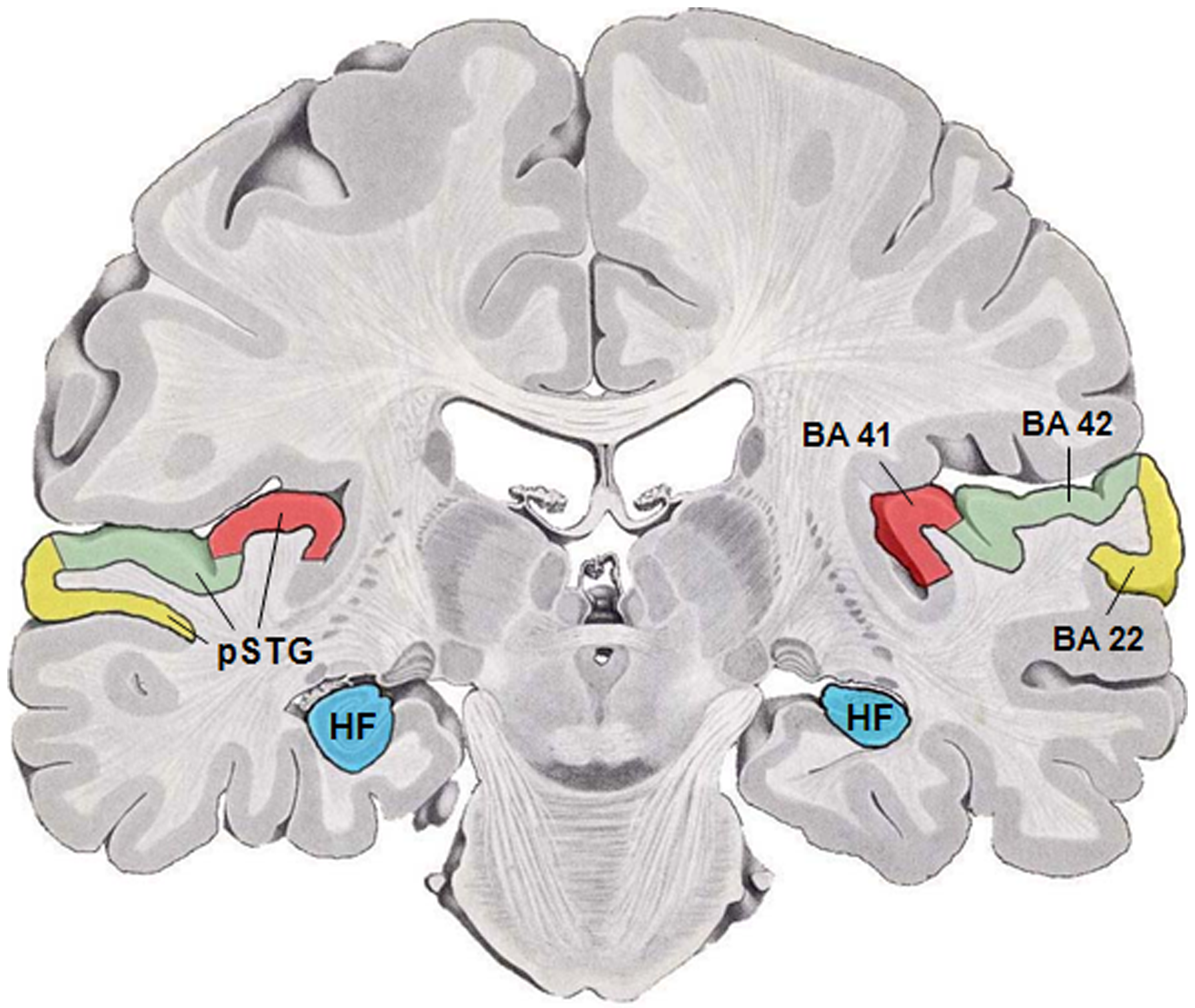
The auditory cortex is the part of the temporal lobe that processes auditory information in humans and many other vertebrates. It is a part of the auditory system, performing basic and higher functions in hearing, such as possible relations to language switching.Cf. Pickles, James O. (2012). ''An Introduction to the Physiology of Hearing'' (4th ed.). Bingley, UK: Emerald Group Publishing Limited, p. 238. It is located bilaterally, roughly at the upper sides of the temporal lobes – in humans, curving down and onto the medial surface, on the superior temporal plane, within the lateral sulcus and comprising parts of the transverse temporal gyri, and the superior temporal gyrus, including the planum polare and planum temporale (roughly Brodmann areas 41 and 42, and partially 22). The auditory cortex takes part in the spectrotemporal, meaning involving time and frequency, analysis of the inputs passed on from the ear. The cortex then filters and passes on the information to ... [...More Info...] [...Related Items...] OR: [Wikipedia] [Google] [Baidu] |
Calcarine Sulcus
The calcarine sulcus (or calcarine fissure) is an anatomical landmark located at the caudal end of the medial surface of the brain of humans and other primates. Its name comes from the Latin "calcar" meaning "spur". It is very deep, and known as a complete sulcus. Structure The calcarine sulcus begins near the occipital pole in two converging rami. It runs forward to a point a little below the splenium of the corpus callosum. Here, it is joined at an acute angle by the medial part of the parieto-occipital sulcus. The anterior part of this sulcus gives rise to the prominence of the calcar avis in the posterior cornu of the lateral ventricle. The cuneus is above the calcarine sulcus, while the lingual gyrus is below it. Development In humans, the calcarine sulcus usually becomes visible between 20 weeks and 28 weeks of gestation. Function The calcarine sulcus is associated with the visual cortex. It is where the primary visual cortex (V1) is concentrated. The central vi ... [...More Info...] [...Related Items...] OR: [Wikipedia] [Google] [Baidu] |
Superior Temporal Gyrus
The superior temporal gyrus (STG) is one of three (sometimes two) gyri in the temporal lobe of the human brain, which is located laterally to the head, situated somewhat above the external ear. The superior temporal gyrus is bounded by: * the lateral sulcus above; * the superior temporal sulcus (not always present or visible) below; * an imaginary line drawn from the preoccipital notch to the lateral sulcus posteriorly. The superior temporal gyrus contains several important structures of the brain, including: * Brodmann areas 41 and 42, marking the location of the auditory cortex, the cortical region responsible for the sensation of sound; * Wernicke's area, Brodmann area 22, an important region for the processing of speech so that it can be understood as language. The superior temporal gyrus contains the auditory cortex, which is responsible for processing sounds. Specific sound frequencies map precisely onto the auditory cortex. This auditory (or tonotopic) map is si ... [...More Info...] [...Related Items...] OR: [Wikipedia] [Google] [Baidu] |
Transverse Temporal Gyrus
The transverse temporal gyrus, also called Heschl's gyrus () or Heschl's convolutions, is a gyrus found in the area of each primary auditory cortex buried within the lateral sulcus of the human brain, occupying Brodmann areas 41 and 42. Transverse temporal gyri are superior to and separated from the planum temporale (cortex involved in language production) by Heschl's sulcus. Transverse temporal gyri are found in varying numbers in both the right and left hemispheres of the brain and one study found that this number is not related to the hemisphere or dominance of hemisphere studied in subjects. Transverse temporal gyri can be viewed in the sagittal plane as either an omega shape (if one gyrus is present) or a heart shape (if two gyri and a sulcus are present). Transverse temporal gyri are the first cortical structures to process incoming auditory information. Anatomically, the transverse temporal gyri are distinct in that they run mediolaterally (toward the center of the brai ... [...More Info...] [...Related Items...] OR: [Wikipedia] [Google] [Baidu] |
Visual Word Form Area
The visual word form area (VWFA) is a functional region of the left fusiform gyrus and surrounding cortex (right-hand side being part of the fusiform face area) that is hypothesized to be involved in identifying words and letters from lower-level shape images, prior to association with phonology or semantics. Because the alphabet is relatively new in human evolution, it is unlikely that this region developed as a result of selection pressures related to word recognition per se; however, this region may be highly specialized for certain types of shapes that occur naturally in the environment and are therefore likely to surface within written language. In addition to word recognition, the VWFA may participate in higher-level processing of word meaning. In 2003, functional imaging experiments raised doubts about whether the VWFA is an actual region. This skepticism has largely disappeared; however, there seems to be much variability in its size. An area that may fall within this ment ... [...More Info...] [...Related Items...] OR: [Wikipedia] [Google] [Baidu] |
Lateralization Of Brain Function
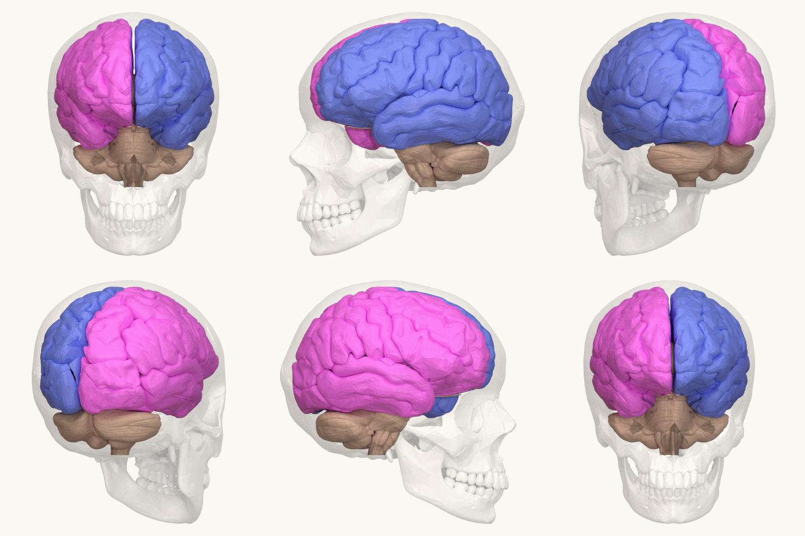
The lateralization of brain function (or hemispheric dominance/ lateralization) is the tendency for some neural functions or cognitive processes to be specialized to one side of the brain or the other. The median longitudinal fissure separates the human brain into two distinct cerebral hemispheres connected by the corpus callosum. Both hemispheres exhibit Brain asymmetry, brain asymmetries in both structure and neuronal network composition associated with specialized function. Lateralization of brain structures has been studied using both healthy and split-brain patients. However, there are numerous counterexamples to each generalization and each human's brain develops differently, leading to unique lateralization in individuals. This is different from specialization, as lateralization refers only to the function of one structure divided between two hemispheres. Specialization is much easier to observe as a trend, since it has a stronger Anthropology, anthropological history. T ... [...More Info...] [...Related Items...] OR: [Wikipedia] [Google] [Baidu] |
Event-related Potential
An event-related potential (ERP) is the measured brain response that is the direct result of a specific sense, sensory, cognition, cognitive, or motor system, motor event. More formally, it is any stereotyped electrophysiology, electrophysiological response to a stimulus. The study of the brain in this way provides a Invasiveness of surgical procedures, noninvasive means of evaluating brain functioning. ERPs are measured by means of electroencephalography (EEG). The magnetoencephalography (MEG) equivalent of ERP is the ERF, or event-related field. Evoked potentials and induced potentials are subtypes of ERPs. History With the discovery of the electroencephalogram (EEG) in 1924, Hans Berger revealed that one could measure the electrical activity of the human brain by placing electrodes on the scalp and amplifying the signal. Changes in voltage can then be plotted over a period of time. He observed that the voltages could be influenced by external events that stimulated the sense ... [...More Info...] [...Related Items...] OR: [Wikipedia] [Google] [Baidu] |
Electroencephalography
Electroencephalography (EEG) is a method to record an electrogram of the spontaneous electrical activity of the brain. The biosignal, bio signals detected by EEG have been shown to represent the postsynaptic potentials of pyramidal neurons in the neocortex and allocortex. It is typically non-invasive, with the EEG electrodes placed along the scalp (commonly called "scalp EEG") using the 10–20 system (EEG), International 10–20 system, or variations of it. Electrocorticography, involving surgical placement of electrodes, is sometimes called Electrocorticography, "intracranial EEG". Clinical interpretation of EEG recordings is most often performed by visual inspection of the tracing or quantitative EEG, quantitative EEG analysis. Voltage fluctuations measured by the EEG bioamplifier, bio amplifier and electrodes allow the evaluation of normal Brain activity and meditation, brain activity. As the electrical activity monitored by EEG originates in neurons in the underlying Huma ... [...More Info...] [...Related Items...] OR: [Wikipedia] [Google] [Baidu] |
Prosody (linguistics)
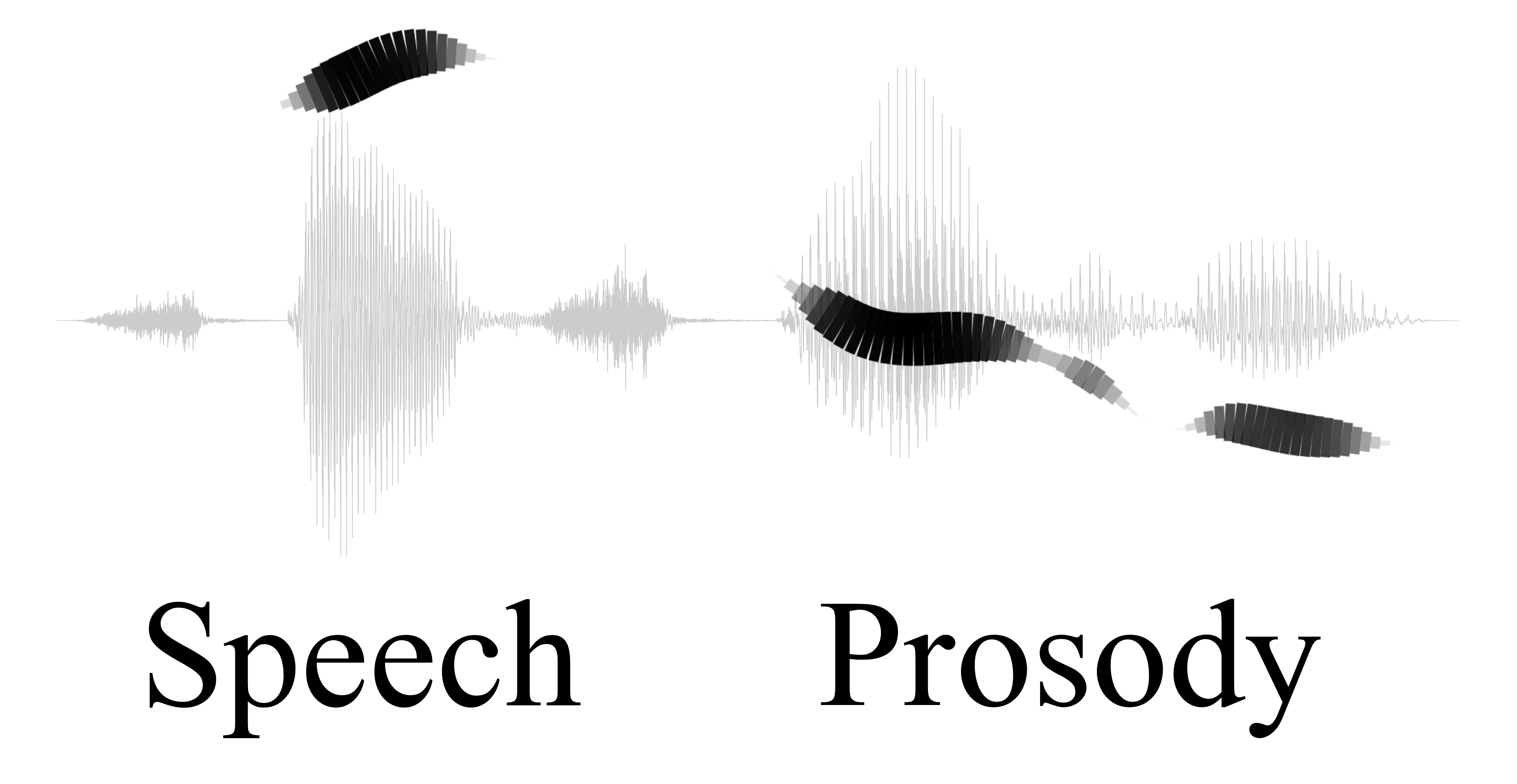
In linguistics, prosody () is the study of elements of speech, including intonation, stress, rhythm and loudness, that occur simultaneously with individual phonetic segments: vowels and consonants. Often, prosody specifically refers to such elements, known as ''suprasegmentals'', when they extend across more than one phonetic segment. Prosody reflects the nuanced emotional features of the speaker or of their utterances: their obvious or underlying emotional state, the form of utterance (statement, question, or command), the presence of irony or sarcasm, certain emphasis on words or morphemes, contrast, focus, and so on. Prosody displays elements of language that are not encoded by grammar, punctuation or choice of vocabulary. Attributes of prosody In the study of prosodic aspects of speech, it is usual to distinguish between auditory measures ( subjective impressions produced in the mind of the listener) and objective measures (physical properties of the sound wave and ... [...More Info...] [...Related Items...] OR: [Wikipedia] [Google] [Baidu] |
Cohesion (linguistics)
Cohesion is the grammar, grammatical and Lexicon, lexical linking within a text or sentence (linguistics), sentence that holds a text together and gives it meaning. It is related to the broader concept of coherence (linguistics), coherence. There are two main types of cohesion: * grammatical cohesion: based on structural content * lexical cohesion: based on lexical content and background knowledge. A cohesive text is created in many different ways. In ''Cohesion in English'', M.A.K. Halliday and Ruqaiya Hasan identify five general categories of cohesive devices that create coherence in texts: reference, ellipsis (narrative device), ellipsis, substitution, lexical cohesion and grammatical conjunction, conjunction. Referencing There are two referential devices that can create cohesion: *anaphora (linguistics), Anaphoric reference occurs when the writer refers back to someone or something that has been previously identified, to avoid repetition. Some examples: replacing "the taxi dri ... [...More Info...] [...Related Items...] OR: [Wikipedia] [Google] [Baidu] |


