|
Quadrants And Regions Of Abdomen
The human abdomen is divided into quadrants and regions by anatomists and physicians for the purposes of study, diagnosis, and treatment. The division into four quadrants allows the localisation of pain and tenderness, scars, lumps, and other items of interest, narrowing in on which organs and tissues may be involved. The quadrants are referred to as the left lower quadrant, left upper quadrant, right upper quadrant and right lower quadrant. These terms are not used in comparative anatomy, since most other animals do not stand erect. The left lower quadrant includes the left iliac fossa and half of the flank. The equivalent in other animals is ''left posterior quadrant''. The left upper quadrant extends from the umbilical plane to the left ribcage. This is the ''left anterior quadrant'' in other animals. The right upper quadrant extends from umbilical plane to the right ribcage. The equivalent in other animals is ''right anterior quadrant''. The right lower quadrant ex ... [...More Info...] [...Related Items...] OR: [Wikipedia] [Google] [Baidu] |
Abdomen
The abdomen (colloquially called the belly, tummy, midriff, tucky or stomach) is the part of the body between the thorax (chest) and pelvis, in humans and in other vertebrates. The abdomen is the front part of the abdominal segment of the torso. The area occupied by the abdomen is called the abdominal cavity. In arthropods it is the posterior tagma of the body; it follows the thorax or cephalothorax. In humans, the abdomen stretches from the thorax at the thoracic diaphragm to the pelvis at the pelvic brim. The pelvic brim stretches from the lumbosacral joint (the intervertebral disc between L5 and S1) to the pubic symphysis and is the edge of the pelvic inlet. The space above this inlet and under the thoracic diaphragm is termed the abdominal cavity. The boundary of the abdominal cavity is the abdominal wall in the front and the peritoneal surface at the rear. In vertebrates, the abdomen is a large body cavity enclosed by the abdominal muscles, at front and to ... [...More Info...] [...Related Items...] OR: [Wikipedia] [Google] [Baidu] |
Body Of Pancreas
The pancreas is an organ of the digestive system and endocrine system of vertebrates. In humans, it is located in the abdomen behind the stomach and functions as a gland. The pancreas is a mixed or heterocrine gland, i.e. it has both an endocrine and a digestive exocrine function. 99% of the pancreas is exocrine and 1% is endocrine. As an endocrine gland, it functions mostly to regulate blood sugar levels, secreting the hormones insulin, glucagon, somatostatin, and pancreatic polypeptide. As a part of the digestive system, it functions as an exocrine gland secreting pancreatic juice into the duodenum through the pancreatic duct. This juice contains bicarbonate, which neutralizes acid entering the duodenum from the stomach; and digestive enzymes, which break down carbohydrates, proteins, and fats in food entering the duodenum from the stomach. Inflammation of the pancreas is known as pancreatitis, with common causes including chronic alcohol use and gallstones. Becau ... [...More Info...] [...Related Items...] OR: [Wikipedia] [Google] [Baidu] |
Left Lobe Of Liver
In human anatomy, the liver is divided grossly into four parts or lobes: the right lobe, the left lobe, the caudate lobe, and the quadrate lobe. Seen from the front – the diaphragmatic surface - the liver is divided into two lobes: the right lobe and the left lobe. Viewed from the underside – the visceral surface, the other two smaller lobes the caudate lobe, and the quadrate lobe are also visible. The two smaller lobes, the caudate lobe and the quadrate lobe, are known as superficial or accessory lobes, and both are located on the underside of the right lobe. The falciform ligament, visible on the front of the liver, makes a superficial division of the right and left lobes of the liver. From the underside, the two additional lobes are located on the right lobe. A line can be imagined running from the left of the vena cava and all the way forward to divide the liver and gallbladder into two halves. This line is called Cantlie's line and is used to mark the division between t ... [...More Info...] [...Related Items...] OR: [Wikipedia] [Google] [Baidu] |
Spleen
The spleen is an organ found in almost all vertebrates. Similar in structure to a large lymph node, it acts primarily as a blood filter. The word spleen comes .σπλήν Henry George Liddell, Robert Scott, ''A Greek-English Lexicon'', on Perseus Digital Library The spleen plays very important roles in regard to red blood cells (erythrocytes) and the . It removes old red blood cells and holds a reserve of blood, which can be valuable in case of [...More Info...] [...Related Items...] OR: [Wikipedia] [Google] [Baidu] |
Stomach
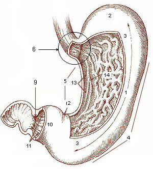
The stomach is a muscular, hollow organ in the gastrointestinal tract of humans and many other animals, including several invertebrates. The stomach has a dilated structure and functions as a vital organ in the digestive system. The stomach is involved in the gastric phase of digestion, following chewing. It performs a chemical breakdown by means of enzymes and hydrochloric acid. In humans and many other animals, the stomach is located between the oesophagus and the small intestine. The stomach secretes digestive enzymes and gastric acid to aid in food digestion. The pyloric sphincter controls the passage of partially digested food (chyme) from the stomach into the duodenum, where peristalsis takes over to move this through the rest of intestines. Structure In the human digestive system, the stomach lies between the oesophagus and the duodenum (the first part of the small intestine). It is in the left upper quadrant of the abdominal cavity. The top of the stomach lies ag ... [...More Info...] [...Related Items...] OR: [Wikipedia] [Google] [Baidu] |
Median Plane
The median plane also called a mid-sagittal plane is used to describe the sagittal plane as it bisects the body vertically through the midline marked by the navel, dividing the body exactly in left and right side. The term parasagittal plane is used to refer to any plane parallel to the sagittal and median plane. It is one of the lines used to define the right upper quadrant of the human abdomen. The midsternal line can be interpreted as a segment of the median plane. File:Sagittal brain MRI.jpg, Median plane magnetic resonance imaging of the head. File:Median plane CT scan of a pregnancy of 37 weeks of gestational age.jpg, Median plane CT scan of a pregnant woman. The fetus (exposed in the coronal plane The coronal plane (also known as the frontal plane) is an anatomical plane that divides the body into dorsal and ventral sections. It is perpendicular to the sagittal and transverse planes. Details The coronal plane is an example of a longitud ...) is 37 weeks of gest ... [...More Info...] [...Related Items...] OR: [Wikipedia] [Google] [Baidu] |
Ureter
The ureters are tubes made of smooth muscle that propel urine from the kidneys to the urinary bladder. In a human adult, the ureters are usually long and around in diameter. The ureter is lined by urothelial cells, a type of transitional epithelium, and has an additional smooth muscle layer that assists with peristalsis in its lowest third. The ureters can be affected by a number of diseases, including urinary tract infections and kidney stone. is when a ureter is narrowed, due to for example chronic inflammation. Congenital abnormalities that affect the ureters can include the development of two ureters on the same side or abnormally placed ureters. Additionally, reflux of urine from the bladder back up the ureters is a condition commonly seen in children. The ureters have been identified for at least two thousand years, with the word "ureter" stemming from the stem relating to urinating and seen in written records since at least the time of Hippocrates. It is, however, ... [...More Info...] [...Related Items...] OR: [Wikipedia] [Google] [Baidu] |
Fallopian Tube
The fallopian tubes, also known as uterine tubes, oviducts or salpinges (singular salpinx), are paired tubes in the human female that stretch from the uterus to the ovaries. The fallopian tubes are part of the female reproductive system. In other mammals they are only called oviducts. Each tube is a muscular hollow organ that is on average between 10 and 14 cm in length, with an external diameter of 1 cm. It has four described parts: the intramural part, isthmus, ampulla, and infundibulum with associated fimbriae. Each tube has two openings a proximal opening nearest and opening to the uterus, and a distal opening furthest and opening to the abdomen. The fallopian tubes are held in place by the mesosalpinx, a part of the broad ligament mesentery that wraps around the tubes. Another part of the broad ligament, the mesovarium suspends the ovaries in place. An egg cell is transported from an ovary to a fallopian tube where it may be fertilized in the ampulla of th ... [...More Info...] [...Related Items...] OR: [Wikipedia] [Google] [Baidu] |
Ovary
The ovary is an organ in the female reproductive system that produces an ovum. When released, this travels down the fallopian tube into the uterus, where it may become fertilized by a sperm. There is an ovary () found on each side of the body. The ovaries also secrete hormones that play a role in the menstrual cycle and fertility. The ovary progresses through many stages beginning in the prenatal period through menopause. It is also an endocrine gland because of the various hormones that it secretes. Structure The ovaries are considered the female gonads. Each ovary is whitish in color and located alongside the lateral wall of the uterus in a region called the ovarian fossa. The ovarian fossa is the region that is bounded by the external iliac artery and in front of the ureter and the internal iliac artery. This area is about 4 cm x 3 cm x 2 cm in size.Daftary, Shirish; Chakravarti, Sudip (2011). Manual of Obstetrics, 3rd Edition. Elsevier. pp. 1-16. . The ... [...More Info...] [...Related Items...] OR: [Wikipedia] [Google] [Baidu] |
Sigmoid Colon
The sigmoid colon (or pelvic colon) is the part of the large intestine that is closest to the rectum and anus. It forms a loop that averages about in length. The loop is typically shaped like a Greek letter sigma (ς) or Latin letter S (thus '' sigma'' + '' -oid''). This part of the colon normally lies within the pelvis, but due to its freedom of movement it is liable to be displaced into the abdominal cavity. Structure The sigmoid colon begins at the superior aperture of the lesser pelvis, where it is continuous with the iliac colon, and passes transversely across the front of the sacrum to the right side of the pelvis. It then curves on itself and turns toward the left to reach the middle line at the level of the third piece of the sacrum, where it bends downward and ends in the rectum. Its function is to expel solid and gaseous waste from the gastrointestinal tract. The curving path it takes toward the anus allows it to store gas in the superior arched portion, enab ... [...More Info...] [...Related Items...] OR: [Wikipedia] [Google] [Baidu] |
Descending Colon
In the anatomy of humans and homologous primates, the descending colon is the part of the colon extending from the left colic flexure to the level of the iliac crest (whereupon it transitions into the sigmoid colon). The function of the descending colon in the digestive system is to store the remains of digested food that will be emptied into the rectum. The descending colon is on the left side of the body (barring any malformations). The term left colon is hypernymous to ''descending colon'' in precise use; many casual mentions of the left colon chiefly concern the descending colon. Structure The descending colon extends from the left colic flexure at the upper left part of the abdomen inferior-ward through the left hypochondrium and lumbar regions, along the outer border of the left kidney, ending at the level of the iliac crest at the lower left part of the abdomen, being contunued thenceforth as the sigmoid colon. It usually retroperitoneal (being lined by peritoneu ... [...More Info...] [...Related Items...] OR: [Wikipedia] [Google] [Baidu] |


