|
Pneumatic Otoscopy
The pneumatic otoscope is the standard tool used in diagnosing otitis media Otitis media is a group of inflammatory diseases of the middle ear. One of the two main types is acute otitis media (AOM), an infection of rapid onset that usually presents with ear pain. In young children this may result in pulling at the ear, .... In addition to the pneumatic (diagnostic) head, a surgical head also is useful. The pneumatic head contains a lens, an enclosed light source, and a nipple for attachment of a rubber bulb and tubing. The head is designed so that when a speculum is attached and fitted snugly into the patient’s external auditory canal, an air-tight chamber is produced. In some cases, the addition of a small sleeve of rubber tubing at the end of the plastic speculum or use of a rubber-tipped speculum helps to avoid trauma and improve the air-tight seal. Gently squeezing and releasing the rubber bulb in rapid succession permits observation of the degree of eardrum mobility in re ... [...More Info...] [...Related Items...] OR: [Wikipedia] [Google] [Baidu] |
Otoscope
An otoscope or auriscope is a medical device which is used to look into the ears. Health care providers use otoscopes to screen for illness during regular check-ups and also to investigate ear symptoms. An otoscope potentially gives a view of the ear canal and tympanic membrane or eardrum. Because the eardrum is the border separating the external ear canal from the middle ear, its characteristics can be indicative of various diseases of the middle ear space. The presence of earwax (cerumen), shed skin, pus, canal skin edema, foreign body, and various ear diseases can obscure any view of the eardrum and thus severely compromise the value of otoscopy done with a common otoscope, but confirm the presence of obstructing symptoms. The most commonly used otoscopes consist of a handle and a head. The head contains a light source and a simple low-power magnifying glass, magnifying lens, typically around 8 diopters (3.00x Mag). The Anatomical terms of location#Proximal and distal ... [...More Info...] [...Related Items...] OR: [Wikipedia] [Google] [Baidu] |
Otitis Media
Otitis media is a group of inflammatory diseases of the middle ear. One of the two main types is acute otitis media (AOM), an infection of rapid onset that usually presents with ear pain. In young children this may result in pulling at the ear, increased crying, and poor sleep. Decreased eating and a fever may also be present. The other main type is otitis media with effusion (OME), typically not associated with symptoms, although occasionally a feeling of fullness is described; it is defined as the presence of non-infectious fluid in the middle ear which may persist for weeks or months often after an episode of acute otitis media. Chronic suppurative otitis media (CSOM) is middle ear inflammation that results in a perforated tympanic membrane with discharge from the ear for more than six weeks. It may be a complication of acute otitis media. Pain is rarely present. All three types of otitis media may be associated with hearing loss. If children with hearing loss due to OME do n ... [...More Info...] [...Related Items...] OR: [Wikipedia] [Google] [Baidu] |
Speculum (medical)
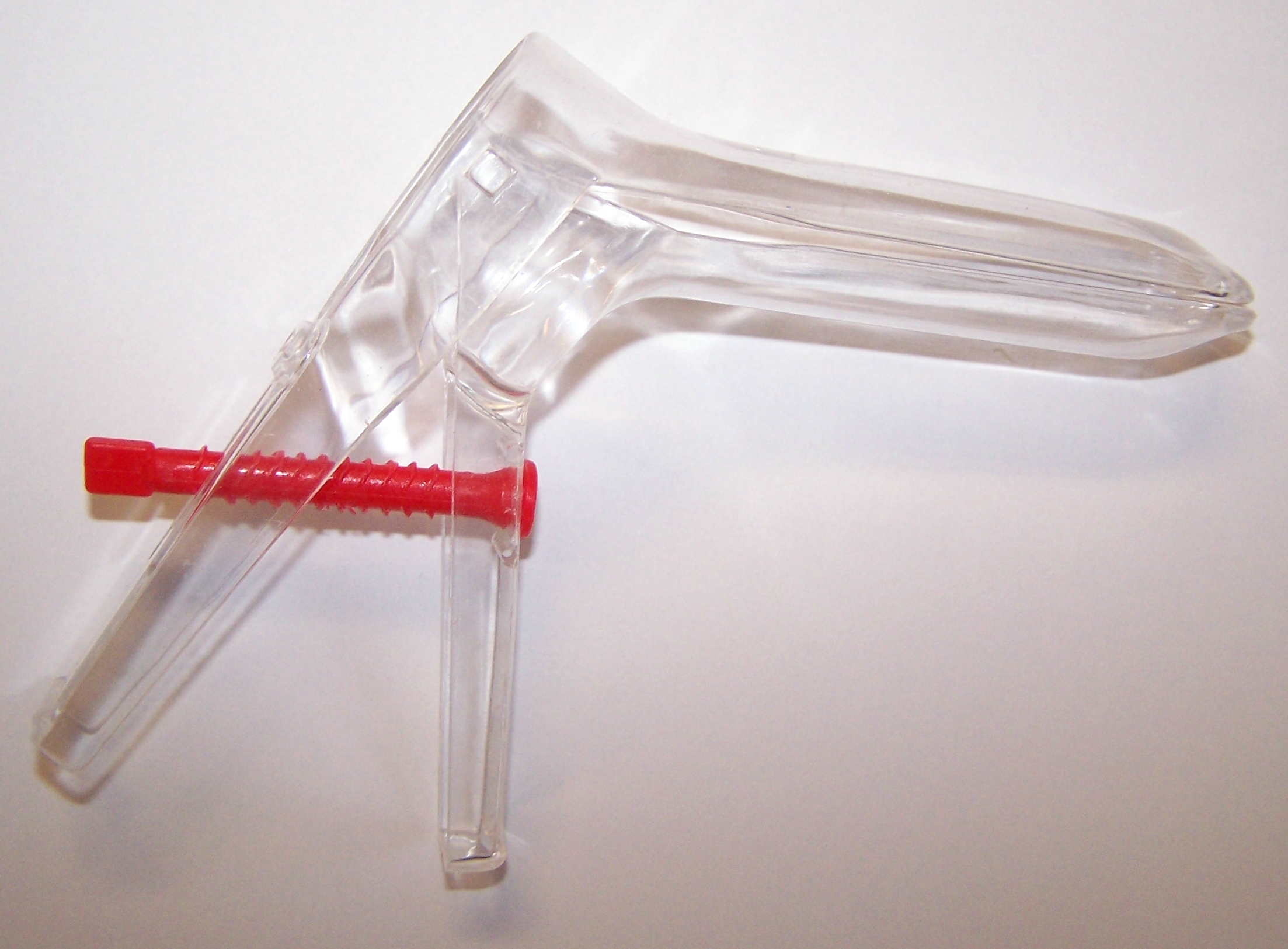
A speculum (Latin for 'mirror'; plural specula or speculums) is a medical tool for investigating body orifices, with a form dependent on the orifice for which it is designed. In old texts, the speculum may also be referred to as a diopter or dioptra. Like an endoscope, a speculum allows a view inside the body; endoscopes, however, tend to have optics while a speculum is intended for direct vision. History Vaginal and anal specula were used by the ancient Greeks and Romans, and speculum artifacts have been found in Pompeii. A vaginal speculum, developed by J. Marion Sims, consists of a hollow cylinder with a rounded end that is divided into two hinged parts, somewhat like the beak of a duck. This speculum is inserted into the vagina to dilate it for examination of the vagina and cervix. The modern vaginal speculum was developed by J. Marion Sims, a plantation doctor in Lancaster County, South Carolina. Between 1845 and 1849, Sims performed dozens of surgeries, without anest ... [...More Info...] [...Related Items...] OR: [Wikipedia] [Google] [Baidu] |
Ear Procedures
An ear is the organ that enables hearing and, in mammals, body balance using the vestibular system. In mammals, the ear is usually described as having three parts—the outer ear, the middle ear and the inner ear. The outer ear consists of the pinna and the ear canal. Since the outer ear is the only visible portion of the ear in most animals, the word "ear" often refers to the external part alone. The middle ear includes the tympanic cavity and the three ossicles. The inner ear sits in the bony labyrinth, and contains structures which are key to several senses: the semicircular canals, which enable balance and eye tracking when moving; the utricle and saccule, which enable balance when stationary; and the cochlea, which enables hearing. The ears of vertebrates are placed somewhat symmetrically on either side of the head, an arrangement that aids sound localisation. The ear develops from the first pharyngeal pouch and six small swellings that develop in the early embryo ... [...More Info...] [...Related Items...] OR: [Wikipedia] [Google] [Baidu] |


