|
Perlia's Nucleus
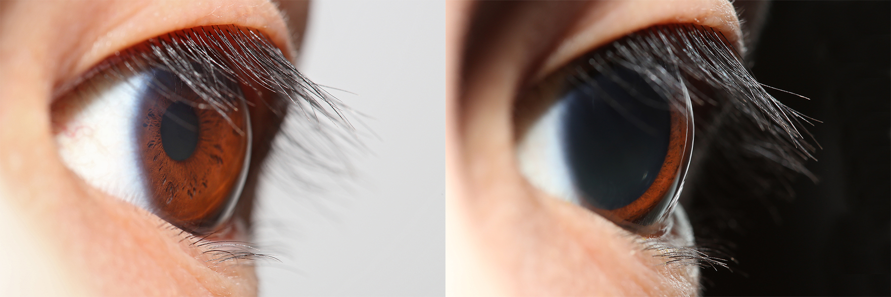
Perlia's nucleus, also known as nucleus of Perlia and abbreviated as NP, is a spindle-shaped Nucleus (neuroanatomy), nucleus located in the Midbrain, mesencephalon, a subdivision of the Edinger-Westphal nucleus situated between the right and left Oculomotor nucleus, oculomotor nuclei. It is implicated in parasympathetic oculomotor functions, possibly including input to the Iris (anatomy), iris and ciliary muscle, ciliary. Perlia's nucleus is believed to be a characteristic found exclusively in animals capable of binocular vision. Moreover, it might be an exclusive characteristic of humans, as indicated by a systematic study of monkey brains, where only 9% exhibited a clear midline group, potentially corresponding to the NP. In 1891, Perlia's nucleus was identified as a central mediator for the convergent movement of the eyes based on clinical findings in ophthalmospegias. It has also recently been attributed an important role in the upward movement or gaze of the eyes. Structure ... [...More Info...] [...Related Items...] OR: [Wikipedia] [Google] [Baidu] |
Nucleus (neuroanatomy)
In neuroanatomy, a nucleus (: nuclei) is a cluster of neurons in the central nervous system, located deep within the cerebral hemispheres and brainstem. The neurons in one nucleus usually have roughly similar connections and functions. Nuclei are connected to other nuclei by tracts, the bundles (fascicles) of axons (nerve fibers) extending from the cell bodies. A nucleus is one of the two most common forms of nerve cell organization, the other being layered structures such as the cerebral cortex or cerebellar cortex. In anatomical sections, a nucleus shows up as a region of gray matter, often bordered by white matter. The vertebrate brain contains hundreds of distinguishable nuclei, varying widely in shape and size. A nucleus may itself have a complex internal structure, with multiple types of neurons arranged in clumps (subnuclei) or layers. The term "nucleus" is in some cases used rather loosely, to mean simply an identifiably distinct group of neurons, even if they are sprea ... [...More Info...] [...Related Items...] OR: [Wikipedia] [Google] [Baidu] |
Motor Neuron
A motor neuron (or motoneuron), also known as efferent neuron is a neuron whose cell body is located in the motor cortex, brainstem or the spinal cord, and whose axon (fiber) projects to the spinal cord or outside of the spinal cord to directly or indirectly control effector organs, mainly muscles and glands. There are two types of motor neuron – upper motor neurons and lower motor neurons. Axons from upper motor neurons synapse onto interneurons in the spinal cord and occasionally directly onto lower motor neurons. The axons from the lower motor neurons are efferent nerve fibers that carry signals from the spinal cord to the effectors. Types of lower motor neurons are alpha motor neurons, beta motor neurons, and gamma motor neurons. A single motor neuron may innervate many muscle fibres and a muscle fibre can undergo many action potentials in the time taken for a single muscle twitch. Innervation takes place at a neuromuscular junction and twitches can become superimpo ... [...More Info...] [...Related Items...] OR: [Wikipedia] [Google] [Baidu] |
Edinger–Westphal Nucleus
The Edinger–Westphal nucleus also called the accessory or visceral oculomotor nerve, is one of the two nuclei of the oculomotor nerve (CN III) located in the midbrain. It receives afferents from both pretectal nuclei (which have in turn received afferents from the optic tract). It contains parasympathetic pre-ganglionic neuron cell bodies that synapse in the ciliary ganglion. It contributes the autonomic, parasympathetic component to the oculomotor nerve (CN III), ultimately providing innervation to the iris sphincter muscle and ciliary muscle to mediate the pupillary light reflex and accommodation, respectively. The Edinger–Westphal nucleus has two parts. The first is of preganglionic fibers (EWpg) that terminate in the ciliary ganglion. The second is of centrally projecting cells (EWcp) that project to a number of brainstem structures. Structure The Edinger–Westphal nucleus refers to the adjacent population of non-preganglionic neurons that do not project to the cilia ... [...More Info...] [...Related Items...] OR: [Wikipedia] [Google] [Baidu] |
Richard Perlia
Richard is a male given name. It originates, via Old French, from Old Frankish and is a compound of the words descending from Proto-Germanic language">Proto-Germanic ''*rīk-'' 'ruler, leader, king' and ''*hardu-'' 'strong, brave, hardy', and it therefore means 'strong in rule'. Nicknames include "Richie", " Dick", "Dickon", " Dickie", "Rich", "Rick", "Rico (name), Rico", " Ricky", and more. Richard is a common English (the name was introduced into England by the Normans), German and French male name. It's also used in many more languages, particularly Germanic, such as Norwegian, Danish, Swedish, Icelandic, and Dutch, as well as other languages including Irish, Scottish, Welsh and Finnish. Richard is cognate with variants of the name in other European languages, such as the Swedish "Rickard", the Portuguese and Spanish "Ricardo" and the Italian "Riccardo" (see comprehensive variant list below). People named Richard Multiple people with the same name * Richard Andersen ( ... [...More Info...] [...Related Items...] OR: [Wikipedia] [Google] [Baidu] |
Somatic Cell
In cellular biology, a somatic cell (), or vegetal cell, is any biological cell forming the body of a multicellular organism other than a gamete, germ cell, gametocyte or undifferentiated stem cell. Somatic cells compose the body of an organism and divide through mitosis. In contrast, gametes derive from meiosis within the germ cells of the germline and they fuse during sexual reproduction. Stem cells also can divide through mitosis, but are different from somatic in that they differentiate into diverse specialized cell types. In mammals, somatic cells make up all the internal organs, skin, bones, blood and connective tissue, while mammalian germ cells give rise to spermatozoa and ova which fuse during fertilization to produce a cell called a zygote, which divides and differentiates into the cells of an embryo. There are approximately 220 types of somatic cell in the human body. Theoretically, these cells are not germ cells (the source of gametes); they transmit their mut ... [...More Info...] [...Related Items...] OR: [Wikipedia] [Google] [Baidu] |
Von Economo Neuron
Von Economo neurons, also called spindle neurons, are a specific class of mammalian cortical neurons characterized by a large spindle-shaped soma (or body) gradually tapering into a single apical axon (the ramification that ''transmits'' signals) in one direction, with only a single dendrite (the ramification that ''receives'' signals) facing opposite. Other cortical neurons tend to have many dendrites, and the bipolar-shaped morphology of von Economo neurons is unique here. Von Economo neurons are found in two very restricted regions in the brains of hominids (humans and other great apes): the anterior cingulate cortex (ACC) and the fronto-insular cortex (FI) (which each make up the salience network). In 2008, they were also found in the dorsolateral prefrontal cortex of humans. Von Economo neurons are also found in the brains of a number of cetaceans, African and Asian elephants, and to a lesser extent in macaque monkeys and raccoons. The appearance of von Economo neur ... [...More Info...] [...Related Items...] OR: [Wikipedia] [Google] [Baidu] |
Bipolar Neuron
A bipolar neuron, or bipolar cell, is a type of neuron characterized by having both an axon and a dendrite extending from the soma (cell body) in opposite directions. These neurons are predominantly found in the retina and olfactory system. The embryological period encompassing weeks seven through eight marks the commencement of bipolar neuron development. Many bipolar cells are specialized sensory neurons (afferent neurons) for the transmission of sense. As such, they are part of the sensory pathways for smell, sight, taste, hearing, touch, balance and proprioception. The other shape classifications of neurons include unipolar, pseudounipolar and multipolar. During embryonic development, pseudounipolar neurons begin as bipolar in shape but become pseudounipolar as they mature. Common examples are the retina bipolar cell, the spiral ganglion and vestibular ganglion of the vestibulocochlear nerve (cranial nerve VIII), the extensive use of bipolar cells to transmit efferent ... [...More Info...] [...Related Items...] OR: [Wikipedia] [Google] [Baidu] |
Multipolar Neuron
A multipolar neuron is a type of neuron that possesses a single axon and many dendrites (and dendritic branches), allowing for the integration of a great deal of information from other neurons. These processes are projections from the neuron cell body. Multipolar neurons constitute the majority of neurons in the central nervous system. They include motor neurons, and also interneurons (relay neurons), which are most commonly found in the cortex of the brain and the spinal cord The spinal cord is a long, thin, tubular structure made up of nervous tissue that extends from the medulla oblongata in the lower brainstem to the lumbar region of the vertebral column (backbone) of vertebrate animals. The center of the spinal c .... Peripherally, multipolar neurons are found in autonomic ganglia. See also * Dogiel cells * Ganglion cell * Purkinje cell * Pyramidal cell Additional images File:Blausen 0672 NeuralTissue.png, Neural tissue References External links DiagramDi ... [...More Info...] [...Related Items...] OR: [Wikipedia] [Google] [Baidu] |
Binocular Vision
Binocular vision is seeing with two eyes. The Field_of_view, field of view that can be surveyed with two eyes is greater than with one eye. To the extent that the visual fields of the two eyes overlap, #Depth, binocular depth can be perceived. This allows objects to be recognized more quickly, camouflage to be detected, spatial relationships to be perceived more quickly and accurately (#Stereopsis, stereopsis) and perception to be less susceptible to optical illusions, optical illusions. In secion #Medical, Medical attention is paid to the occurrence, defects and sharpness of binocular vision. In section #Biological, Biological the occurrence of binocular vision in animals is described. Geometric terms When the left eye (LE) and the right eye (RE) observe two objects X and Y, the following concepts are important:Krol J.D.(1982),"Perceptual ghosts in stereopsis, a ghosly problem in binocular vision", PhD thesis ISBN 90-9000382-7.Koenderink J.J.;van Doorn A.J. (1976) "Geometry of ... [...More Info...] [...Related Items...] OR: [Wikipedia] [Google] [Baidu] |
Midbrain
The midbrain or mesencephalon is the uppermost portion of the brainstem connecting the diencephalon and cerebrum with the pons. It consists of the cerebral peduncles, tegmentum, and tectum. It is functionally associated with vision, hearing, motor control, sleep and wakefulness, arousal (alertness), and temperature regulation.Breedlove, Watson, & Rosenzweig. Biological Psychology, 6th Edition, 2010, pp. 45-46 The name ''mesencephalon'' comes from the Greek ''mesos'', "middle", and ''enkephalos'', "brain". Structure The midbrain is the shortest segment of the brainstem, measuring less than 2cm in length. It is situated mostly in the posterior cranial fossa, with its superior part extending above the tentorial notch. The principal regions of the midbrain are the tectum, the cerebral aqueduct, tegmentum, and the cerebral peduncles. Rostral and caudal, Rostrally the midbrain adjoins the diencephalon (thalamus, hypothalamus, etc.), while Rostral and caudal, cau ... [...More Info...] [...Related Items...] OR: [Wikipedia] [Google] [Baidu] |
Ciliary Muscle
The ciliary muscle is an intrinsic muscle of the eye formed as a ring of smooth muscleSchachar, Ronald A. (2012). "Anatomy and Physiology." (Chapter 4) . in the eye's middle layer, the uvea ( vascular layer). It controls accommodation for viewing objects at varying distances and regulates the flow of aqueous humor into Schlemm's canal. It also changes the shape of the lens within the eye but not the size of the pupil which is carried out by the sphincter pupillae muscle and dilator pupillae. The ciliary muscle, pupillary sphincter muscle and pupillary dilator muscle sometimes are called intrinsic ocular muscles or intraocular muscles. Structure Development The ciliary muscle develops from mesenchyme within the choroid and is considered a cranial neural crest derivative. Nerve supply The ciliary muscle receives parasympathetic fibers from the short ciliary nerves that arise from the ciliary ganglion. The parasympathetic postganglionic fibers are part of cranial n ... [...More Info...] [...Related Items...] OR: [Wikipedia] [Google] [Baidu] |
Iris (anatomy)
The iris (: irides or irises) is a thin, annular structure in the eye in most mammals and birds that is responsible for controlling the diameter and size of the pupil, and thus the amount of light reaching the retina. In optical terms, the pupil is the eye's aperture, while the iris is the diaphragm (optics), diaphragm. Eye color is defined by the iris. Etymology The word "iris" is derived from the Greek word for "rainbow", also Iris (mythology), its goddess plus messenger of the gods in the ''Iliad'', because of the many eye color, colours of this eye part. Structure The iris consists of two layers: the front pigmented Wikt:fibrovascular, fibrovascular layer known as a stroma of iris, stroma and, behind the stroma, pigmented epithelial cells. The stroma is connected to a sphincter muscle (sphincter pupillae), which contracts the pupil in a circular motion, and a set of dilator muscles (dilator pupillae), which pull the iris radially to enlarge the pupil, pulling it in folds. ... [...More Info...] [...Related Items...] OR: [Wikipedia] [Google] [Baidu] |


