|
Parathyroid Hormone 1 Receptor
Parathyroid hormone/parathyroid hormone-related peptide receptor, also known as parathyroid hormone 1 receptor (PTH1R), is a protein that in humans is encoded by the ''PTH1R'' gene. PTH1R functions as a receptor for parathyroid hormone ( PTH) and for parathyroid hormone-related protein (PTHrP), also called parathyroid hormone-like hormone (PTHLH). Function This "classical" PTH receptor is expressed in high levels in bone and kidney and regulates calcium ion homeostasis through activation of adenylate cyclase and phospholipase C. In bone, it is expressed on the surface of osteoblasts. When the receptor is activated through PTH binding, osteoblasts express RANKL (Receptor Activator of Nuclear Factor kB Ligand), which binds to RANK (Receptor Activator of Nuclear Factor kB) on osteoclasts. This turns on osteoclasts to ultimately increase the resorption rate. Mechanism It is a member of the secretin family of G protein-coupled receptors. The activity of this receptor is mediated by ... [...More Info...] [...Related Items...] OR: [Wikipedia] [Google] [Baidu] |
Protein
Proteins are large biomolecules and macromolecules that comprise one or more long chains of amino acid residue (biochemistry), residues. Proteins perform a vast array of functions within organisms, including Enzyme catalysis, catalysing metabolic reactions, DNA replication, Cell signaling, responding to stimuli, providing Cytoskeleton, structure to cells and Fibrous protein, organisms, and Intracellular transport, transporting molecules from one location to another. Proteins differ from one another primarily in their sequence of amino acids, which is dictated by the Nucleic acid sequence, nucleotide sequence of their genes, and which usually results in protein folding into a specific Protein structure, 3D structure that determines its activity. A linear chain of amino acid residues is called a polypeptide. A protein contains at least one long polypeptide. Short polypeptides, containing less than 20–30 residues, are rarely considered to be proteins and are commonly called pep ... [...More Info...] [...Related Items...] OR: [Wikipedia] [Google] [Baidu] |
G Protein-coupled Receptor
G protein-coupled receptors (GPCRs), also known as seven-(pass)-transmembrane domain receptors, 7TM receptors, heptahelical receptors, serpentine receptors, and G protein-linked receptors (GPLR), form a large group of evolutionarily related proteins that are cell surface receptors that detect molecules outside the cell and activate cellular responses. They are coupled with G proteins. They pass through the cell membrane seven times in the form of six loops (three extracellular loops interacting with ligand molecules, three intracellular loops interacting with G proteins, an N-terminal extracellular region and a C-terminal intracellular region) of amino acid residues, which is why they are sometimes referred to as seven-transmembrane receptors. Text was copied from this source, which is available under Attribution 2.5 Generic (CC BY 2.5) licence/ref> Ligands can bind either to the extracellular N-terminus and loops (e.g. glutamate receptors) or to the binding site wi ... [...More Info...] [...Related Items...] OR: [Wikipedia] [Google] [Baidu] |
Sodium-hydrogen Antiporter 3 Regulator 1
Sodium-hydrogen antiporter 3 regulator 1 (SLC9A3R1) is a human protein. It is a regulator of Sodium-hydrogen antiporter 3 and is encoded by the gene ''SLC9A3R1''. It is also known as ERM Binding Protein 50 (EBP50) or Na+/H+ Exchanger Regulatory Factor (NHERF1). It is believed to interact via long-range allostery, involving significant protein dynamics. Mechanism Members of the ezrin (VIL2; MIM 123900)-radixin (RDX; MIM 179410)-moesin (MSN; MIM 309845) (ERM) protein family are highly concentrated in the apical aspect of polarized epithelial cells. These cells are studded with microvilli containing bundles of actin filaments, which must attach to the membrane to assemble and maintain the microvilli. The ERM proteins, together with merlin, the NF2 (MIM 607379) gene product, are thought to be linkers between integral membrane and cytoskeletal proteins, and they bind directly to actin in vitro. Actin cytoskeleton reorganization requires the activation of a sodium/hydrogen exchanger ... [...More Info...] [...Related Items...] OR: [Wikipedia] [Google] [Baidu] |
Sodium-hydrogen Exchange Regulatory Cofactor 2
Sodium-hydrogen exchange regulatory cofactor NHE-RF2 (NHERF-2) also known as tyrosine kinase activator protein 1 (TKA-1) or SRY-interacting protein 1 (SIP-1) is a protein that in humans is encoded by the ''SLC9A3R2'' (solute carrier family 9 isoform A3 regulatory factor 2) gene. NHERF-2 is a scaffold protein that connects plasma membrane proteins with members of the ezrin/moesin/radixin family and thereby helps to link them to the actin cytoskeleton and to regulate their surface expression. It is necessary for cAMP-mediated phosphorylation and inhibition of SLC9A3. In addition, it may also act as scaffold protein in the nucleus. Function This regulatory protein (factor) interacts with a sodium/hydrogen exchanger NHE3 ( SLC9A3) in the brush border membrane of the proximal tubule, small intestine, and colon that plays a major role in transepithelial sodium absorption. SLC9A3R2, as well as SLC9A3R1 and protein kinase A phosphorylation, may play a role in NHE3 regulation. Int ... [...More Info...] [...Related Items...] OR: [Wikipedia] [Google] [Baidu] |
Enchondromatosis
Enchondromatosis is a form of osteochondrodysplasia characterized by a proliferation of enchondromas. Ollier disease can be considered a synonym for enchondromatosis. Maffucci syndrome Maffucci syndrome is a very Rare disease, rare disorder in which multiple benign tumors of cartilage develop within the bones (such tumors are known as enchondromas). The tumors most commonly appear in the bones of the hands, feet, and limbs, causi ... is enchondromatosis with hemangiomatosis. References External links Osseous and chondromatous neoplasia {{neoplasm-stub ... [...More Info...] [...Related Items...] OR: [Wikipedia] [Google] [Baidu] |
Chondrodysplasia Blomstrand
Chondrodysplasia Blomstrand is a rare genetic disorder characterized by a mutation of the parathyroid hormone receptor, leading to the absence of a functional PTHR1. This condition causes abnormal ossification of the endocrine system The endocrine system is a messenger system in an organism comprising feedback loops of hormones that are released by internal glands directly into the circulatory system and that target and regulate distant Organ (biology), organs. In vertebrat ... and intermembranous tissues, along with accelerated skeletal maturation. References External links Endocrine diseases {{endocrine-disease-stub ... [...More Info...] [...Related Items...] OR: [Wikipedia] [Google] [Baidu] |
Jansen's Metaphyseal Chondrodysplasia
Jansen's metaphyseal chondrodysplasia (JMC) is a disease that results from Ligand (biochemistry), ligand-independent activation of the type 1 (PTH1R) of the parathyroid hormone receptor, due to one of three reported mutations (activating mutation). JMC is extremely rare, and as of 2007 there are fewer than 20 reported cases worldwide. There are only 2 known families, from Dubai and Texas, in which the disease was passed from mother to daughter (Texas), and from a mother to her 2 sons (Dubai). Presentation & Genetics Blood levels of parathyroid hormone (PTH) are undetectable, but the mutation in the PTH1R leads to auto-activation of the signaling as though the hormone PTH is present. Severe JMC produces a dwarfing phenotype, or short stature. Examination of the bone reveals normal epiphyseal plates but disorganized metaphyseal regions. Hypercalcemia (elevated levels of calcium in the blood) and hypophosphatemia (reduced blood levels of phosphate), and elevated urinary calcium and ... [...More Info...] [...Related Items...] OR: [Wikipedia] [Google] [Baidu] |
Second Messenger
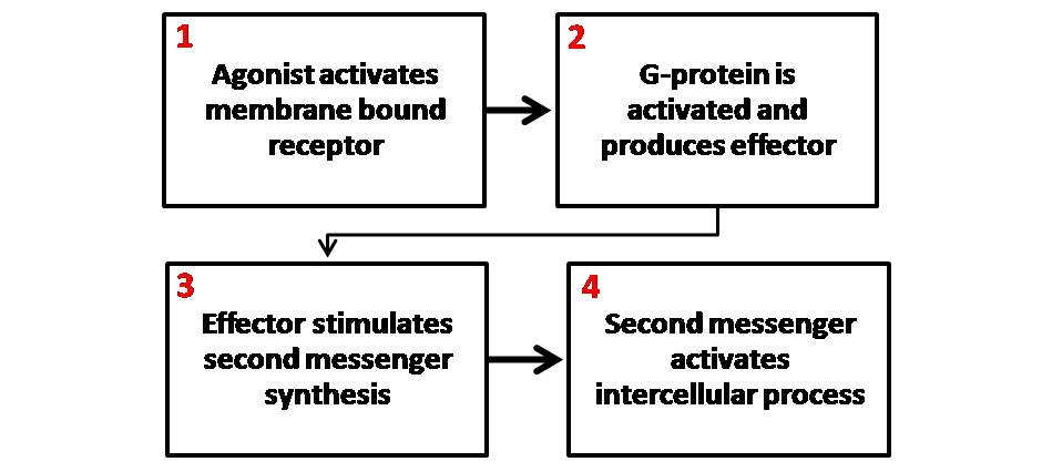
Second messengers are intracellular signaling molecules released by the cell in response to exposure to extracellular signaling molecules—the first messengers. (Intercellular signals, a non-local form of cell signaling, encompassing both first messengers and second messengers, are classified as autocrine, juxtacrine, paracrine, and endocrine depending on the range of the signal.) Second messengers trigger physiological changes at cellular level such as proliferation, differentiation, migration, survival, apoptosis and depolarization. They are one of the triggers of intracellular signal transduction cascades. Examples of second messenger molecules include cyclic AMP, cyclic GMP, inositol triphosphate, diacylglycerol, and calcium. First messengers are extracellular factors, often hormones or neurotransmitters, such as epinephrine, growth hormone, and serotonin. Because peptide hormones and neurotransmitters typically are biochemically hydrophilic molecules, these first mess ... [...More Info...] [...Related Items...] OR: [Wikipedia] [Google] [Baidu] |
Phosphatidylinositol
Phosphatidylinositol or inositol phospholipid is a biomolecule. It was initially called "inosite" when it was discovered by Léon Maquenne and Johann Joseph von Scherer in the late 19th century. It was discovered in bacteria but later also found in eukaryotes, and was found to be a signaling molecule. The biomolecule can exist in 9 different isomers. It is a lipid which contains a phosphate group, two fatty acid chains, and one inositol sugar molecule. Typically, the phosphate group has a negative charge (at physiological pH values). As a result, the molecule is amphiphilic. The production of the molecule is limited to the endoplasmic reticulum. History of phospatidylinositol Phosphatidylinositol (PI) and its derivatives have a rich history dating back to their discovery by Johann Joseph von Scherer and Léon Maquenne in the late 19th century. Initially known as " inosite" based on its sweet taste, the isolation and characterization of inositol laid the groundwork for und ... [...More Info...] [...Related Items...] OR: [Wikipedia] [Google] [Baidu] |
Adenylyl Cyclase
Adenylate cyclase (EC 4.6.1.1, also commonly known as adenyl cyclase and adenylyl cyclase, abbreviated AC) is an enzyme with systematic name ATP diphosphate-lyase (cyclizing; 3′,5′-cyclic-AMP-forming). It catalyzes the following reaction: :ATP = 3′,5′-cyclic AMP + diphosphate It has key regulatory roles in essentially all Cell (biology), cells. It is the most Polyphyly, polyphyletic known enzyme: six distinct classes have been described, all catalysis, catalyzing the same reaction but representing unrelated gene Gene family, families with no known Sequence homology, sequence or structural homology. The best known class of adenylyl cyclases is class III or AC-III (Roman numerals are used for classes). AC-III occurs widely in eukaryotes and has important roles in many human Tissue (biology), tissues. All classes of adenylyl cyclase Catalysis, catalyse the conversion of adenosine triphosphate (ATP) to cyclic adenosine monophosphate, 3',5'-cyclic AMP (cAMP) and pyrophosphate ... [...More Info...] [...Related Items...] OR: [Wikipedia] [Google] [Baidu] |
Gs Alpha Subunit
The Gs alpha subunit (Gαs, Gsα) is a subunit of the heterotrimeric G protein Gs that stimulates the cAMP-dependent pathway by activating adenylyl cyclase. Gsα is a GTPase that functions as a cellular signaling protein. Gsα is the founding member of one of the four families of heterotrimeric G proteins, defined by the alpha subunits they contain: the Gαs family, Gαi/Gαo family, Gαq family, and Gα12/Gα13 family. The Gs-family has only two members: the other member is Golf, named for its predominant expression in the olfactory system. In humans, Gsα is encoded by the GNAS complex locus, while Golfα is encoded by the GNAL gene. Function The general function of Gs is to activate intracellular signaling pathways in response to activation of cell surface G protein-coupled receptors (GPCRs). GPCRs function as part of a three-component system of receptor-transducer-effector. The transducer in this system is a heterotrimeric G protein, composed of three subunits: a G ... [...More Info...] [...Related Items...] OR: [Wikipedia] [Google] [Baidu] |


