|
Olivocerebellar Tract
The olivocerebellar tract, also known as olivocerebellar fibers, are neural fibers which originate at the olivary nucleus and pass out through the hilum and decussate with those from the opposite olive in the raphe nucleus, then as internal arcuate fibers they pass partly through and partly around the opposite olive and enter the inferior peduncle to be distributed to the cerebellar hemisphere The cerebellum consists of three parts, a median and two lateral, which are continuous with each other, and are substantially the same in structure. The median portion is constricted, and is called the vermis, from its annulated appearance which ... of the opposite side from which they arise. They terminate directly on Purkinje cells as the climbing fiber input system.Eccles J.C, Llinas R, and Sasaki. Excitation of cerebellar Purkinje cells by the climbing fibers. Nature 203: 245-246, 1964 References External links * Cerebellar connections {{Neuroanatomy-stub ... [...More Info...] [...Related Items...] OR: [Wikipedia] [Google] [Baidu] |
Medulla Oblongata
The medulla oblongata or simply medulla is a long stem-like structure which makes up the lower part of the brainstem. It is anterior and partially inferior to the cerebellum. It is a cone-shaped neuronal mass responsible for autonomic (involuntary) functions, ranging from vomiting to sneezing. The medulla contains the cardiovascular center, the respiratory center, vomiting and vasomotor centers, responsible for the autonomic functions of breathing, heart rate and blood pressure as well as the sleep–wake cycle. "Medulla" is from Latin, ‘pith or marrow’. And "oblongata" is from Latin, ‘lengthened or longish or elongated'. During embryonic development, the medulla oblongata develops from the myelencephalon. The myelencephalon is a secondary brain vesicle which forms during the maturation of the rhombencephalon, also referred to as the hindbrain. The bulb is an archaic term for the medulla oblongata. In modern clinical usage, the word bulbar (as in bulbar palsy) is r ... [...More Info...] [...Related Items...] OR: [Wikipedia] [Google] [Baidu] |
Olivary Nucleus
The olivary bodies or simply olives (Latin ''oliva'' and ''olivae'', singular and plural, respectively) are a pair of prominent oval structures on either side of the medullary pyramids in the medulla, the lower portion of the brainstem. They contain the olivary nuclei. Structure Each olivary body is located on the anterior surface of the medulla lateral to the pyramid, from which it is separated by the antero-lateral sulcus and the fibers of the hypoglossal nerve. Behind (dorsally), it is separated from the postero-lateral sulcus by the ventral spinocerebellar fasciculus. In the depression between the upper end of the olive and the pons lies the vestibulocochlear nerve. In humans, it measures about 1.25 cm in length, and between its upper end and the pons there is a slight depression to which the roots of the facial nerve are attached. The external arcuate fibers wind across the lower part of the pyramid and olive and enter the inferior peduncle. Olivary nuclei The olive ... [...More Info...] [...Related Items...] OR: [Wikipedia] [Google] [Baidu] |
Hippocampus Proper
The hippocampal subfields are four subfields CA1, CA2, CA3, and CA4 that make up the structure of the hippocampus. Regions described in the hippocampus are the head, body, and tail, and other hippocampal subfields include the dentate gyrus, the presubiculum, and the subiculum. The CA subfields use the initials of Hippocampus#Name, cornu ammonis, an earlier name of the hippocampus. Structure There are four hippocampal subfields, in the hippocampus proper which form a neural circuit called the trisynaptic circuit. CA1 CA1 is the first region in the hippocampal circuit, from which a significant output pathway goes to layer V of the entorhinal cortex. The main output of CA1 is to the subiculum. CA2 CA2 is a small region located between CA1 and CA3. It receives some input from layer II of the entorhinal cortex via the perforant path. Its pyramidal cells are more like those in CA3 than those in CA1. It is often ignored due to its small size. CA3 CA3 receives input from the mossy fi ... [...More Info...] [...Related Items...] OR: [Wikipedia] [Google] [Baidu] |
Decussate
Decussation is used in biological contexts to describe a crossing (due to the shape of the Roman numeral for ten, an uppercase 'X' (), ). In Latin anatomical terms, the form is used, e.g. . Similarly, the anatomical term chiasma is named after the Greek uppercase 'Χ' ( chi). Whereas a decussation refers to a crossing within the central nervous system, various kinds of crossings in the peripheral nervous system are called chiasma. Examples include: * In the brain, where nerve fibers obliquely cross from one lateral side of the brain to the other, that is to say they cross at a level other than their origin. See for examples decussation of pyramids and sensory decussation. In neuroanatomy, the term ''chiasma'' is reserved for crossing of- or within nerves such as in the optic chiasm. * In botanical leaf taxology, the word ''decussate'' describes an opposite pattern of leaves which has successive pairs at right angles to each other (i.e. rotated 90 degrees along the stem w ... [...More Info...] [...Related Items...] OR: [Wikipedia] [Google] [Baidu] |
Raphe Nucleus
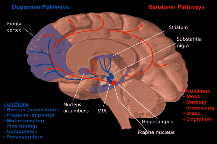
The raphe nuclei (, "seam") are a moderate-size cluster of nuclei found in the brain stem. They have 5-HT1 receptors which are coupled with Gi/Go-protein-inhibiting adenyl cyclase. They function as autoreceptors in the brain and decrease the release of serotonin. The anxiolytic drug Buspirone acts as partial agonist against these receptors. Selective serotonin reuptake inhibitor (SSRI) antidepressants are believed to act in these nuclei, as well as at their targets. Anatomy The raphe nuclei are traditionally considered to be the medial portion of the reticular formation, and appear as a ridge of cells in the center and most medial portion of the brain stem. In order from caudal to rostral, the raphe nuclei are known as the '' nucleus raphe obscurus'', the '' nucleus raphe pallidus'', the ''nucleus raphe magnus'', the '' nucleus raphe pontis'', the '' median raphe nucleus'', ''dorsal raphe nucleus'', ''caudal linear nucleus''. In the first systematic examination of the raphe ... [...More Info...] [...Related Items...] OR: [Wikipedia] [Google] [Baidu] |
Internal Arcuate Fibers
In neuroanatomy, the internal arcuate fibers or internal arcuate tract are the axons of second-order sensory neurons that compose the gracile and cuneate nuclei of the medulla oblongata. These second-order neurons begin in the gracile and cuneate nuclei in the medulla. They receive input from first-order sensory neurons, which provide sensation to many areas of the body and have cell bodies in the dorsal root ganglia of the dorsal root of the spinal nerves. Upon decussation (crossing over) from one side of the medulla to the other, also known as the sensory decussation, they are then called the medial lemniscus. The internal arcuate fibers are part of the second-order neurons of the posterior column-medial lemniscus system, and are important for relaying the sensation of fine touch and proprioception to the thalamus and ultimately to the cerebral cortex The cerebral cortex, also known as the cerebral mantle, is the outer layer of neural tissue of the cerebrum of the brai ... [...More Info...] [...Related Items...] OR: [Wikipedia] [Google] [Baidu] |
Inferior Peduncle
The inferior cerebellar peduncle is formed by fibers of the restiform body that join with fibers from the much smaller juxtarestiform body. The inferior cerebellar peduncle is the smallest of the three cerebellar peduncles. The upper part of the posterior district of the medulla oblongata is occupied by the inferior cerebellar peduncle, a thick rope-like strand situated between the lower part of the fourth ventricle and the roots of the glossopharyngeal and vagus nerves. Each cerebellar inferior peduncle connects the spinal cord and medulla oblongata with the cerebellum, and comprises the juxtarestiform body and restiform body. Important fibers running through the inferior cerebellar peduncle include the dorsal spinocerebellar tract and axons from the inferior olivary nucleus, among others. Function The inferior cerebellar peduncle carries many types of input and output fibers that are mainly concerned with integrating proprioceptive sensory input with motor vestibular functi ... [...More Info...] [...Related Items...] OR: [Wikipedia] [Google] [Baidu] |
Cerebellar Hemisphere
The cerebellum consists of three parts, a median and two lateral, which are continuous with each other, and are substantially the same in structure. The median portion is constricted, and is called the vermis, from its annulated appearance which it owes to the transverse ridges and furrows upon it; the lateral expanded portions are named the hemispheres. Sections *The "intermediate hemisphere" is also known as the " spinocerebellum". *The "lateral hemisphere" is also known as the " pontocerebellum". *The lateral hemisphere is considered the portion of the cerebellum to develop most recently. Additional images File:Cerebellar hemisphere -- animation.gif, Animation. File:Cerebellar hemisphere --- animation.gif, Close up animation. File:Cerebellar hemisphere by Sanjoy Sanyal.webmsd.webm, Dissection video (45 s). Demonstrating the two cerebellar hemispheres. File:Human cerebellum anterior view description.JPG, Human cerebellum anterior view description (Cerebellar hemisphere is #8 ... [...More Info...] [...Related Items...] OR: [Wikipedia] [Google] [Baidu] |

