|
Navicular Bone
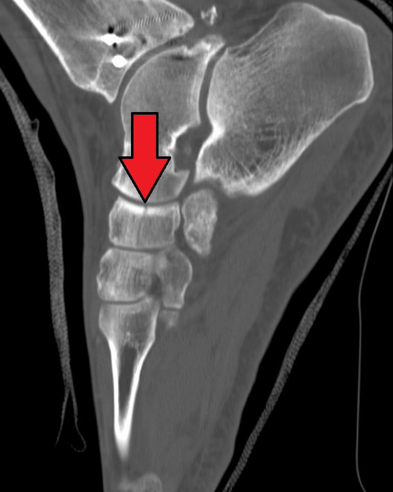
The navicular bone is a small bone found in the feet of most mammals. Human anatomy The navicular bone in humans is one of the tarsus (skeleton), tarsal bones, found in the foot. Its name derives from the human bone's resemblance to a small boat, caused by the strongly concave Anatomical terms of location#Proximal and distal, proximal joint, articular surface. The term ''navicular bone'' or ''hand navicular bone'' was formerly used for the scaphoid bone, one of the Carpal bones, carpal bones of the wrist. The navicular bone in humans is located on the Anatomical terms of location#Relative directions, medial side of the foot, and articulates proximally with the Talus bone, talus, Anatomical terms of location#Relative directions, distally with the three cuneiform bones, and Anatomical terms of location#Relative directions, laterally with the Cuboid bone, cuboid. It is the last of the foot bones to start ossification and does not tend to do so until the end of the third year in ... [...More Info...] [...Related Items...] OR: [Wikipedia] [Google] [Baidu] |
Foot
The foot (: feet) is an anatomical structure found in many vertebrates. It is the terminal portion of a limb which bears weight and allows locomotion. In many animals with feet, the foot is an organ at the terminal part of the leg made up of one or more segments or bones, generally including claws and/or nails. Etymology The word "foot", in the sense of meaning the "terminal part of the leg of a vertebrate animal" comes from Old English ''fot'', from Proto-Germanic *''fot'' (source also of Old Frisian ''fot'', Old Saxon ''fot'', Old Norse ''fotr'', Danish ''fod'', Swedish ''fot'', Dutch ''voet'', Old High German ''fuoz'', German ''Fuß'', Gothic ''fotus'', all meaning "foot"), from PIE root *''ped-'' "foot". The plural form ''feet'' is an instance of i-mutation. Structure The human foot is a strong and complex mechanical structure containing 26 bones, 33 joints (20 of which are actively articulated), and more than a hundred muscles, tendons, and ligaments.Podiatry Chan ... [...More Info...] [...Related Items...] OR: [Wikipedia] [Google] [Baidu] |
Sesamoid
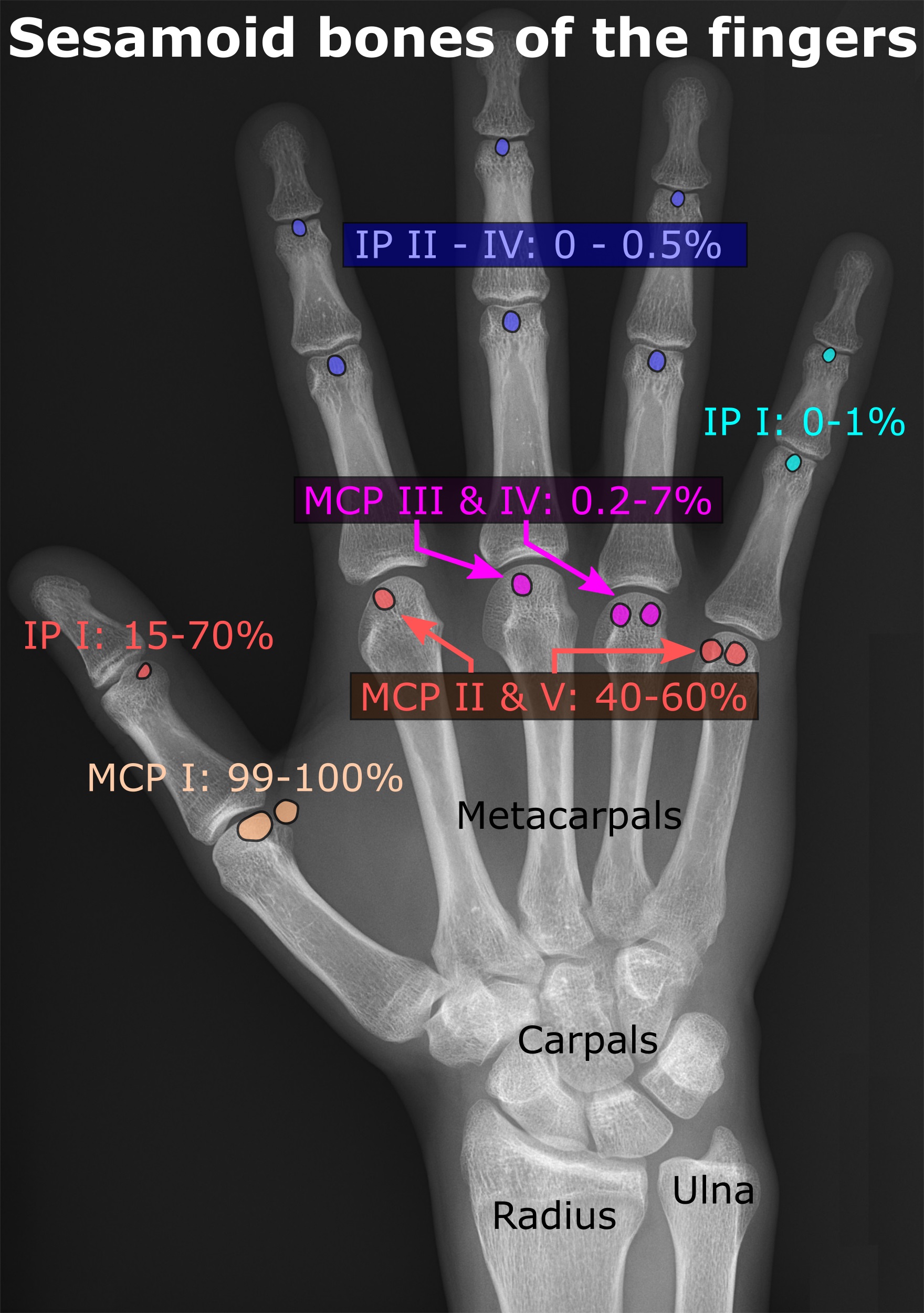
In anatomy, a sesamoid bone () is a bone embedded within a tendon or a muscle. Its name is derived from the Greek word for 'sesame seed', indicating the small size of most sesamoids. Often, these bones form in response to strain, or can be present as a normal variant. The patella is the largest sesamoid bone in the body. Sesamoids act like pulleys, providing a smooth surface for tendons to slide over, increasing the tendon's ability to transmit muscular forces. Structure Sesamoid bones can be found on joints throughout the human body, including: * In the knee—the patella (within the quadriceps tendon). This is the largest sesamoid bone. * In the hand—two sesamoid bones are commonly found in the distal portions of the first metacarpal bone (within the tendons of adductor pollicis and flexor pollicis brevis). There is also commonly a sesamoid bone in distal portions of the second metacarpal bone and fifth metacarpal bone. * In the wrist—The pisiform of the wrist is a ... [...More Info...] [...Related Items...] OR: [Wikipedia] [Google] [Baidu] |
Horse Anatomy
Equine anatomy encompasses the gross and microscopic anatomy of horses, ponies and other equids, including donkeys, mules and zebras. While all anatomical features of equids are described in the same terms as for other animals by the International Committee on Veterinary Gross Anatomical Nomenclature in the book '' Nomina Anatomica Veterinaria'', there are many horse-specific colloquial terms used by equestrians. External anatomy * Back: the area where the saddle sits, beginning at the end of the withers, extending to the last thoracic vertebrae (colloquially includes the loin or "coupling", though technically incorrect usage) * Barrel: the body of the horse, enclosing the rib cage and the major internal organs * Buttock: the part of the hindquarters behind the thighs and below the root of the tail * Cannon or cannon bone: the area between the knee or hock and the fetlock joint, sometimes called the "shin" of the horse, though technically it is the third metacarpal * Chestn ... [...More Info...] [...Related Items...] OR: [Wikipedia] [Google] [Baidu] |
Skeletal System
A skeleton is the structural frame that supports the body of most animals. There are several types of skeletons, including the exoskeleton, which is a rigid outer shell that holds up an organism's shape; the endoskeleton, a rigid internal frame to which the organs and soft tissues attach; and the hydroskeleton, a flexible internal structure supported by the hydrostatic pressure of body fluids. Vertebrates are animals with an endoskeleton centered around an axial vertebral column, and their skeletons are typically composed of bones and cartilages. Invertebrates are other animals that lack a vertebral column, and their skeletons vary, including hard-shelled exoskeleton (arthropods and most molluscs), plated internal shells (e.g. cuttlebones in some cephalopods) or rods (e.g. ossicles in echinoderms), hydrostatically supported body cavities (most), and spicules (sponges). Cartilage is a rigid connective tissue that is found in the skeletal systems of vertebrates and invert ... [...More Info...] [...Related Items...] OR: [Wikipedia] [Google] [Baidu] |
Terms For Anatomical Location
Standard anatomical terms of location are used to describe unambiguously the anatomy of humans and other animals. The terms, typically derived from Latin or Greek language, Greek roots, describe something in its standard anatomical position. This position provides a definition of what is at the front ("anterior"), behind ("posterior") and so on. As part of defining and describing terms, the body is described through the use of anatomical planes and anatomical axes, axes. The meaning of terms that are used can change depending on whether a vertebrate is a biped or a quadruped, due to the difference in the neuraxis, or if an invertebrate is a non-bilaterian. A non-bilaterian has no anterior or posterior surface for example but can still have a descriptor used such as proximal or distal in relation to a body part that is nearest to, or furthest from its middle. International organisations have determined vocabularies that are often used as standards for subdisciplines of anatomy. ... [...More Info...] [...Related Items...] OR: [Wikipedia] [Google] [Baidu] |
Bone
A bone is a rigid organ that constitutes part of the skeleton in most vertebrate animals. Bones protect the various other organs of the body, produce red and white blood cells, store minerals, provide structure and support for the body, and enable mobility. Bones come in a variety of shapes and sizes and have complex internal and external structures. They are lightweight yet strong and hard and serve multiple functions. Bone tissue (osseous tissue), which is also called bone in the uncountable sense of that word, is hard tissue, a type of specialised connective tissue. It has a honeycomb-like matrix internally, which helps to give the bone rigidity. Bone tissue is made up of different types of bone cells. Osteoblasts and osteocytes are involved in the formation and mineralisation of bone; osteoclasts are involved in the resorption of bone tissue. Modified (flattened) osteoblasts become the lining cells that form a protective layer on the bone surface. The mine ... [...More Info...] [...Related Items...] OR: [Wikipedia] [Google] [Baidu] |
Radiographic
Radiography is an imaging technique using X-rays, gamma rays, or similar ionizing radiation and non-ionizing radiation to view the internal form of an object. Applications of radiography include medical ("diagnostic" radiography and "therapeutic radiography") and industrial radiography. Similar techniques are used in airport security, (where "body scanners" generally use backscatter X-ray). To create an image in conventional radiography, a beam of X-rays is produced by an X-ray generator and it is projected towards the object. A certain amount of the X-rays or other radiation are absorbed by the object, dependent on the object's density and structural composition. The X-rays that pass through the object are captured behind the object by a detector (either photographic film or a digital detector). The generation of flat two-dimensional images by this technique is called projectional radiography. In computed tomography (CT scanning), an X-ray source and its associated detectors ... [...More Info...] [...Related Items...] OR: [Wikipedia] [Google] [Baidu] |
Eponymous
An eponym is a noun after which or for which someone or something is, or is believed to be, named. Adjectives derived from the word ''eponym'' include ''eponymous'' and ''eponymic''. Eponyms are commonly used for time periods, places, innovations, biological nomenclature, astronomical objects, works of art and media, and tribal names. Various orthographic conventions are used for eponyms. Usage of the word The term ''eponym'' functions in multiple related ways, all based on an explicit relationship between two named things. ''Eponym'' may refer to a person or, less commonly, a place or thing for which someone or something is, or is believed to be, named. ''Eponym'' may also refer to someone or something named after, or believed to be named after, a person or, less commonly, a place or thing. A person, place, or thing named after a particular person share an eponymous relationship. In this way, Elizabeth I of England is the eponym of the Elizabethan era, but the Elizabethan ... [...More Info...] [...Related Items...] OR: [Wikipedia] [Google] [Baidu] |
Hock (zoology)
The hock, tarsus or uncommonly gambrel, is the region formed by the tarsal bones connecting the tibia and metatarsus of a digitigrade or unguligrade quadrupedal mammal, such as a horse, cat, or dog. This joint may include articulations between tarsal bones and the fibula in some species (such as cats), while in others the fibula has been greatly reduced and is only found as a vestigial remnant fused to the distal portion of the tibia (as in horses). It is the anatomical homologue of the ankle of the human foot. While homologous joints occur in other tetrapods, the term is generally restricted to mammals, particularly long-legged domesticated species. Horse The terms ''tarsus'' and ''hock'' refer to the region between the gaskin (crus) and cannon regions (metatarsus), which includes the bones, joints, and soft tissues of the area. The hock is especially important in equine anatomy, due to the great strain it receives when the horse is worked. Jumping, quick turns or s ... [...More Info...] [...Related Items...] OR: [Wikipedia] [Google] [Baidu] |
Phalanx Bones
The phalanges (: phalanx ) are digital bones in the hands and feet of most vertebrates. In primates, the thumbs and big toes have two phalanges while the other digits have three phalanges. The phalanges are classed as long bones. Structure The phalanges are the bones that make up the fingers of the hand and the toes of the foot. There are 56 phalanges in the human body, with fourteen on each hand and foot. Three phalanges are present on each finger and toe, with the exception of the thumb and big toe, which possess only two. The middle and far phalanges of the fifth toes are often fused together (symphalangism). The phalanges of the hand are commonly known as the finger bones. The phalanges of the foot differ from the hand in that they are often shorter and more compressed, especially in the proximal phalanges, those closest to the torso. A phalanx is named according to whether it is proximal, middle, or distal and its associated finger or toe. The proximal phalang ... [...More Info...] [...Related Items...] OR: [Wikipedia] [Google] [Baidu] |
Equine Forelimb Anatomy
The limbs of the horse are structures made of dozens of bones, joints, muscles, tendons, and ligaments that support the weight of the equine body. They include three apparatuses: the suspensory apparatus, which carries much of the weight, prevents overextension of the joint and absorbs shock, the stay apparatus, which locks major joints in the limbs, allowing horses to remain standing while relaxed or asleep, and the reciprocal apparatus, which causes the hock to follow the motions of the stifle. The limbs play a major part in the movement of the horse, with the legs performing the functions of absorbing impact, bearing weight, and providing thrust. In general, the majority of the weight is borne by the front legs, while the rear legs provide propulsion. The hooves are also important structures, providing support, traction and shock absorption, and containing structures that provide blood flow through the lower leg. As the horse developed as a cursorial animal, with a primary ... [...More Info...] [...Related Items...] OR: [Wikipedia] [Google] [Baidu] |
Horse Hoof
A horse hoof is the lower extremity of each leg of a horse, the part that makes contact with the ground and carries the weight of the animal. It is both hard and flexible. It is a complex structure surrounding the distal Phalanx bones, phalanx of the 3rd digit (digit III of the basic pentadactyl limb of vertebrates, evolved into a single weight-bearing digit in horses) of each of the four limbs, which is covered by soft tissue and keratinised (cornified) matter. Anatomy The hoof is made up of two parts. The outer part, called the hoof capsule, is composed of various cornified specialized structures. The inner, living part of the hoof, is made up of soft tissues and bone. The cornified materials of the hoof capsule differ in structure and properties. Dorsally, it covers, protects, and supports P3 (also known as the coffin bone, pedal bone, or PIII). Palmarly/plantarly, it covers and protects specialised soft tissues, such as tendons, ligaments, fibro-fatty and/or fibrocartilagin ... [...More Info...] [...Related Items...] OR: [Wikipedia] [Google] [Baidu] |


