|
Lower Extremity Of Tibia
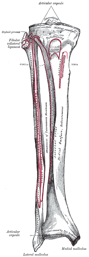
The tibia (; ), also known as the shinbone or shankbone, is the larger, stronger, and anterior (frontal) of the two bones in the leg below the knee in vertebrates (the other being the fibula, behind and to the outside of the tibia); it connects the knee with the ankle. The tibia is found on the medial side of the leg next to the fibula and closer to the median plane. The tibia is connected to the fibula by the interosseous membrane of leg, forming a type of fibrous joint called a syndesmosis with very little movement. The tibia is named for the flute ''tibia''. It is the second largest bone in the human body, after the femur. The leg bones are the strongest long bones as they support the rest of the body. Structure In human anatomy, the tibia is the second largest bone next to the femur. As in other vertebrates the tibia is one of two bones in the lower leg, the other being the fibula, and is a component of the knee and ankle joints. The ossification or formation of the bone st ... [...More Info...] [...Related Items...] OR: [Wikipedia] [Google] [Baidu] |
Knee
In humans and other primates, the knee joins the thigh with the leg and consists of two joints: one between the femur and tibia (tibiofemoral joint), and one between the femur and patella (patellofemoral joint). It is the largest joint in the human body. The knee is a modified hinge joint, which permits flexion and extension as well as slight internal and external rotation. The knee is vulnerable to injury and to the development of osteoarthritis. It is often termed a ''compound joint'' having tibiofemoral and patellofemoral components. (The fibular collateral ligament is often considered with tibiofemoral components.) Structure The knee is a modified hinge joint, a type of synovial joint, which is composed of three functional compartments: the patellofemoral articulation, consisting of the patella, or "kneecap", and the patellar groove on the front of the femur through which it slides; and the medial and lateral tibiofemoral articulations linking the femur, or thigh ... [...More Info...] [...Related Items...] OR: [Wikipedia] [Google] [Baidu] |
Long Bone
The long bones are those that are longer than they are wide. They are one of five types of bones: long, short, flat, irregular and sesamoid. Long bones, especially the femur and tibia, are subjected to most of the load during daily activities and they are crucial for skeletal mobility. They grow primarily by elongation of the diaphysis, with an epiphysis at each end of the growing bone. The ends of epiphyses are covered with hyaline cartilage ("articular cartilage"). The longitudinal growth of long bones is a result of endochondral ossification at the epiphyseal plate. Bone growth in length is stimulated by the production of growth hormone (GH), a secretion of the anterior lobe of the pituitary gland. The long bone category includes the femora, tibiae, and fibulae of the legs; the humeri, radii, and ulnae of the arms; metacarpals and metatarsals of the hands and feet, the phalanges of the fingers and toes, and the clavicles or collar bones. The long bones of the hum ... [...More Info...] [...Related Items...] OR: [Wikipedia] [Google] [Baidu] |
Lateral Condyle Of Tibia
The lateral condyle is the lateral portion of the upper extremity of tibia. It serves as the insertion for the biceps femoris muscle (small slip). Most of the tendon of the biceps femoris inserts on the fibula The fibula or calf bone is a leg bone on the lateral side of the tibia, to which it is connected above and below. It is the smaller of the two bones and, in proportion to its length, the most slender of all the long bones. Its upper extremity .... See also * Gerdy's tubercle * Medial condyle of tibia Additional images File:Gray258.png, Bones of the right leg. Anterior surface. File:Slide2bib.JPG, Right knee in extension. Deep dissection. Posterior view. File:Slide2cocc.JPG, Right knee in extension. Deep dissection. Posterior view. References External links * * * () Bones of the lower limb Tibia {{musculoskeletal-stub ... [...More Info...] [...Related Items...] OR: [Wikipedia] [Google] [Baidu] |
Medial Condyle Of Tibia
The medial condyle is the medial (or inner) portion of the upper extremity of tibia. It is the site of insertion for the semimembranosus muscle. See also * Lateral condyle of tibia * Medial collateral ligament The medial collateral ligament (MCL), or tibial collateral ligament (TCL), is one of the four major ligaments of the knee. It is on the medial (inner) side of the knee joint in humans and other primates. Its primary function is to resist outwar ... Additional images File:Gray258.png, Bones of the right leg. Anterior surface. File:Gray259.png, Bones of the right leg. Posterior surface. File:Slide2bib.JPG, Right knee in extension. Deep dissection. Posterior view. File:Slide2cocc.JPG, Right knee in extension. Deep dissection. Posterior view. References External links * * * () Bones of the lower limb Tibia {{musculoskeletal-stub ... [...More Info...] [...Related Items...] OR: [Wikipedia] [Google] [Baidu] |
Knee Diagram
In humans and other primates, the knee joins the thigh with the leg and consists of two joints: one between the femur and tibia (tibiofemoral joint), and one between the femur and patella (patellofemoral joint). It is the largest joint in the human body. The knee is a modified hinge joint, which permits flexion and extension as well as slight internal and external rotation. The knee is vulnerable to injury and to the development of osteoarthritis. It is often termed a ''compound joint'' having tibiofemoral and patellofemoral components. (The fibular collateral ligament is often considered with tibiofemoral components.) Structure The knee is a modified hinge joint, a type of synovial joint, which is composed of three functional compartments: the patellofemoral articulation, consisting of the patella, or "kneecap", and the patellar groove on the front of the femur through which it slides; and the medial and lateral tibiofemoral articulations linking the femur, or thigh bone ... [...More Info...] [...Related Items...] OR: [Wikipedia] [Google] [Baidu] |
Gray257
Grey (more common in British English) or gray (more common in American English) is an intermediate color between black and white. It is a neutral or achromatic color, meaning literally that it is "without color", because it can be composed of black and white. It is the color of a cloud-covered sky, of ash and of lead. The first recorded use of ''grey'' as a color name in the English language was in 700 CE.Maerz and Paul ''A Dictionary of Color'' New York:1930 McGraw-Hill Page 196 ''Grey'' is the dominant spelling in European and Commonwealth English, while ''gray'' has been the preferred spelling in American English; both spellings are valid in both varieties of English. In Europe and North America, surveys show that grey is the color most commonly associated with neutrality, conformity, boredom, uncertainty, old age, indifference, and modesty. Only one percent of respondents chose it as their favorite color. Etymology ''Grey'' comes from the Middle English or ... [...More Info...] [...Related Items...] OR: [Wikipedia] [Google] [Baidu] |
Human Foot
The foot ( : feet) is an anatomical structure found in many vertebrates. It is the terminal portion of a limb which bears weight and allows locomotion. In many animals with feet, the foot is a separate organ at the terminal part of the leg made up of one or more segments or bones, generally including claws or nails. Etymology The word "foot", in the sense of meaning the "terminal part of the leg of a vertebrate animal" comes from "Old English fot "foot," from Proto-Germanic *fot (source also of Old Frisian fot, Old Saxon fot, Old Norse fotr, Danish fod, Swedish fot, Dutch voet, Old High German fuoz, German Fuß, Gothic fotus "foot"), from PIE root *ped- "foot". The "plural form feet is an instance of i-mutation." Structure The human foot is a strong and complex mechanical structure containing 26 bones, 33 joints (20 of which are actively articulated), and more than a hundred muscles, tendons, and ligaments.Podiatry Channel, ''Anatomy of the foot and ankle'' The joints ... [...More Info...] [...Related Items...] OR: [Wikipedia] [Google] [Baidu] |
Lower Extremity Of Tibia
The tibia (; ), also known as the shinbone or shankbone, is the larger, stronger, and anterior (frontal) of the two bones in the leg below the knee in vertebrates (the other being the fibula, behind and to the outside of the tibia); it connects the knee with the ankle. The tibia is found on the medial side of the leg next to the fibula and closer to the median plane. The tibia is connected to the fibula by the interosseous membrane of leg, forming a type of fibrous joint called a syndesmosis with very little movement. The tibia is named for the flute ''tibia''. It is the second largest bone in the human body, after the femur. The leg bones are the strongest long bones as they support the rest of the body. Structure In human anatomy, the tibia is the second largest bone next to the femur. As in other vertebrates the tibia is one of two bones in the lower leg, the other being the fibula, and is a component of the knee and ankle joints. The ossification or formation of the bone st ... [...More Info...] [...Related Items...] OR: [Wikipedia] [Google] [Baidu] |
Thigh
In human anatomy, the thigh is the area between the hip ( pelvis) and the knee. Anatomically, it is part of the lower limb. The single bone in the thigh is called the femur. This bone is very thick and strong (due to the high proportion of bone tissue), and forms a ball and socket joint at the hip, and a modified hinge joint at the knee. Structure Bones The femur is the only bone in the thigh and serves as an attachment site for all muscles in the thigh. The head of the femur articulates with the acetabulum in the pelvic bone forming the hip joint, while the distal part of the femur articulates with the tibia and patella forming the knee. By most measures, the femur is the strongest bone in the body. The femur is also the longest bone in the body. The femur is categorised as a long bone and comprises a diaphysis, the shaft (or body) and two epiphysis or extremities that articulate with adjacent bones in the hip and knee. Muscular compartments In cross-secti ... [...More Info...] [...Related Items...] OR: [Wikipedia] [Google] [Baidu] |
Upper Extremity Of Tibia
The tibia (; ), also known as the shinbone or shankbone, is the larger, stronger, and anterior (frontal) of the two bones in the leg below the knee in vertebrates (the other being the fibula, behind and to the outside of the tibia); it connects the knee with the ankle. The tibia is found on the medial side of the leg next to the fibula and closer to the median plane. The tibia is connected to the fibula by the interosseous membrane of leg, forming a type of fibrous joint called a syndesmosis with very little movement. The tibia is named for the flute ''tibia''. It is the second largest bone in the human body, after the femur. The leg bones are the strongest long bones as they support the rest of the body. Structure In human anatomy, the tibia is the second largest bone next to the femur. As in other vertebrates the tibia is one of two bones in the lower leg, the other being the fibula, and is a component of the knee and ankle joints. The ossification or formation of the bon ... [...More Info...] [...Related Items...] OR: [Wikipedia] [Google] [Baidu] |
Body Of Tibia
The tibia (; ), also known as the shinbone or shankbone, is the larger, stronger, and anterior (frontal) of the two bones in the leg below the knee in vertebrates (the other being the fibula, behind and to the outside of the tibia); it connects the knee with the ankle. The tibia is found on the medial side of the leg next to the fibula and closer to the median plane. The tibia is connected to the fibula by the interosseous membrane of leg, forming a type of fibrous joint called a syndesmosis with very little movement. The tibia is named for the flute '' tibia''. It is the second largest bone in the human body, after the femur. The leg bones are the strongest long bones as they support the rest of the body. Structure In human anatomy, the tibia is the second largest bone next to the femur. As in other vertebrates the tibia is one of two bones in the lower leg, the other being the fibula, and is a component of the knee and ankle joints. The ossification or formation of th ... [...More Info...] [...Related Items...] OR: [Wikipedia] [Google] [Baidu] |
Epiphysis
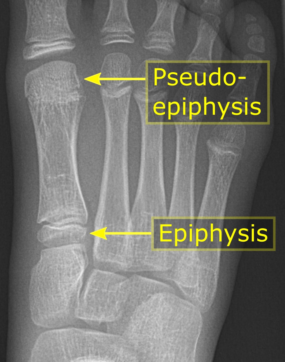
The epiphysis () is the rounded end of a long bone, at its joint with adjacent bone(s). Between the epiphysis and diaphysis (the long midsection of the long bone) lies the metaphysis, including the epiphyseal plate (growth plate). At the joint, the epiphysis is covered with articular cartilage; below that covering is a zone similar to the epiphyseal plate, known as subchondral bone. The epiphysis is filled with red bone marrow, which produces erythrocytes (red blood cells). Structure There are four types of epiphysis: # Pressure epiphysis: The region of the long bone that forms the joint is a pressure epiphysis (e.g. the head of the femur, part of the hip joint complex). Pressure epiphyses assist in transmitting the weight of the human body and are the regions of the bone that are under pressure during movement or locomotion. Another example of a pressure epiphysis is the head of the humerus which is part of the shoulder complex. condyles of femur and tibia also comes un ... [...More Info...] [...Related Items...] OR: [Wikipedia] [Google] [Baidu] |


.jpg)
