|
List Of Skin Diseases
Many skin conditions affect the human integumentary system—the organ system covering the entire surface of the body and composed of skin, hair, nails, and related muscle and glands. The major function of this system is as a barrier against the external environment. The skin weighs an average of four kilograms, covers an area of two square metres, and is made of three distinct layers: the epidermis, dermis, and subcutaneous tissue. The two main types of human skin are: glabrous skin, the hairless skin on the palms and soles (also referred to as the "palmoplantar" surfaces), and hair-bearing skin.Burns, Tony; ''et al''. (2006) ''Rook's Textbook of Dermatology CD-ROM''. Wiley-Blackwell. . Within the latter type, the hairs occur in structures called pilosebaceous units, each with hair follicle, sebaceous gland, and associated arrector pili muscle. In the embryo, the epidermis, hair, and glands form from the ectoderm, which is chemically influenced by the underlying mesoderm that ... [...More Info...] [...Related Items...] OR: [Wikipedia] [Google] [Baidu] |
Arrector Pili
The arrector pili muscles, also known as hair erector muscles, are small muscles attached to hair follicles in mammals. Contraction of these muscles causes the hairs to stand on end, known colloquially as goose bumps (piloerection). Structure Each arrector pili is composed of a bundle of smooth muscle fibres which attach to several follicles (a follicular unit). Each is innervated by the sympathetic division of the autonomic nervous system. The muscle attaches to the follicular stem cell niche in the follicular bulge, splitting at their deep end to encircle the follicle. Function The contraction of the muscle is involuntary. Stresses such as cold, fear etc. may stimulate the sympathetic nervous system, and thus cause muscle contraction. Thermal insulation Contraction of arrector pili muscles have a principal function in the majority of mammals of providing thermal insulation. Air becomes trapped between the erect hairs, helping the animal retain heat. Self defence Erec ... [...More Info...] [...Related Items...] OR: [Wikipedia] [Google] [Baidu] |
Melanocyte
Melanocytes are melanin-producing neural crest-derived cells located in the bottom layer (the stratum basale) of the skin's epidermis, the middle layer of the eye (the uvea), the inner ear, vaginal epithelium, meninges, bones, and heart. Melanin is a dark pigment primarily responsible for skin color. Once synthesized, melanin is contained in special organelles called melanosomes which can be transported to nearby keratinocytes to induce pigmentation. Thus darker skin tones have more melanosomes present than lighter skin tones. Functionally, melanin serves as protection against UV radiation. Melanocytes also have a role in the immune system. Function Through a process called melanogenesis, melanocytes produce melanin, which is a pigment found in the skin, eyes, hair, nasal cavity, and inner ear. This melanogenesis leads to a long-lasting pigmentation, which is in contrast to the pigmentation that originates from oxidation of already-existing melanin. There a ... [...More Info...] [...Related Items...] OR: [Wikipedia] [Google] [Baidu] |
Keratinocyte
Keratinocytes are the primary type of cell found in the epidermis, the outermost layer of the skin. In humans, they constitute 90% of epidermal skin cells. Basal cells in the basal layer (''stratum basale'') of the skin are sometimes referred to as basal keratinocytes. Keratinocytes form a barrier against environmental damage by heat, UV radiation, water loss, pathogenic bacteria, fungi, parasites, and viruses. A number of structural proteins, enzymes, lipids, and antimicrobial peptides contribute to maintain the important barrier function of the skin. Keratinocytes differentiate from epidermal stem cells in the lower part of the epidermis and migrate towards the surface, finally becoming corneocytes and eventually be shed off, which happens every 40 to 56 days in humans. Function The primary function of keratinocytes is the formation of a barrier against environmental damage by heat, UV radiation, water loss, pathogenic bacteria, fungi, parasites, and viruses. Pathogens ... [...More Info...] [...Related Items...] OR: [Wikipedia] [Google] [Baidu] |
Diffusion
Diffusion is the net movement of anything (for example, atoms, ions, molecules, energy) generally from a region of higher concentration to a region of lower concentration. Diffusion is driven by a gradient in Gibbs free energy or chemical potential. It is possible to diffuse "uphill" from a region of lower concentration to a region of higher concentration, like in spinodal decomposition. The concept of diffusion is widely used in many fields, including physics ( particle diffusion), chemistry, biology, sociology, economics, and finance (diffusion of people, ideas, and price values). The central idea of diffusion, however, is common to all of these: a substance or collection undergoing diffusion spreads out from a point or location at which there is a higher concentration of that substance or collection. A gradient is the change in the value of a quantity, for example, concentration, pressure, or temperature with the change in another variable, usually distance. A chan ... [...More Info...] [...Related Items...] OR: [Wikipedia] [Google] [Baidu] |
Stratum Germinativum
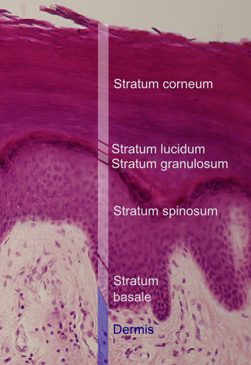
The ''stratum basale'' (basal layer, sometimes referred to as ''stratum germinativum'') is the deepest layer of the five layers of the epidermis, the external covering of skin in mammals. The ''stratum basale'' is a single layer of columnar or cuboidal basal cells. The cells are attached to each other and to the overlying stratum spinosum cells by desmosomes and hemidesmosomes. The nucleus is large, ovoid and occupies most of the cell. Some basal cells can act like stem cells with the ability to divide and produce new cells, and these are sometimes called basal keratinocyte stem cells. Others serve to anchor the epidermis glabrous skin (hairless), and hyper-proliferative epidermis (from a skin disease).McGrath, J.A.; Eady, R.A.; Pope, F.M. (2004). ''Rook's Textbook of Dermatology'' (Seventh Edition). Blackwell Publishing. Pages 3.7. . They divide to form the keratinocytes of the stratum spinosum, which migrate superficially. Other types of cells found within the ''stratum basa ... [...More Info...] [...Related Items...] OR: [Wikipedia] [Google] [Baidu] |
Stratum Spinosum
The stratum spinosum (or spinous layer/prickle cell layer) is a layer of the epidermis found between the stratum granulosum and stratum basale. This layer is composed of polyhedral keratinocytes. These are joined with desmosomes. Their spiny (Latin, spinosum) appearance is due to shrinking of the microfilaments between desmosomes that occurs when stained with H&E. Keratinization begins in the stratum spinosum, although the actual keratinocytes begin in the stratum basale. They have large pale-staining nuclei as they are active in synthesizing fibrilar proteins, known as cytokeratin, which build up within the cells aggregating together forming tonofibrils. The tonofibrils go on to form the desmosomes, which allow for strong connections to form between adjacent keratinocytes. The stratum spinosum also contains Langerhans cells. Clinical significance Diffuse hyperplasia of the stratum spinosum is termed acanthosis The epidermis is the outermost of the three layers that co ... [...More Info...] [...Related Items...] OR: [Wikipedia] [Google] [Baidu] |
Stratum Granulosum
The stratum granulosum (or granular layer) is a thin layer of cells in the epidermis lying above the stratum spinosum and below the stratum corneum ( stratum lucidum on the soles and palms).James, William; Berger, Timothy; Elston, Dirk (2005) ''Andrews' Diseases of the Skin: Clinical Dermatology'' (10th ed.). Saunders. Page 2. . Keratinocytes migrating from the underlying stratum spinosum become known as granular cells in this layer. These cells contain keratohyalin granules, which are filled with histidine- and cysteine-rich proteins that appear to bind the keratin filaments together. Therefore, the main function of keratohyalin granules is to bind intermediate keratin filaments together.Marks, James G; Miller, Jeffery (2006). ''Lookingbill and Marks' Principles of Dermatology'' (4th ed.). Elsevier Inc. Page 7. . At the transition between this layer and the stratum corneum, cells secrete lamellar bodies (containing lipids and proteins) into the extracellular space. This results ... [...More Info...] [...Related Items...] OR: [Wikipedia] [Google] [Baidu] |
Stratum Lucidum
The stratum lucidum (Latin, 'clear layer') is a thin, clear layer of dead skin cells in the epidermis named for its translucent appearance under a microscope. It is readily visible by light microscopy only in areas of thick skin, which are found on the palms of the hands and the soles of the feet. Located between the stratum granulosum and stratum corneum layers, it is composed of three to five layers of dead, flattened keratinocytes.McGrath, J.A.; Eady, R.A.; Pope, F.M. (2004). ''Rook's Textbook of Dermatology'' (Seventh Edition). Blackwell Publishing. Pages 3.8. .Tortora, Gerard; Derrickson, Bryan; Principles of Anatomy and Physiology (2009)152 John Wiley & Sons Inc, Hoboken, NJ . The keratinocytes of the stratum lucidum do not feature distinct boundaries and are filled with eleidin, an intermediate form of keratin. They are surrounded by an oily substance that is the result of the exocytosis of lamellar bodies accumulated while the keratinocytes are moving through the stratu ... [...More Info...] [...Related Items...] OR: [Wikipedia] [Google] [Baidu] |
Stratum Corneum
The stratum corneum (Latin for 'horny layer') is the outermost layer of the epidermis. The human stratum corneum comprises several levels of flattened corneocytes that are divided into two layers: the ''stratum disjunctum'' and ''stratum compactum''. The skin's protective acid mantle and lipid barrier sit on top of the stratum disjunctum. The stratum disjunctum is the uppermost and loosest layer of skin. The stratum compactum is the comparatively deeper, more compacted and more cohesive part of the stratum corneum. The corneocytes of the stratum disjunctum are larger, more rigid and more hydrophobic than that of the stratum compactum. The stratum corneum is the dead tissue that performs protective and adaptive physiological functions including mechanical shear, impact resistance, water flux and hydration regulation, microbial proliferation and invasion regulation, initiation of inflammation through cytokine activation and dendritic cell activity, and selective permeability to e ... [...More Info...] [...Related Items...] OR: [Wikipedia] [Google] [Baidu] |
Stratum
In geology and related fields, a stratum ( : strata) is a layer of rock or sediment characterized by certain lithologic properties or attributes that distinguish it from adjacent layers from which it is separated by visible surfaces known as either '' bedding surfaces'' or ''bedding planes''.Salvador, A. ed., 1994. ''International stratigraphic guide: a guide to stratigraphic classification, terminology, and procedure. 2nd ed.'' Boulder, Colorado, The Geological Society of America, Inc., 215 pp. . Prior to the publication of the International Stratigraphic Guide, older publications have defined a stratum as either being either equivalent to a single bed or composed of a number of beds; as a layer greater than 1 cm in thickness and constituting a part of a bed; or a general term that includes both ''bed'' and '' lamina''.Neuendorf, K.K.E., Mehl, Jr., J.P., and Jackson, J.A. , eds., 2005. ''Glossary of Geology'' 5th ed. Alexandria, Virginia, American Geological Institute. 779 pp. ... [...More Info...] [...Related Items...] OR: [Wikipedia] [Google] [Baidu] |
Epithelium
Epithelium or epithelial tissue is one of the four basic types of animal tissue, along with connective tissue, muscle tissue and nervous tissue. It is a thin, continuous, protective layer of compactly packed cells with a little intercellular matrix. Epithelial tissues line the outer surfaces of organs and blood vessels throughout the body, as well as the inner surfaces of cavities in many internal organs. An example is the epidermis, the outermost layer of the skin. There are three principal shapes of epithelial cell: squamous (scaly), columnar, and cuboidal. These can be arranged in a singular layer of cells as simple epithelium, either squamous, columnar, or cuboidal, or in layers of two or more cells deep as stratified (layered), or ''compound'', either squamous, columnar or cuboidal. In some tissues, a layer of columnar cells may appear to be stratified due to the placement of the nuclei. This sort of tissue is called pseudostratified. All glands are made up of epit ... [...More Info...] [...Related Items...] OR: [Wikipedia] [Google] [Baidu] |


