|
Lesser Occipital Nerve
The lesser occipital nerve (or small occipital nerve) is a cutaneous spinal nerve of the cervical plexus. It arises from second cervical (spinal) nerve (C2) (along with the greater occipital nerve). It innervates the skin of the back of the upper neck and of the scalp posterior to the ear. Structure Origin It arises from the (lateral branch of the ventral ramus) of cervical spinal nerve C2; it (sources differ) receives or may also receive fibres from cervical spinal nerve C3. It originates between the atlas, and axis. The lesser occipital nerve is one of the four cutaneous branches of the cervical plexus. Course and relations It curves around the accessory nerve (CN XI) to come to course anterior to it. It then curves around and ascends along the posterior border of the sternocleidomastoid muscle; rarely, it may pierce the muscle. Near the cranium, it perforates the deep cervical fascia. It is continued upwards along the scalp posterior to the auricle. It divides in ... [...More Info...] [...Related Items...] OR: [Wikipedia] [Google] [Baidu] |
Cervical Plexus
The cervical plexus is a nerve plexus of the anterior rami of the first (i.e. upper-most) four cervical spinal nerves C1-C4. The cervical plexus provides motor innervation to some muscles of the neck, and the diaphragm; it provides sensory innervation to parts of the head, neck, and chest. Anatomy They are located laterally to the transverse processes between prevertebral muscles from the medial side and vertebral (m. scalenus, m. levator scapulae, m. splenius cervicis) from lateral side. There is anastomosis with accessory nerve, hypoglossal nerve and sympathetic trunk. It is located in the neck, deep to the sternocleidomastoid muscle. The branches of the cervical plexus emerge from the posterior triangle at the nerve point, a point which lies midway on the posterior border of the sternocleidomastoid. Relations The cervical plexus is situated deep to the sternocleidomastoid muscle, internal jugular vein, and deep cervical fascia. It is situated anterior to the mid ... [...More Info...] [...Related Items...] OR: [Wikipedia] [Google] [Baidu] |
Accessory Nerve
The accessory nerve, also known as the eleventh cranial nerve, cranial nerve XI, or simply CN XI, is a cranial nerve that supplies the sternocleidomastoid and trapezius muscles. It is classified as the eleventh of twelve pairs of cranial nerves because part of it was formerly believed to originate in the brain. The sternocleidomastoid muscle tilts and rotates the head, whereas the trapezius muscle, connecting to the scapula, acts to shrug the shoulder. Traditional descriptions of the accessory nerve divide it into a spinal part and a cranial part. The cranial component rapidly joins the vagus nerve, and there is ongoing debate about whether the cranial part should be considered part of the accessory nerve proper. Consequently, the term "accessory nerve" usually refers only to nerve supplying the sternocleidomastoid and trapezius muscles, also called the spinal accessory nerve. Strength testing of these muscles can be measured during a neurological examination to assess func ... [...More Info...] [...Related Items...] OR: [Wikipedia] [Google] [Baidu] |
Occipital Neuralgia
Occipital neuralgia (ON) is a painful condition affecting the posterior head in the distributions of the greater occipital nerve (GON), lesser occipital nerve (LON), third occipital nerve (TON), or a combination of the three. It is paroxysmal, lasting from seconds to minutes, and often consists of lancinating pain that directly results from the pathology of one of these nerves. It is paramount that physicians understand the differential diagnosis for this condition and specific diagnostic criteria. There are multiple treatment modalities, several of which have well-established efficacy in treating this condition. Text was copied from this source, which is available under Creative Commons Attribution 4.0 International License Signs and symptoms Patients presenting with a headache originating at the posterior skull base should be evaluated for ON. This condition typically presents as a paroxysmal, lancinating or stabbing pain lasting from seconds to minutes, and therefore a continu ... [...More Info...] [...Related Items...] OR: [Wikipedia] [Google] [Baidu] |
Posterior Auricular Branch Of The Facial
The posterior auricular nerve is a nerve of the head. It is a branch of the facial nerve (CN VII). It communicates with branches from the vagus nerve, the great auricular nerve, and the lesser occipital nerve. Its auricular branch supplies the posterior auricular muscle, the intrinsic muscles of the auricle, and gives sensation to the auricle. Its occipital branch supplies the occipitalis muscle. Structure The posterior auricular nerve arises from the facial nerve (CN VII). It is the first branch outside of the skull. This origin is close to the stylomastoid foramen. It runs upward in front of the mastoid process. It is joined by a branch from the auricular branch of the vagus nerve (CN X). It communicates with the posterior branch of the great auricular nerve, as well as with the lesser occipital nerve. As it ascends between the external acoustic meatus and mastoid process it divides into auricular and occipital branches. * The ''auricular branch'' travels to the poster ... [...More Info...] [...Related Items...] OR: [Wikipedia] [Google] [Baidu] |
Great Auricular Nerve
The great auricular nerve is a Cutaneous nerve, cutaneous (sensory) nerve of the head. It originates from the second and third spinal nerve, cervical (spinal) nerves (C2-C3) of the cervical plexus. It provides sensory innervation to the skin over the parotid gland and the Mastoid part of the temporal bone, mastoid process, parts of the outer ear, and to the parotid gland and its parotid fascia, fascia. Pain resulting from parotitis is caused by an impingement on the great auricular nerve. Structure The great auricular nerve is the largest of the ascending branches of the cervical plexus. Origin It arises from the second and third cervical (spinal) nerves (C2-C3), with the predominant contribution coming from C2. Course and relations The great auricular nerve is a large trunk that ascends almost vertically over the sternocleidomastoid. It winds around the posterior border of the sternocleidomastoid muscle, then perforates the deep fascia before ascending alongside the exter ... [...More Info...] [...Related Items...] OR: [Wikipedia] [Google] [Baidu] |
Great Auricular
The great auricular nerve is a cutaneous (sensory) nerve of the head. It originates from the second and third cervical (spinal) nerves (C2-C3) of the cervical plexus. It provides sensory innervation to the skin over the parotid gland and the mastoid process, parts of the outer ear, and to the parotid gland and its fascia. Pain resulting from parotitis is caused by an impingement on the great auricular nerve. Structure The great auricular nerve is the largest of the ascending branches of the cervical plexus. Origin It arises from the second and third cervical (spinal) nerves (C2-C3), with the predominant contribution coming from C2. Course and relations The great auricular nerve is a large trunk that ascends almost vertically over the sternocleidomastoid. It winds around the posterior border of the sternocleidomastoid muscle, then perforates the deep fascia before ascending alongside the external jugular vein upon that sternocleidomastoid muscle beneath the platysma mus ... [...More Info...] [...Related Items...] OR: [Wikipedia] [Google] [Baidu] |
Auricle (anatomy)
The auricle or auricula is the visible part of the ear that is outside the head. It is also called the pinna (Latin for 'wing' or ' fin', : pinnae), a term that is used more in zoology. Structure The diagram shows the shape and location of most of these components: * ''antihelix'' forms a 'Y' shape where the upper parts are: ** ''Superior crus'' (to the left of the ''fossa triangularis'' in the diagram) ** ''Inferior crus'' (to the right of the ''fossa triangularis'' in the diagram) * ''Antitragus'' is below the ''tragus'' * ''Aperture'' is the entrance to the ear canal * ''Auricular sulcus'' is the depression behind the ear next to the head * ''Concha'' is the hollow next to the ear canal * Conchal angle is the angle that the back of the ''concha'' makes with the side of the head * ''Crus'' of the helix is just above the ''tragus'' * ''Cymba conchae'' is the narrowest end of the ''concha'' * External auditory meatus is the ear canal * ''Fossa triangularis'' is the depression ... [...More Info...] [...Related Items...] OR: [Wikipedia] [Google] [Baidu] |
Deep Fascia
Deep fascia (or investing fascia) is a fascia, a layer of dense connective tissue that can surround individual muscles and groups of muscles to separate into fascial compartments. This fibrous connective tissue interpenetrates and surrounds the muscles, bones, nerves, and blood vessels of the body. It provides connection and communication in the form of aponeuroses, ligaments, tendons, retinaculum, retinacula, joint capsules, and septum, septa. The deep fasciae envelop all bone (periosteum and endosteum); cartilage (perichondrium), and blood vessels (tunica externa) and become specialized in muscles (epimysium, perimysium, and endomysium) and nerves (epineurium, perineurium, and endoneurium). The high density of collagen fibers gives the deep fascia its strength and integrity. The amount of elastin fiber determines how much extensibility and resilience it will have. Examples Examples include: * Fascia lata * Deep fascia of leg * Brachial fascia * Buck's fascia Fascial dynamics D ... [...More Info...] [...Related Items...] OR: [Wikipedia] [Google] [Baidu] |
Sternocleidomastoid Muscle
The sternocleidomastoid muscle is one of the largest and most superficial cervical muscles. The primary actions of the muscle are rotation of the head to the opposite side and Anatomical terms of motion#Flexion and extension, flexion of the neck. The sternocleidomastoid is innervated by the accessory nerve. Etymology and location It is given the name ''sternocleidomastoid'' because it originates at the manubrium of the Human sternum, sternum (''sterno-'') and the clavicle (''cleido-'') and has an Insertion (anatomy), insertion at the mastoid process of the temporal bone of the human skull, skull. Structure The sternocleidomastoid muscle originates from two locations: the manubrium of the Human sternum, sternum and the clavicle, hence it is said to have two heads: sternal head and clavicular head. It travels obliquely across the side of the neck and inserts at the mastoid process of the temporal bone of the human skull, skull by a thin aponeurosis. The sternocleidomastoid is thick ... [...More Info...] [...Related Items...] OR: [Wikipedia] [Google] [Baidu] |
Axis (anatomy)
In anatomy, the axis (from Latin ''axis'', "axle") is the second cervical vertebra (C2) of the spine, immediately inferior to the atlas, upon which the head rests. The spinal cord passes through the axis. The defining feature of the axis is its strong bony protrusion known as the dens, which rises from the superior aspect of the bone. Structure The body is deeper in front or in the back and is prolonged downward anteriorly to overlap the upper and front part of the third vertebra. It presents a median longitudinal ridge in front, separating two lateral depressions for the attachment of the longus colli muscles. Dens The dens, also called the odontoid process, or the peg, is the most pronounced projecting feature of the axis. The dens exhibits a slight constriction where it joins the main body of the vertebra. The condition where the dens is separated from the body of the axis is called ''os odontoideum'' and may cause nerve and circulation compression syndrome. On its an ... [...More Info...] [...Related Items...] OR: [Wikipedia] [Google] [Baidu] |
Cervical Plexus
The cervical plexus is a nerve plexus of the anterior rami of the first (i.e. upper-most) four cervical spinal nerves C1-C4. The cervical plexus provides motor innervation to some muscles of the neck, and the diaphragm; it provides sensory innervation to parts of the head, neck, and chest. Anatomy They are located laterally to the transverse processes between prevertebral muscles from the medial side and vertebral (m. scalenus, m. levator scapulae, m. splenius cervicis) from lateral side. There is anastomosis with accessory nerve, hypoglossal nerve and sympathetic trunk. It is located in the neck, deep to the sternocleidomastoid muscle. The branches of the cervical plexus emerge from the posterior triangle at the nerve point, a point which lies midway on the posterior border of the sternocleidomastoid. Relations The cervical plexus is situated deep to the sternocleidomastoid muscle, internal jugular vein, and deep cervical fascia. It is situated anterior to the mid ... [...More Info...] [...Related Items...] OR: [Wikipedia] [Google] [Baidu] |
Atlas (anatomy)
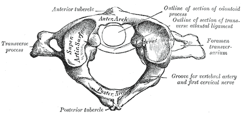
In anatomy, the atlas (C1) is the most superior (first) cervical vertebra of the spine and is located in the neck. The bone is named for Atlas of Greek mythology, just as Atlas bore the weight of the heavens, the first cervical vertebra supports the head. However, the term atlas was first used by the ancient Romans for the seventh cervical vertebra (C7) due to its suitability for supporting burdens. In Greek mythology, Atlas was condemned to bear the weight of the heavens as punishment for rebelling against Zeus. Ancient depictions of Atlas show the globe of the heavens resting at the base of his neck, on C7. Sometime around 1522, anatomists decided to call the first cervical vertebra the atlas. Scholars believe that by switching the designation atlas from the seventh to the first cervical vertebra Renaissance anatomists were commenting that the point of man's burden had shifted from his shoulders to his head—that man's true burden was not a physical load, but rather, his m ... [...More Info...] [...Related Items...] OR: [Wikipedia] [Google] [Baidu] |


