|
Kienböck's Disease
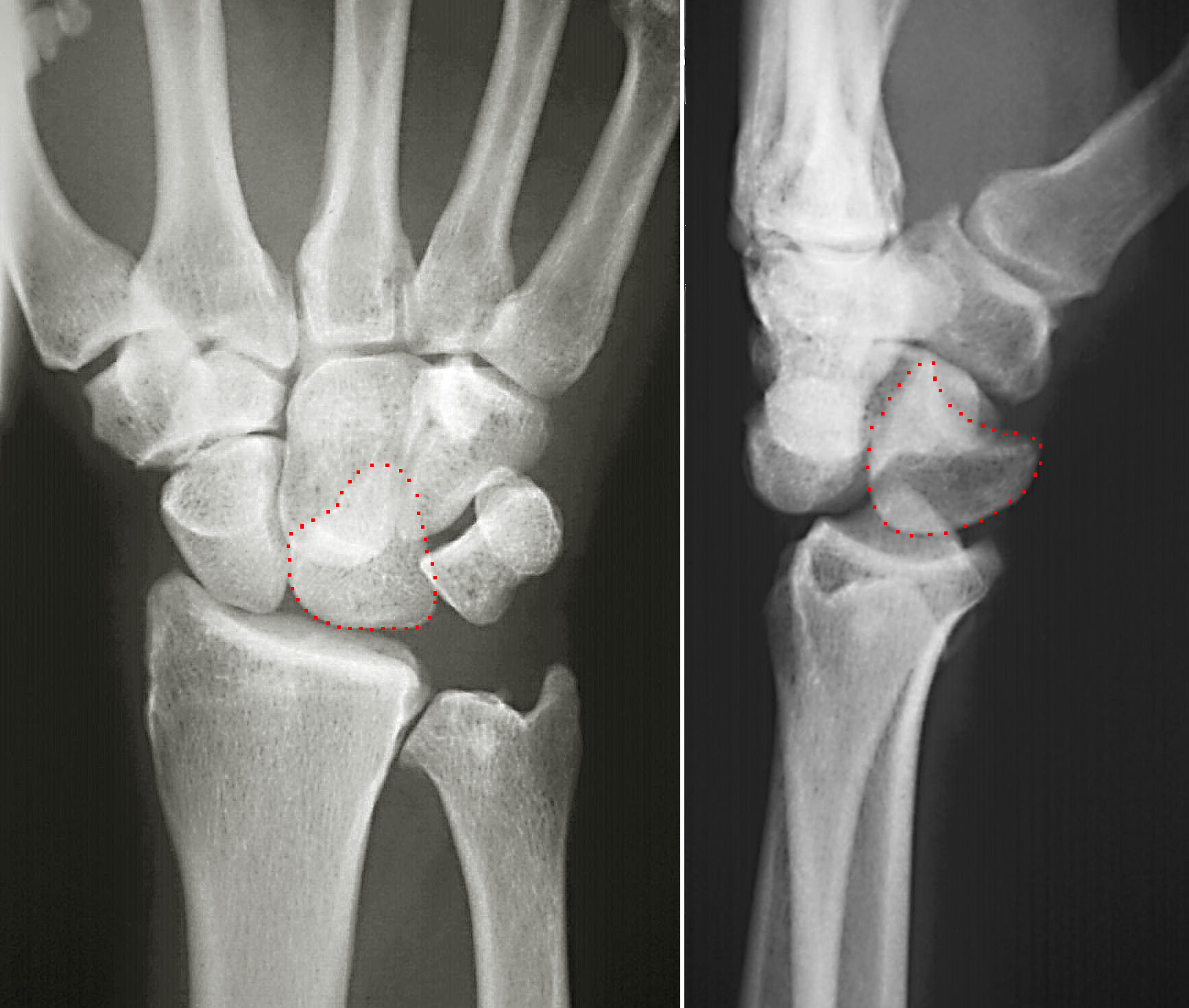
Kienböck's disease is a disorder of the wrist. It is named for Dr. Robert Kienböck, a radiologist in Vienna, Austria who described osteomalacia of the lunate in 1910. It is breakdown of the lunate bone, a carpal bone in the wrist that articulates with the radius in the forearm. Specifically, Kienböck's disease is another name for avascular necrosis (death and fracture of bone tissue due to interruption of blood supply) with fragmentation and collapse of the lunate. This has classically been attributed to arterial disruption, but may also occur after events that produce venous congestion with elevated interosseous pressure. Cause The exact cause of Kienböck's is not known, though there are thought to be a number of factors predisposing a person to Kienböck's. Although there is no evidence that Kienböck's disease is inherited, it is possible that unidentified genetic factors could contribute to the development of the condition. Studies have found a correlation between having ... [...More Info...] [...Related Items...] OR: [Wikipedia] [Google] [Baidu] |
Scaphoid Bone
The scaphoid bone is one of the carpal bones of the wrist. It is situated between the hand and forearm on the thumb side of the wrist (also called the lateral or radial side). It forms the radial border of the carpal tunnel. The scaphoid bone is the largest bone of the proximal row of wrist bones, its long axis being from above downward, lateralward, and forward. It is approximately the size and shape of a medium cashew. Structure The scaphoid is situated between the proximal and distal rows of carpal bones. It is located on the radial side of the wrist, and articulates with the radius, lunate, trapezoid, trapezium, and capitate. Over 80% of the bone is covered in articular cartilage. Bone The palmar surface of the scaphoid is concave, and forming a distal tubercle, giving attachment to the transverse carpal ligament. The proximal surface is triangular, smooth and convex. The lateral surface is narrow and gives attachment to the radial collateral ligament. The medial surfac ... [...More Info...] [...Related Items...] OR: [Wikipedia] [Google] [Baidu] |
Lunate Bone
The lunate bone (semilunar bone) is a carpal bone in the human hand. It is distinguished by its deep concavity and crescentic outline. It is situated in the center of the proximal row carpal bones, which lie between the ulna and radius and the hand. The lunate carpal bone is situated between the lateral scaphoid bone and medial triquetral bone. Structure The lunate is a crescent-shaped carpal bone found within the hand. The lunate is found within the proximal row of carpal bones. Proximally, it abuts the radius. Laterally, it articulates with the scaphoid bone, medially with the triquetral bone, and distally with the capitate bone. The lunate also articulates on its distal and medial surface with the hamate bone. The lunate is stabilised by a medial ligament to the scaphoid bone and a lateral ligament to the triquetral bone. Ligaments between the radius and carpal bone also stabilise the position of the lunate, as does its position in the lunate fossa of the radius. Bone The ... [...More Info...] [...Related Items...] OR: [Wikipedia] [Google] [Baidu] |
Skeletal Disorders
Bone disease refers to the medical conditions which affect the bone. Terminology A bone disease is also called an "osteopathy", but because the term osteopathy is often used to refer to an alternative health-care philosophy, use of the term can cause some confusion. Bone and cartilage disorders Osteochondrodysplasia is a general term for a disorder of the development of bone and cartilage. List A * Ambe * Avascular necrosis or Osteonecrosis * Arthritis B * Bone spur (Osteophytes) C * Craniosynostosis * Coffin–Lowry syndrome * Copenhagen disease F * Fibrodysplasia ossificans progressiva * Fibrous dysplasia * Fong disease (or Nail–patella syndrome) * Fracture G * Giant cell tumor of bone * Greenstick fracture * Gout H * Hypophosphatasia * Hereditary multiple exostoses K * Klippel–Feil syndrome M * Metabolic bone disease * Multiple myeloma N * Nail–patella syndrome O * Osteitis * Osteitis deformans (or Paget's disease of bone) * Osteitis f ... [...More Info...] [...Related Items...] OR: [Wikipedia] [Google] [Baidu] |
Ulna
The ulna (''pl''. ulnae or ulnas) is a long bone found in the forearm that stretches from the elbow to the smallest finger, and when in anatomical position, is found on the medial side of the forearm. That is, the ulna is on the same side of the forearm as the little finger. It runs parallel to the radius, the other long bone in the forearm. The ulna is usually slightly longer than the radius, but the radius is thicker. Therefore, the radius is considered to be the larger of the two. Structure The ulna is a long bone found in the forearm that stretches from the elbow to the smallest finger, and when in anatomical position, is found on the medial side of the forearm. It is broader close to the elbow, and narrows as it approaches the wrist. Close to the elbow, the ulna has a bony process, the olecranon process, a hook-like structure that fits into the olecranon fossa of the humerus. This prevents hyperextension and forms a hinge joint with the trochlea of the humerus. Ther ... [...More Info...] [...Related Items...] OR: [Wikipedia] [Google] [Baidu] |
External Fixator
External fixation is a surgical treatment wherein rods are screwed into bone and exit the body to be attached to a stabilizing structure on the outside of the body. It is an alternative to internal fixation, where the components used to provide stability are positioned entirely within the patient's body. It is used to stabilize bone and soft tissues at a distance from the operative or injury focus. They provide unobstructed access to the relevant skeletal and soft tissue structures for their initial assessment and also for secondary interventions needed to restore bony continuity and a functional soft tissue cover. Indications # Stabilization of severe open fractures # Stabilization of infected nonunions # Correction of extremity malalignments and length discrepancies # Initial stabilization of soft tissue and bony disruption in poly trauma patients (damage control orthopaedics) # Closed fracture with associated severe soft tissue injuries # Severely comminuted diaphyseal and per ... [...More Info...] [...Related Items...] OR: [Wikipedia] [Google] [Baidu] |
Bone Graft
Bone grafting is a surgical procedure that replaces missing bone in order to repair bone fractures that are extremely complex, pose a significant health risk to the patient, or fail to heal properly. Some small or acute fractures can be cured without bone grafting, but the risk is greater for large fractures like compound fractures. Bone generally has the ability to regenerate completely but requires a very small fracture space or some sort of scaffold to do so. Bone grafts may be autologous (bone harvested from the patient's own body, often from the iliac crest), allograft (cadaveric bone usually obtained from a bone bank), or synthetic (often made of hydroxyapatite or other naturally occurring and biocompatible substances) with similar mechanical properties to bone. Most bone grafts are expected to be resorbed and replaced as the natural bone heals over a few months' time. The principles involved in successful bone grafts include osteoconduction (guiding the reparative growt ... [...More Info...] [...Related Items...] OR: [Wikipedia] [Google] [Baidu] |
Revascularization
In medical and surgical therapy, revascularization is the restoration of perfusion to a body part or organ that has had ischemia. It is typically accomplished by surgical means. Vascular bypass and angioplasty are the two primary means of revascularization. The term derives from the prefix re-, in this case meaning "restoration" and vasculature, which refers to the circulatory structures of an organ. It is often combined with "urgent" to form urgent vascularization. Revascularization involves a thorough analysis and diagnosis and treatment of the existing diseased vasculature of the affected organ, and can be aided by the use of different imaging modalities such as magnetic resonance imaging, PET scan, CT scan, and X-ray fluoroscopy. Applications For coronary artery disease (ischemic heart disease), coronary artery bypass surgery and percutaneous coronary intervention (coronary balloon angioplasty) are the two primary means of revascularization. When those cannot be done ... [...More Info...] [...Related Items...] OR: [Wikipedia] [Google] [Baidu] |
Avascular Necrosis
Avascular necrosis (AVN), also called osteonecrosis or bone infarction, is death of bone tissue due to interruption of the blood supply. Early on, there may be no symptoms. Gradually joint pain may develop which may limit the ability to move. Complications may include collapse of the bone or nearby joint surface. Risk factors include bone fractures, joint dislocations, alcoholism, and the use of high-dose steroids. The condition may also occur without any clear reason. The most commonly affected bone is the femur. Other relatively common sites include the upper arm bone, knee, shoulder, and ankle. Diagnosis is typically by medical imaging such as X-ray, CT scan, or MRI. Rarely biopsy may be used. Treatments may include medication, not walking on the affected leg, stretching, and surgery. Most of the time surgery is eventually required and may include core decompression, osteotomy, bone grafts, or joint replacement. About 15,000 cases occur per year in the United States. ... [...More Info...] [...Related Items...] OR: [Wikipedia] [Google] [Baidu] |
Radius (bone)
The radius or radial bone is one of the two large bones of the forearm, the other being the ulna. It extends from the lateral side of the elbow to the thumb side of the wrist and runs parallel to the ulna. The ulna is usually slightly longer than the radius, but the radius is thicker. Therefore the radius is considered to be the larger of the two. It is a long bone, prism-shaped and slightly curved longitudinally. The radius is part of two joints: the elbow and the wrist. At the elbow, it joins with the capitulum of the humerus, and in a separate region, with the ulna at the radial notch. At the wrist, the radius forms a joint with the ulna bone. The corresponding bone in the lower leg is the fibula. Structure The long narrow medullary cavity is enclosed in a strong wall of compact bone. It is thickest along the interosseous border and thinnest at the extremities, same over the cup-shaped articular surface (fovea) of the head. The trabeculae of the spongy ti ... [...More Info...] [...Related Items...] OR: [Wikipedia] [Google] [Baidu] |
Carpal Bones
The carpal bones are the eight small bones that make up the wrist (or carpus) that connects the hand to the forearm. The term "carpus" is derived from the Latin carpus and the Greek καρπός (karpós), meaning "wrist". In human anatomy, the main role of the wrist is to facilitate effective positioning of the hand and powerful use of the extensors and flexors of the forearm, and the mobility of individual carpal bones increase the freedom of movements at the wrist.Kingston 2000, pp 126-127 In tetrapods, the carpus is the sole cluster of bones in the wrist between the radius and ulna and the metacarpus. The bones of the carpus do not belong to individual fingers (or toes in quadrupeds), whereas those of the metacarpus do. The corresponding part of the foot is the tarsus. The carpal bones allow the wrist to move and rotate vertically. Structure Bones The eight carpal bones may be conceptually organized as either two transverse rows, or three longitudinal columns. When co ... [...More Info...] [...Related Items...] OR: [Wikipedia] [Google] [Baidu] |
Osteomalacia
Osteomalacia is a disease characterized by the softening of the bones caused by impaired bone metabolism primarily due to inadequate levels of available phosphate, calcium, and vitamin D, or because of resorption of calcium. The impairment of bone metabolism causes inadequate bone mineralization. Osteomalacia in children is known as rickets, and because of this, use of the term "osteomalacia" is often restricted to the milder, adult form of the disease. Signs and symptoms can include diffuse body pains, muscle weakness, and fragility of the bones. In addition to low systemic levels of circulating mineral ions (for example, caused by vitamin D deficiency or renal phosphate wasting) that result in decreased bone and tooth mineralization, accumulation of mineralization-inhibiting proteins and peptides (such as osteopontin and ASARM peptides), and small inhibitory molecules (such as pyrophosphate), can occur in the extracellular matrix of bones and teeth, contributing locally to cause ... [...More Info...] [...Related Items...] OR: [Wikipedia] [Google] [Baidu] |


