|
Inferior Thoracic Aperture
The superior thoracic aperture, also known as the thoracic outlet, or thoracic inlet refers to the opening at the top of the thoracic cavity. It is also clinically referred to as the thoracic outlet, in the case of thoracic outlet syndrome. A lower thoracic opening is the ''inferior thoracic aperture''. Structure The superior thoracic aperture is essentially a hole surrounded by a bony ring, through which several vital structures pass. It is bounded by: the first thoracic vertebra (T1) ''posteriorly''; the first pair of ribs ''laterally'', forming lateral C-shaped curves posterior to anterior; and the costal cartilage of the first rib and the superior border of the manubrium ''anteriorly''. Dimensions The adult thoracic outlet is around 6.5 cm antero-posteriorly and 11 cm transversely. Because of the obliquity of the first pair of ribs, the aperture slopes antero-inferiorly. Relations The clavicle articulates with the manubrium to form the anterior border of the thoracic out ... [...More Info...] [...Related Items...] OR: [Wikipedia] [Google] [Baidu] |
Thoracic Cavity
The thoracic cavity (or chest cavity) is the chamber of the body of vertebrates that is protected by the thoracic wall (rib cage and associated skin, muscle, and fascia). The central compartment of the thoracic cavity is the mediastinum. There are two openings of the thoracic cavity, a superior thoracic aperture known as the thoracic inlet and a lower inferior thoracic aperture known as the thoracic outlet. The thoracic cavity includes the tendons as well as the cardiovascular system which could be damaged from injury to the back, spine or the neck. Structure Structures within the thoracic cavity include: * structures of the cardiovascular system, including the heart and great vessels, which include the thoracic aorta, the pulmonary artery and all its branches, the superior and inferior vena cava, the pulmonary veins, and the azygos vein * structures of the respiratory system, including the diaphragm, trachea, bronchi and lungs * structures of the digestive system, includi ... [...More Info...] [...Related Items...] OR: [Wikipedia] [Google] [Baidu] |
Phrenic Nerve
The phrenic nerve is a mixed motor/sensory nerve which originates from the C3-C5 spinal nerves in the neck. The nerve is important for breathing because it provides exclusive motor control of the diaphragm, the primary muscle of respiration. In humans, the right and left phrenic nerves are primarily supplied by the C4 spinal nerve, but there is also contribution from the C3 and C5 spinal nerves. From its origin in the neck, the nerve travels downward into the chest to pass between the heart and lungs towards the diaphragm. In addition to motor fibers, the phrenic nerve contains sensory fibers, which receive input from the central tendon of the diaphragm and the mediastinal pleura, as well as some sympathetic nerve fibers. Although the nerve receives contributions from nerves roots of the cervical plexus and the brachial plexus, it is usually considered separate from either plexus. The name of the nerve comes from Ancient Greek ''phren'' 'diaphragm'. Structure The phreni ... [...More Info...] [...Related Items...] OR: [Wikipedia] [Google] [Baidu] |
Lungs
The lungs are the primary organs of the respiratory system in humans and most other animals, including some snails and a small number of fish. In mammals and most other vertebrates, two lungs are located near the backbone on either side of the heart. Their function in the respiratory system is to extract oxygen from the air and transfer it into the bloodstream, and to release carbon dioxide from the bloodstream into the atmosphere, in a process of gas exchange. Respiration is driven by different muscular systems in different species. Mammals, reptiles and birds use their different muscles to support and foster breathing. In earlier tetrapods, air was driven into the lungs by the pharyngeal muscles via buccal pumping, a mechanism still seen in amphibians. In humans, the main muscle of respiration that drives breathing is the diaphragm. The lungs also provide airflow that makes vocal sounds including human speech possible. Humans have two lungs, one on the l ... [...More Info...] [...Related Items...] OR: [Wikipedia] [Google] [Baidu] |
Prevertebral Fascia
The prevertebral fascia (or prevertebral layer of cervical fascia) is a fascia in the neck. Variations In some literature, the prevertebral fascia also includes the other fascial layers extending around the vertebral column and enclosing all muscles laterally and posteriorly to it. However, in this article, it is assumed to be as marked in the corresponding picture. Location The prevertebral fascia extends medially behind the carotid vessels, where it assists in forming their sheath, and passes in front of the prevertebral muscles. The prevertebral fascia is fixed above to the base of the skull, and below it extends behind the esophagus into the posterior mediastinal cavity of the thorax. It descends in front of the longus colli muscles. The prevertebral fascia is prolonged downward and laterally behind the carotid vessels and in front of the scalene muscles. It forms a sheath for the brachial nerves, subclavian artery, and subclavian vein in the posterior triangle of th ... [...More Info...] [...Related Items...] OR: [Wikipedia] [Google] [Baidu] |
Thyroid
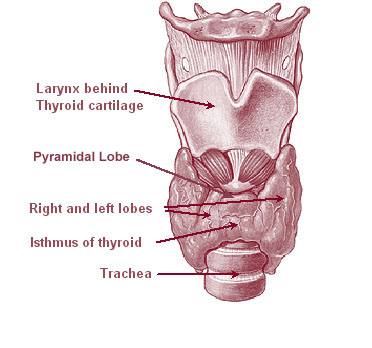
The thyroid, or thyroid gland, is an endocrine gland in vertebrates. In humans it is in the neck and consists of two connected lobes. The lower two thirds of the lobes are connected by a thin band of tissue called the thyroid isthmus. The thyroid is located at the front of the neck, below the Adam's apple. Microscopically, the functional unit of the thyroid gland is the spherical thyroid follicle, lined with follicular cells (thyrocytes), and occasional parafollicular cells that surround a lumen containing colloid. The thyroid gland secretes three hormones: the two thyroid hormones triiodothyronine (T3) and thyroxine (T4)and a peptide hormone, calcitonin. The thyroid hormones influence the metabolic rate and protein synthesis, and in children, growth and development. Calcitonin plays a role in calcium homeostasis. Secretion of the two thyroid hormones is regulated by thyroid-stimulating hormone (TSH), which is secreted from the anterior pituitary gland. TSH is regula ... [...More Info...] [...Related Items...] OR: [Wikipedia] [Google] [Baidu] |
Lymph
Lymph (from Latin, , meaning "water") is the fluid that flows through the lymphatic system, a system composed of lymph vessels (channels) and intervening lymph nodes whose function, like the venous system, is to return fluid from the tissues to be recirculated. At the origin of the fluid-return process, interstitial fluid—the fluid between the cells in all body tissues—enters the lymph capillaries. This lymphatic fluid is then transported via progressively larger lymphatic vessels through lymph nodes, where substances are removed by tissue lymphocytes and circulating lymphocytes are added to the fluid, before emptying ultimately into the right or the left subclavian vein, where it mixes with central venous blood. Because it is derived from interstitial fluid, with which blood and surrounding cells continually exchange substances, lymph undergoes continual change in composition. It is generally similar to blood plasma, which is the fluid component of blood. Lymph return ... [...More Info...] [...Related Items...] OR: [Wikipedia] [Google] [Baidu] |
Lymph Node
A lymph node, or lymph gland, is a kidney-shaped organ of the lymphatic system and the adaptive immune system. A large number of lymph nodes are linked throughout the body by the lymphatic vessels. They are major sites of lymphocytes that include B and T cells. Lymph nodes are important for the proper functioning of the immune system, acting as filters for foreign particles including cancer cells, but have no detoxification function. In the lymphatic system a lymph node is a secondary lymphoid organ. A lymph node is enclosed in a fibrous capsule and is made up of an outer cortex and an inner medulla. Lymph nodes become inflamed or enlarged in various diseases, which may range from trivial throat infections to life-threatening cancers. The condition of lymph nodes is very important in cancer staging, which decides the treatment to be used and determines the prognosis. Lymphadenopathy refers to glands that are enlarged or swollen. When inflamed or enlarged, lymph nodes can be ... [...More Info...] [...Related Items...] OR: [Wikipedia] [Google] [Baidu] |
Subclavian Vein
The subclavian vein is a paired large vein, one on either side of the body, that is responsible for draining blood from the upper extremities, allowing this blood to return to the heart. The left subclavian vein plays a key role in the absorption of lipids, by allowing products that have been carried by lymph in the thoracic duct to enter the bloodstream. The diameter of the subclavian veins is approximately 1–2 cm, depending on the individual. Structure Each subclavian vein is a continuation of the axillary vein and runs from the outer border of the first rib to the medial border of anterior scalene muscle. From here it joins with the internal jugular vein to form the brachiocephalic vein (also known as "innominate vein"). The angle of union is termed the venous angle. The subclavian vein follows the subclavian artery and is separated from the subclavian artery by the insertion of anterior scalene. Thus, the subclavian vein lies anterior to the anterior scalene while ... [...More Info...] [...Related Items...] OR: [Wikipedia] [Google] [Baidu] |
Brachiocephalic Vein
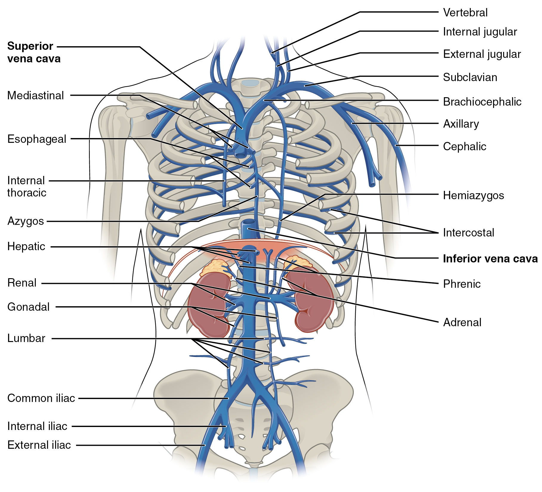
The left and right brachiocephalic veins (previously called innominate veins) are major veins in the upper chest, formed by the union of each corresponding internal jugular vein and subclavian vein. This is at the level of the sternoclavicular joint. The left brachiocephalic vein is nearly always longer than the right. These veins merge to form the superior vena cava, a great vessel, posterior to the junction of the first costal cartilage with the manubrium of the sternum. The brachiocephalic veins are the major veins returning blood to the superior vena cava. Tributaries The brachiocephalic vein is formed by the confluence of the subclavian and internal jugular veins. In addition it receives drainage from: * Left and right internal thoracic vein (Also called internal mammary veins): drain into the inferior border of their corresponding vein * Left and right inferior thyroid veins: drain into the superior aspect of their corresponding veins near the confluence * Left and rig ... [...More Info...] [...Related Items...] OR: [Wikipedia] [Google] [Baidu] |
Internal Jugular Vein
The internal jugular vein is a paired jugular vein that collects blood from the brain and the superficial parts of the face and neck. This vein runs in the carotid sheath with the common carotid artery and vagus nerve. It begins in the posterior compartment of the jugular foramen, at the base of the skull. It is somewhat dilated at its origin, which is called the ''superior bulb''. This vein also has a common trunk into which drains the anterior branch of the retromandibular vein, the facial vein, and the lingual vein. It runs down the side of the neck in a vertical direction, being at one end lateral to the internal carotid artery, and then lateral to the common carotid artery, and at the root of the neck, it unites with the subclavian vein to form the brachiocephalic vein (innominate vein); a little above its termination is a second dilation, the ''inferior bulb''. Above, it lies upon the rectus capitis lateralis, behind the internal carotid artery and the nerves pas ... [...More Info...] [...Related Items...] OR: [Wikipedia] [Google] [Baidu] |
Subclavian Artery
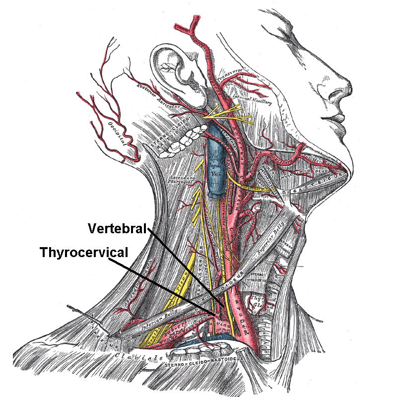
In human anatomy, the subclavian arteries are paired major arteries of the upper thorax, below the clavicle. They receive blood from the aortic arch. The left subclavian artery supplies blood to the left arm and the right subclavian artery supplies blood to the right arm, with some branches supplying the head and thorax. On the left side of the body, the subclavian comes directly off the aortic arch, while on the right side it arises from the relatively short brachiocephalic artery when it bifurcates into the subclavian and the right common carotid artery. The usual branches of the subclavian on both sides of the body are the vertebral artery, the internal thoracic artery, the thyrocervical trunk, the costocervical trunk and the dorsal scapular artery, which may branch off the transverse cervical artery, which is a branch of the thyrocervical trunk. The subclavian becomes the axillary artery at the lateral border of the first rib. Structure From its origin, the subclavian ... [...More Info...] [...Related Items...] OR: [Wikipedia] [Google] [Baidu] |
Common Carotid Artery
In anatomy, the left and right common carotid arteries (carotids) ( in Merriam-Webster Online Dictionary '.) are arteries that supply the head and neck with ; they divide in the neck to form the external and internal carotid arteries. [...More Info...] [...Related Items...] OR: [Wikipedia] [Google] [Baidu] |


.jpg)


