|
Hepatogastric Ligament
The hepatogastric ligament or gastrohepatic ligament connects the liver to the lesser curvature of the stomach. It contains the right and the left gastric arteries. In the abdominal cavity it separates the greater and lesser sac The lesser sac, also known as the omental bursa, is a part of the peritoneal cavity that is formed by the lesser and greater omentum. Usually found in mammals, it is connected with the greater sac via the omental foramen or ''Foramen of Wins ...s on the right. It is sometimes cut during surgery in order to access the lesser sac. The hepatogastric ligament consists of a dense cranial portion and the caudal portion termed the ''pars flaccida''. Additional images File:Slide6ddd.JPG, Hepatogastric ligament References External links * - "Abdominal Cavity - The Lesser Omentum" * Abdomen {{ligament-stub ... [...More Info...] [...Related Items...] OR: [Wikipedia] [Google] [Baidu] |
Hepatoduodenal Ligament
The hepatoduodenal ligament is the portion of the lesser omentum extending between the porta hepatis of the liver and the superior part of the duodenum. Running inside it are the following structures collectively known as the portal triad: * hepatic artery proper * portal vein * common bile duct Manual compression of the hepatoduodenal ligament during surgery is known as the Pringle manoeuvre. The cystoduodenal ligament is also found in the lesser omentum and is distinct from both the hepatoduodenal and hepatogastric ligaments. The cystoduodenal ligament is an abnormal peritoneal fold that attaches the duodenum to the gallbladder, representing a rare variation in the anatomy of the lesser sac and its foramen. Another variation sometimes present at the duodenal termination of the hepatoduodenal ligament is the duodenorenal ligament which passes to the front of the right kidney The kidneys are two reddish-brown bean-shaped organs found in vertebrates. They are located ... [...More Info...] [...Related Items...] OR: [Wikipedia] [Google] [Baidu] |
Liver
The liver is a major organ only found in vertebrates which performs many essential biological functions such as detoxification of the organism, and the synthesis of proteins and biochemicals necessary for digestion and growth. In humans, it is located in the right upper quadrant of the abdomen, below the diaphragm. Its other roles in metabolism include the regulation of glycogen storage, decomposition of red blood cells, and the production of hormones. The liver is an accessory digestive organ that produces bile, an alkaline fluid containing cholesterol and bile acids, which helps the breakdown of fat. The gallbladder, a small pouch that sits just under the liver, stores bile produced by the liver which is later moved to the small intestine to complete digestion. The liver's highly specialized tissue, consisting mostly of hepatocytes, regulates a wide variety of high-volume biochemical reactions, including the synthesis and breakdown of small and complex molecule ... [...More Info...] [...Related Items...] OR: [Wikipedia] [Google] [Baidu] |
Stomach
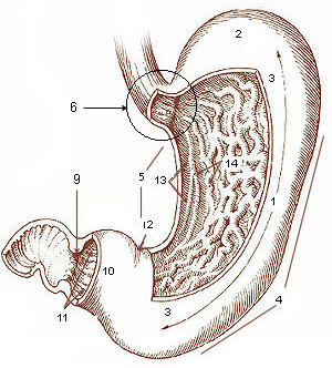
The stomach is a muscular, hollow organ in the gastrointestinal tract of humans and many other animals, including several invertebrates. The stomach has a dilated structure and functions as a vital organ in the digestive system. The stomach is involved in the gastric phase of digestion, following chewing. It performs a chemical breakdown by means of enzymes and hydrochloric acid. In humans and many other animals, the stomach is located between the oesophagus and the small intestine. The stomach secretes digestive enzymes and gastric acid to aid in food digestion. The pyloric sphincter controls the passage of partially digested food (chyme) from the stomach into the duodenum, where peristalsis takes over to move this through the rest of intestines. Structure In the human digestive system, the stomach lies between the oesophagus and the duodenum (the first part of the small intestine). It is in the left upper quadrant of the abdominal cavity. The top of the stomach lies ag ... [...More Info...] [...Related Items...] OR: [Wikipedia] [Google] [Baidu] |
Curvatures Of The Stomach
The curvatures of the stomach refer to the greater and lesser curvatures. The greater curvature of the stomach is four or five times as long as the lesser curvature. Greater curvature The greater curvature of the stomach forms the lower left or lateral border of the stomach. Surface Starting from the cardiac orifice at the incisura cardiaca, it forms an arch backward, upward, and to the left; the highest point of the convexity is on a level with the sixth left costal cartilage. From this level it may be followed downward and forward, with a slight convexity to the left as low as the cartilage of the ninth rib; it then turns to the right, to the end of the pylorus. Directly opposite the incisura angularis of the lesser curvature the greater curvature presents a dilatation, which is the left extremity of the pyloric part; this dilatation is limited on the right by a slight groove, the sulcus intermedius, which is about 2.5 cm, from the duodenopyloric constriction. Th ... [...More Info...] [...Related Items...] OR: [Wikipedia] [Google] [Baidu] |
Right Gastric Artery
The right gastric artery arises, in most cases (53% of cases), from the proper hepatic artery, descends to the pyloric end of the stomach, and passes from right to left along its lesser curvature, supplying it with branches, and anastomosing with the left gastric artery. It can also arise from the region of division of the common hepatic artery The common hepatic artery is a short blood vessel that supplies oxygenated blood to the liver, pylorus of the stomach, duodenum, pancreas, and gallbladder. It arises from the celiac artery and has the following branches: Additional images ... (20% of cases), the left branch of the hepatic artery (15% of cases), the gastroduodenal artery (8% of cases), and most rarely, the common hepatic artery itself (4% of cases). Additional images File:Gray532.png, The celiac artery and its branches; the liver has been raised, and the lesser omentum and anterior layer of the greater omentum removed. File:Slide14fff.JPG, Right gastric arter ... [...More Info...] [...Related Items...] OR: [Wikipedia] [Google] [Baidu] |
Left Gastric Artery
In human anatomy, the left gastric artery arises from the celiac artery and runs along the superior portion of the lesser curvature of the stomach. Branches also supply the lower esophagus. The left gastric artery anastomoses with the right gastric artery, which runs right to left. Important to note is that the esophageal branch of the left gastric artery ascends and passes through the esophageal hiatus. Clinical significance In terms of disease, the left gastric artery may be involved in peptic ulcer disease: if an ulcer erodes through the stomach mucosa into a branch of the artery, this can cause massive blood loss into the stomach, which may result in such symptoms as hematemesis or melaena. Additional images File:Stomach blood supply.svg, Blood supply to the stomach: left and right gastric artery, left and right gastro-omental artery and short gastric artery The short gastric arteries consist of from five to seven small branches, which arise from the end of the sp ... [...More Info...] [...Related Items...] OR: [Wikipedia] [Google] [Baidu] |
Abdominal Cavity
The abdominal cavity is a large body cavity in humans and many other animals that contains many organs. It is a part of the abdominopelvic cavity. It is located below the thoracic cavity, and above the pelvic cavity. Its dome-shaped roof is the thoracic diaphragm, a thin sheet of muscle under the lungs, and its floor is the pelvic inlet, opening into the pelvis. Structure Organs Organs of the abdominal cavity include the stomach, liver, gallbladder, spleen, pancreas, small intestine, kidneys, large intestine, and adrenal glands. Peritoneum The abdominal cavity is lined with a protective membrane termed the peritoneum. The inside wall is covered by the parietal peritoneum. The kidneys are located behind the peritoneum, in the retroperitoneum, outside the abdominal cavity. The viscera are also covered by visceral peritoneum. Between the visceral and parietal peritoneum is the peritoneal cavity, which is a potential space. It contains a serous fluid called peritone ... [...More Info...] [...Related Items...] OR: [Wikipedia] [Google] [Baidu] |
Greater Sac
In human anatomy, the greater sac, also known as the general cavity (of the abdomen) or peritoneum of the peritoneal cavity proper, is the cavity in the abdomen that is inside the peritoneum but outside the lesser sac. It is connected with the lesser sac via the omental foramen, also known as the ''foramen of Winslow'' or ''epiploic foramen'', which is anteriorly bounded by the portal triad – portal vein, hepatic artery, and common bile duct The common bile duct, sometimes abbreviated as CBD, is a duct in the gastrointestinal tract of organisms that have a gallbladder. It is formed by the confluence of the common hepatic duct and cystic duct and terminates by uniting with pancrea .... Additional images File:Gray989.png, Schematic figure of the bursa omentalis, etc. Human embryo of eight weeks. File:Gray990.png, Diagrams to illustrate the development of the greater omentum and transverse mesocolon. See also External links * * Diagram at ccccd.edu {{Peritoneum ... [...More Info...] [...Related Items...] OR: [Wikipedia] [Google] [Baidu] |
Lesser Sac
The lesser sac, also known as the omental bursa, is a part of the peritoneal cavity that is formed by the lesser and greater omentum. Usually found in mammals, it is connected with the greater sac via the omental foramen or ''Foramen of Winslow''. In mammals, it is common for the lesser sac to contain considerable amounts of fat. Anatomic margins ;Anterior margin: listed from the top-to-bottom margin: Quadrate lobe of the liver, lesser omentum, stomach, gastrocolic ligament ;Lateral margin: listed from the most anterior to the most posterior margin: Gastrosplenic ligament, spleen, phrenicosplenic ligament ;Posterior margin: Left kidney and adrenal gland, pancreas ;Inferior margin: Greater omentum ;Superior margin: Liver If any of the marginal structures rupture their contents could leak into the lesser sac. If the stomach were to rupture on its anterior side though the leak would collect in the greater sac. The lesser sac is formed during embryogenesis from an infoldi ... [...More Info...] [...Related Items...] OR: [Wikipedia] [Google] [Baidu] |


