|
Human Skull
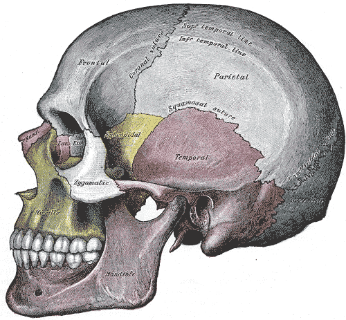
The skull, or cranium, is typically a bony enclosure around the brain of a vertebrate. In some fish, and amphibians, the skull is of cartilage. The skull is at the head end of the vertebrate. In the human, the skull comprises two prominent parts: the neurocranium and the facial skeleton, which evolved from the first pharyngeal arch. The skull forms the frontmost portion of the axial skeleton and is a product of cephalization and vesicular enlargement of the brain, with several special senses structures such as the eyes, ears, nose, tongue and, in fish, specialized tactile organs such as barbels near the mouth. The skull is composed of three types of bone: cranial bones, facial bones and ossicles, which is made up of a number of fused flat and irregular bones. The cranial bones are joined at firm fibrous junctions called sutures and contains many foramina, fossae, processes, and sinuses. In zoology, the openings in the skull are called fenestrae, the ... [...More Info...] [...Related Items...] OR: [Wikipedia] [Google] [Baidu] |
Skeletal System
A skeleton is the structural frame that supports the body of most animals. There are several types of skeletons, including the exoskeleton, which is a rigid outer shell that holds up an organism's shape; the endoskeleton, a rigid internal frame to which the organs and soft tissues attach; and the hydroskeleton, a flexible internal structure supported by the hydrostatic pressure of body fluids. Vertebrates are animals with an endoskeleton centered around an axial vertebral column, and their skeletons are typically composed of bones and cartilages. Invertebrates are other animals that lack a vertebral column, and their skeletons vary, including hard-shelled exoskeleton (arthropods and most molluscs), plated internal shells (e.g. cuttlebones in some cephalopods) or rods (e.g. ossicles in echinoderms), hydrostatically supported body cavities (most), and spicules (sponges). Cartilage is a rigid connective tissue that is found in the skeletal systems of vertebrates and invert ... [...More Info...] [...Related Items...] OR: [Wikipedia] [Google] [Baidu] |
Special Senses
In medicine and anatomy, the special senses are the senses that have specialized organs devoted to them: * vision (the eye) * hearing and balance (the ear, which includes the auditory system and vestibular system) * smell (the nose) * taste (the tongue) The distinction between special and general senses is used to classify nerve fibers running to and from the central nervous system – information from special senses is carried in special somatic afferents and special visceral afferents. In contrast, the other sense, touch, is a somatic sense which does not have a specialized organ but comes from all over the body, most noticeably the skin but also the internal organs (viscera). Touch includes mechanoreception (pressure, vibration and proprioception), pain (nociception) and heat (thermoception), and such information is carried in general somatic afferents and general visceral afferents. Vision Visual perception is the ability to interpret the surrounding environment usi ... [...More Info...] [...Related Items...] OR: [Wikipedia] [Google] [Baidu] |
Fossa (anatomy)
In anatomy, a fossa (; : fossae ( or ); ) is a depression or hollow, usually in a bone, such as the hypophyseal fossa (the depression in the sphenoid bone).Venieratos D, Anagnostopoulou S, Garidou A., A new morphometric method for the sella turcica and the hypophyseal fossa and its clinical relevance.;Folia Morphol (Warsz). 2005 Nov;64(4):240-7. Some examples include: In the skull: * Cranial fossa ** Anterior cranial fossa ** Middle cranial fossa *** Interpeduncular fossa ** Posterior cranial fossa * Hypophyseal fossa * Temporal bone fossa ** Mandibular fossa ** Jugular fossa * Infratemporal fossa * Pterygopalatine fossa * Pterygoid fossa * Lacrimal fossa ** Fossa for lacrimal gland ** Fossa for lacrimal sac * Scaphoid fossa * Condyloid fossa * Rhomboid fossa In the mandible: * Retromolar fossa In the torso: * Fossa ovalis (heart) * Infraclavicular fossa * Pyriform fossa * Substernal fossa * Iliac fossa * Ovarian fossa * Paravesical fossa * Coccygeal f ... [...More Info...] [...Related Items...] OR: [Wikipedia] [Google] [Baidu] |
Foramina
In anatomy and osteology, a foramen (; : foramina, or foramens ; ) is an opening or enclosed gap within the dense connective tissue (bones and deep fasciae) of extant and extinct amniote animals, typically to allow passage of nerves, arteries, veins or other soft tissue structures (e.g. muscle tendon) from one body compartment to another. Skull The skulls of vertebrates have foramina through which nerves, arteries, veins, and other structures pass. The human skull has many foramina, collectively referred to as the cranial foramina. Spine Within the vertebral column (spine) of vertebrates, including the human spine, each bone has an opening at both its top and bottom to allow nerves, arteries, veins, etc. to pass through. Other * Apical foramen, the hole at the tip of the root of a tooth * Foramen ovale (heart), a hole between the venous and arterial sides of the fetal heart * Transverse foramen, one of a pair of openings in each cervical vertebra, in which ... [...More Info...] [...Related Items...] OR: [Wikipedia] [Google] [Baidu] |
Suture (joint)
In anatomy, fibrous joints are joints connected by fibrous tissue, consisting mainly of collagen. These are fixed joints where bones are united by a layer of white fibrous tissue of varying thickness. In the skull, the joints between the bones are called sutures. Such immovable joints are also referred to as synarthroses. Types Most fibrous joints are also called "fixed" or "immovable". These joints have no joint cavity and are connected via fibrous connective tissue. * Sutures: The skull bones are connected by fibrous joints called '' sutures''. In fetal skulls, the sutures are wide to allow slight movement during birth. They later become rigid ( synarthrodial). * Syndesmosis: Some of the long bones in the body such as the radius and ulna in the forearm are joined by a '' syndesmosis'' (along the interosseous membrane). Syndemoses are slightly moveable ( amphiarthrodial). The distal tibiofibular joint is another example. * A '' gomphosis'' is a joint between the roo ... [...More Info...] [...Related Items...] OR: [Wikipedia] [Google] [Baidu] |
Irregular Bone
The irregular bones are bones which, from their peculiar form, cannot be grouped as long, short, flat or sesamoid bones. Irregular bones serve various purposes in the body, such as protection of nervous tissue (such as the vertebrae protect the spinal cord), affording multiple anchor points for skeletal muscle attachment (as with the sacrum), and maintaining pharynx and trachea support, and tongue attachment (such as the hyoid bone). They consist of cancellous tissue enclosed within a thin layer of compact bone. Irregular bones can also be used for joining all parts of the spinal column together. The spine is the place in the human body where the most irregular bones can be found. There are, in all, 33 irregular bones found here. The irregular bones are: the vertebrae, sacrum, coccyx, temporal, sphenoid, ethmoid, zygomatic, maxilla, mandible, palatine, inferior nasal concha The inferior nasal concha (inferior turbinated bone or inferior turbinal/turbinate) is one of ... [...More Info...] [...Related Items...] OR: [Wikipedia] [Google] [Baidu] |
Flat Bone
Flat bones are bones whose principal function is either extensive protection or the provision of broad surfaces for muscular attachment. These bones are expanded into broad, flat plates,'' Gray's Anatomy'' (1918). (See infobox) as in the cranium ( skull), the ilium, ischium, and pubis (pelvis), sternum and the rib cage. The flat bones are: the occipital, parietal, frontal, nasal, lacrimal, vomer, sternum, ribs, and scapulae. These bones are composed of two thin layers of compact bone enclosing between them a variable quantity of cancellous bone, which is the location of red bone marrow. In an adult, most red blood cells are formed in flat bones. In the cranial bones, the layers of compact tissue are familiarly known as the tables of the skull; the outer one is thick and tough; the inner is thin, dense, and brittle, and hence is termed the vitreous (glass-like) table. The intervening cancellous tissue is called the diploë, and this, in the nasal region of the skul ... [...More Info...] [...Related Items...] OR: [Wikipedia] [Google] [Baidu] |
Ossicle
The ossicles (also called auditory ossicles) are three irregular bones in the middle ear of humans and other mammals, and are among the smallest bones in the human body. Although the term "ossicle" literally means "tiny bone" (from Latin ''ossiculum'') and may refer to any small bone throughout the body, it typically refers specifically to the malleus, incus and stapes ("hammer, anvil, and stirrup") of the middle ear. The auditory ossicles serve as a kinematic chain to transmit and amplify (intensity (physics), intensify) sound vibrations collected from the air by the ear drum to the fluid-filled labyrinth (inner ear), labyrinth (cochlea). The absence or pathology of the auditory ossicles would constitute a moderate-to-severe conductive hearing loss. Structure The ossicles are, in order from the eardrum to the inner ear (from superficial to deep): the malleus, incus, and stapes, terms that in Latin are translated as "the hammer, anvil, and stirrup". * The malleus () articulat ... [...More Info...] [...Related Items...] OR: [Wikipedia] [Google] [Baidu] |
Facial Bone
The facial skeleton comprises the ''facial bones'' that may attach to build a portion of the skull. The remainder of the skull is the neurocranium. In human anatomy and development, the facial skeleton is sometimes called the ''membranous viscerocranium'', which comprises the mandible and dermatocranial elements that are not part of the braincase. Structure In the human skull, the facial skeleton consists of fourteen bones in the face: * Inferior turbinal (2) * Lacrimal bones (2) * Mandible * Maxilla (2) * Nasal bones (2) * Palatine bones (2) * Vomer * Zygomatic bones (2) Variations Elements of the ''cartilaginous viscerocranium'' (i.e., splanchnocranial elements), such as the hyoid bone, are sometimes considered part of the facial skeleton. The ethmoid bone (or a part of it) and also the sphenoid bone are sometimes included, but otherwise considered part of the neurocranium. Because the maxillary bones are fused, they are often collectively listed as only one bone. The mandi ... [...More Info...] [...Related Items...] OR: [Wikipedia] [Google] [Baidu] |
Encyclopædia Britannica
The is a general knowledge, general-knowledge English-language encyclopaedia. It has been published by Encyclopædia Britannica, Inc. since 1768, although the company has changed ownership seven times. The 2010 version of the 15th edition, which spans 32 volumes and 32,640 pages, was the last printed edition. Since 2016, it has been published exclusively as an online encyclopedia, online encyclopaedia. Printed for 244 years, the ''Britannica'' was the longest-running in-print encyclopaedia in the English language. It was first published between 1768 and 1771 in Edinburgh, Scotland, in three volumes. The encyclopaedia grew in size; the second edition was 10 volumes, and by its fourth edition (1801–1810), it had expanded to 20 volumes. Its rising stature as a scholarly work helped recruit eminent contributors, and the 9th (1875–1889) and Encyclopædia Britannica Eleventh Edition, 11th editions (1911) are landmark encyclopaedias for scholarship and literary ... [...More Info...] [...Related Items...] OR: [Wikipedia] [Google] [Baidu] |
Barbel (zoology)
In fish anatomy and turtle anatomy, a barbel is a slender, whisker like sensory organ near the mouth (sometimes called whiskers or tendrils). Fish that have barbels include the catfish, the carp, the goatfish, the hagfish, the sturgeon, the zebrafish, the black dragonfish and some species of shark such as the sawshark. Barbels house the taste buds of such fish and are used to search for food in murky water. The word ''barbel'' comes from Latin ''barbula'' 'little beard'. Barbels are sometimes erroneously referred to as '' barbs'', which are found in bird feathers for flight. Barbels may be located in a variety of locations on the head of a fish. "Maxillary barbels" refers to barbels on either side of the mouth. Barbels may also be nasal, extending from the nostrils. Also, barbels are often mandibular or mental, being located on the chin The chin is the forward pointed part of the anterior mandible (List_of_human_anatomical_regions#Regions, mental region) below the low ... [...More Info...] [...Related Items...] OR: [Wikipedia] [Google] [Baidu] |
Organ (biology)
In a multicellular organism, an organ is a collection of Tissue (biology), tissues joined in a structural unit to serve a common function. In the biological organization, hierarchy of life, an organ lies between Tissue (biology), tissue and an organ system. Tissues are formed from same type Cell (biology), cells to act together in a function. Tissues of different types combine to form an organ which has a specific function. The Gastrointestinal tract, intestinal wall for example is formed by epithelial tissue and smooth muscle tissue. Two or more organs working together in the execution of a specific body function form an organ system, also called a biological system or body system. An organ's tissues can be broadly categorized as parenchyma, the functional tissue, and stroma (tissue), stroma, the structural tissue with supportive, connective, or ancillary functions. For example, the gland's tissue that makes the hormones is the parenchyma, whereas the stroma includes the nerve t ... [...More Info...] [...Related Items...] OR: [Wikipedia] [Google] [Baidu] |


