|
HEENT Examination
A HEENT examination is a portion of a physical examination that principally concerns the head, eyes, ears, nose, and throat. Steps * IPPA ** Inspection of scars or skin changes ** Palpation of temporomandibular joint, thyroid, and lymph nodes ** Percussion may involve the skin above the frontal sinuses and paranasal sinuses to detect any signs of pain ** Auscultation for carotid bruits * Tests specific to HEENT examination ** Eyes: eye examination and acuity (including ophthalmoscope) ** Ears: hearing examination and evaluation of tympanic membrane (TM) (otoscope used in evaluation of ears, nose, and mouth) A neurological examination is usually considered separate from the HEENT evaluation, although there can be some overlap in some cases. Sample write-up References {{Medical records and physical exam Medical terminology ... [...More Info...] [...Related Items...] OR: [Wikipedia] [Google] [Baidu] |
Physical Examination
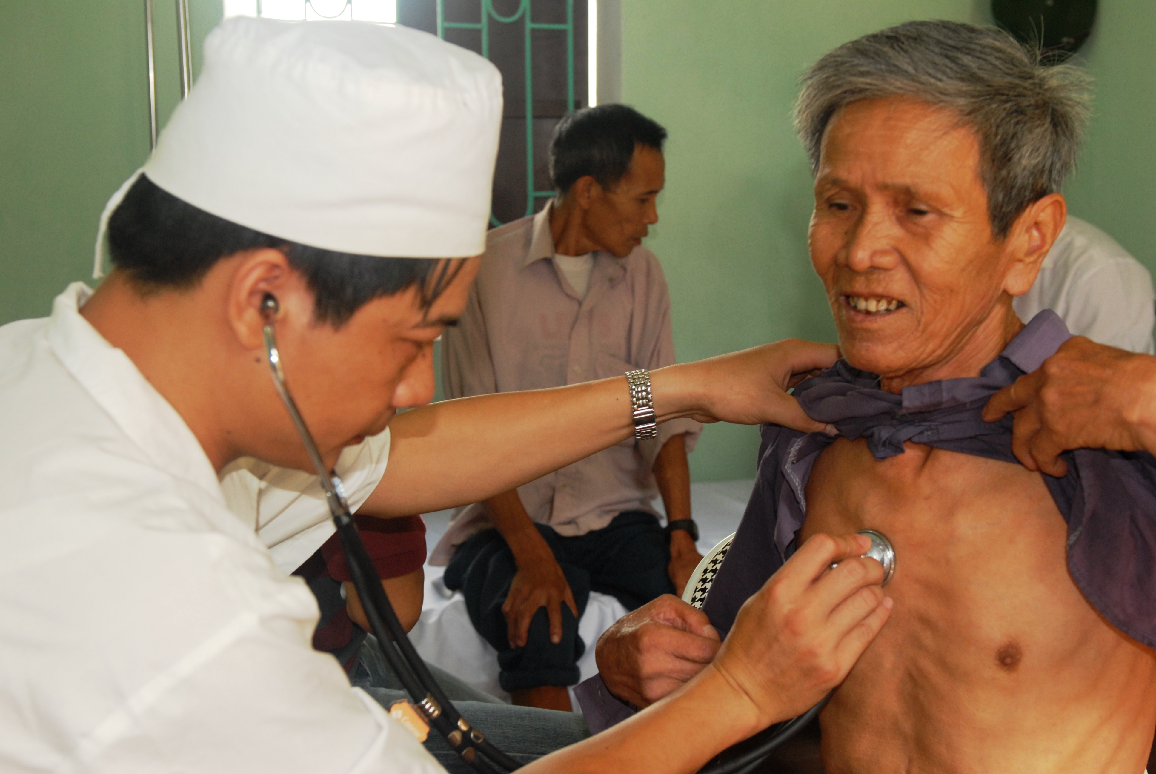
In a physical examination, medical examination, clinical examination, or medical checkup, a medical practitioner examines a patient for any possible medical signs or symptoms of a Disease, medical condition. It generally consists of a series of questions about the patient's medical history followed by an examination based on the reported symptoms. Together, the medical history and the physical examination help to determine a medical diagnosis, diagnosis and devise the treatment plan. These data then become part of the medical record. Types Routine The ''routine physical'', also known as ''general medical examination'', ''periodic health evaluation'', ''annual physical'', ''comprehensive medical exam'', ''general health check'', ''preventive health examination'', ''medical check-up'', or simply ''medical'', is a physical examination performed on an asymptomatic patient for medical screening purposes. These are normally performed by a pediatrician, family practice physician, ... [...More Info...] [...Related Items...] OR: [Wikipedia] [Google] [Baidu] |
Mucous Membrane
A mucous membrane or mucosa is a membrane that lines various cavities in the body of an organism and covers the surface of internal organs. It consists of one or more layers of epithelial cells overlying a layer of loose connective tissue. It is mostly of endodermal origin and is continuous with the skin at body openings such as the eyes, eyelids, ears, inside the nose, inside the mouth, lips, the genital areas, the urethral opening and the anus. Some mucous membranes secrete mucus, a thick protective fluid. The function of the membrane is to stop pathogens and dirt from entering the body and to prevent bodily tissues from becoming dehydrated. Structure The mucosa is composed of one or more layers of epithelial cells that secrete mucus, and an underlying lamina propria of loose connective tissue. The type of cells and type of mucus secreted vary from organ to organ and each can differ along a given tract. Mucous membranes line the digestive, respiratory and rep ... [...More Info...] [...Related Items...] OR: [Wikipedia] [Google] [Baidu] |
Mouth
A mouth also referred to as the oral is the body orifice through which many animals ingest food and animal communication#Auditory, vocalize. The body cavity immediately behind the mouth opening, known as the oral cavity (or in Latin), is also the first part of the alimentary canal, which leads to the pharynx and the gullet. In tetrapod vertebrates, the mouth is bounded on the outside by the lips and cheeks — thus the oral cavity is also known as the buccal cavity (from Latin ', meaning "cheek") — and contains the tongue on the inside. Except for some groups like birds and lissamphibians, vertebrates usually have teeth in their mouths, although some fish species have pharyngeal teeth instead of oral teeth. Most bilaterian phylum, phyla, including arthropods, molluscs and chordates, have a two-opening gut tube with a mouth at one end and an anus at the other. Which end forms first in ontogeny is a criterion used to classify bilaterian animals into protostomes and deuterostomes ... [...More Info...] [...Related Items...] OR: [Wikipedia] [Google] [Baidu] |
Exudate
An exudate is a fluid released by an organism through pores or a wound, a process known as exuding or exudation. ''Exudate'' is derived from ''exude'' 'to ooze' from Latin language, Latin 'to (ooze out) sweat' (' 'out' and ' 'to sweat'). Medicine An exudate is any fluid that filters from the circulatory system into lesions or areas of inflammation. It can be a pus-like or clear fluid. When an injury occurs, leaving skin exposed, it leaks out of the blood vessels and into nearby tissues. The fluid is composed of serum (blood), serum, fibrin, and White blood cell, leukocytes. Exudate may ooze from cuts or from areas of infection or inflammation. Types * Purulent or suppurative exudate consists of plasma with both active and dead neutrophils, fibrinogen, and necrotic parenchymal cells. This kind of exudate is consistent with more severe infections, and is commonly referred to as pus. * Fibrinous exudate is composed mainly of fibrinogen and fibrin. It is characteristic of Rheuma ... [...More Info...] [...Related Items...] OR: [Wikipedia] [Google] [Baidu] |
Erythema
Erythema (, ) is redness of the skin or mucous membranes, caused by hyperemia (increased blood flow) in superficial capillaries. It occurs with any skin injury, infection, or inflammation. Examples of erythema not associated with pathology include nervous blushes. Types * Erythema ab igne * Erythema chronicum migrans * Erythema induratum * Erythema infectiosum (or fifth disease) * Erythema marginatum * Erythema migrans * Erythema multiforme (EM) * Erythema nodosum * Erythema toxicum * Erythema elevatum diutinum * Erythema gyratum repens * Keratolytic winter erythema * Palmar erythema Causes It can be caused by infection, massage, electrical treatment, acne medication, allergies, exercise, solar radiation (sunburn), photosensitization, acute radiation syndrome, mercury toxicity, blister agents, niacin administration, or waxing and tweezing of the hairs—any of which can cause the affected capillaries to dilate, resulting in redness. Erythema is a common ... [...More Info...] [...Related Items...] OR: [Wikipedia] [Google] [Baidu] |
Oropharynx
The pharynx (: pharynges) is the part of the throat behind the mouth and nasal cavity, and above the esophagus and trachea (the tubes going down to the stomach and the lungs respectively). It is found in vertebrates and invertebrates, though its structure varies across species. The pharynx carries food to the esophagus and air to the larynx. The flap of cartilage called the epiglottis stops food from entering the larynx. In humans, the pharynx is part of the digestive system and the conducting zone of the respiratory system. (The conducting zone—which also includes the nostrils of the nose, the larynx, trachea, bronchi, and bronchioles—filters, warms, and moistens air and conducts it into the lungs). The human pharynx is conventionally divided into three sections: the nasopharynx, oropharynx, and laryngopharynx (hypopharynx). In humans, two sets of pharyngeal muscles form the pharynx and determine the shape of its lumen. They are arranged as an inner layer of longitud ... [...More Info...] [...Related Items...] OR: [Wikipedia] [Google] [Baidu] |
Throat
In vertebrate anatomy, the throat is the front part of the neck, internally positioned in front of the vertebrae. It contains the Human pharynx, pharynx and larynx. An important section of it is the epiglottis, separating the esophagus from the trachea (windpipe), preventing food and drinks being inhaled into the lungs. The throat contains various blood vessels, pharyngeal muscles, the adenoid, nasopharyngeal tonsil, the tonsils, the palatine uvula, the trachea, the esophagus, and the vocal cords. Mammal throats consist of two bones, the hyoid bone and the clavicle. The "throat" is sometimes thought to be synonymous for the Fauces (throat), fauces. It works with the mouth, ears and nose, as well as a number of other parts of the body. Its pharynx is connected to the mouth, allowing speech to occur, and food and liquid to pass down the throat. It is joined to the nose by the nasopharynx at the top of the throat, and to the ear by its Eustachian tube. The throat's trachea carries in ... [...More Info...] [...Related Items...] OR: [Wikipedia] [Google] [Baidu] |
Nasal Congestion
Nasal congestion is the partial or complete blockage of nasal passages, leading to impaired nasal breathing, usually due to membranes lining the nose becoming swollen from inflammation of blood vessels. Background In about 85% of cases, nasal congestion leads to mouth breathing rather than nasal breathing. According to Jason Turowski, MD of the Cleveland Clinic, "we are designed to breathe through our noses from birth—it's the way humans have evolved." This is referred to as " obligate nasal breathing." Nasal congestion can interfere with hearing and speech. Significant congestion may interfere with sleep, cause snoring, and can be associated with sleep apnea or upper airway resistance syndrome. In children, nasal congestion from enlarged adenoids has caused chronic sleep apnea with insufficient oxygen levels and hypoxia. The problem usually resolves after surgery to remove the adenoids and tonsils, however the problem often relapses later in life due to craniofacial ... [...More Info...] [...Related Items...] OR: [Wikipedia] [Google] [Baidu] |
Human Nose
The human nose is the first organ of the respiratory system. It is also the principal organ in the olfactory system. The shape of the nose is determined by the nasal bones and the nasal cartilages, including the nasal septum, which separates the nostrils and divides the nasal cavity into two. The nose has an important function in breathing. The nasal mucosa lining the nasal cavity and the paranasal sinuses carries out the necessary conditioning of inhaled air by warming and moistening it. Nasal conchae, shell-like bones in the walls of the cavities, play a major part in this process. Filtering of the air by nasal hair in the nostrils prevents large particles from entering the lungs. Sneezing is a reflex to expel unwanted particles from the nose that irritate the mucosal lining. Sneezing can Transmission (medicine), transmit infections, because aerosols are created in which the Respiratory droplets, droplets can harbour pathogens. Another major function of the nose is olfactio ... [...More Info...] [...Related Items...] OR: [Wikipedia] [Google] [Baidu] |
Eardrum
In the anatomy of humans and various other tetrapods, the eardrum, also called the tympanic membrane or myringa, is a thin, cone-shaped membrane that separates the external ear from the middle ear. Its function is to transmit changes in pressure of sound from the air to the ossicles inside the middle ear, and thence to the oval window in the fluid-filled cochlea. The ear thereby converts and amplifies vibration in the air to vibration in cochlear fluid. The malleus bone bridges the gap between the eardrum and the other ossicles. Rupture or perforation of the eardrum can lead to conductive hearing loss. Collapse or retraction of the eardrum can cause conductive hearing loss or cholesteatoma. Structure Orientation and relations The tympanic membrane is oriented obliquely in the anteroposterior, mediolateral, and superoinferior planes. Consequently, its superoposterior end lies lateral to its anteroinferior end. Anatomically, it relates superiorly to the middle crani ... [...More Info...] [...Related Items...] OR: [Wikipedia] [Google] [Baidu] |
Papilledema
Papilledema or papilloedema is optic disc swelling that is caused by increased intracranial pressure due to any cause. The swelling is usually bilateral and can occur over a period of hours to weeks. Unilateral presentation is extremely rare. In intracranial hypertension, the optic disc swelling most commonly occurs bilaterally. When papilledema is found on fundoscopy, further evaluation is warranted because vision loss can result if the underlying condition is not treated. Further evaluation with a CT scan or MRI of the brain and/or spine is usually done. Recent research has shown that point-of-care ultrasound can be used to measure optic nerve sheath diameter for detection of increased intracranial pressure and shows good diagnostic test accuracy compared to CT. Thus, if there is a question of papilledema on fundoscopic examination or if the optic disc cannot be adequately visualized, ultrasound can be used to rapidly assess for increased intracranial pressure and help direct f ... [...More Info...] [...Related Items...] OR: [Wikipedia] [Google] [Baidu] |


