|
First Pharyngeal Arch
The pharyngeal arches, also known as visceral arches'','' are structures seen in the embryonic development of vertebrates that are recognisable precursors for many structures. In fish, the arches are known as the branchial arches, or gill arches. In the human embryo, the arches are first seen during the fourth week of development. They appear as a series of outpouchings of mesoderm on both sides of the developing pharynx. The vasculature of the pharyngeal arches is known as the aortic arches. In fish, the branchial arches support the gills. Structure In vertebrates, the pharyngeal arches are derived from all three germ layers (the primary layers of cells that form during embryogenesis). Neural crest cells enter these arches where they contribute to features of the skull and facial skeleton such as bone and cartilage. However, the existence of pharyngeal structures before neural crest cells evolved is indicated by the existence of neural crest-independent mechanisms of pharyn ... [...More Info...] [...Related Items...] OR: [Wikipedia] [Google] [Baidu] |
Pharyngeal Pouch (embryology)
In the embryonic development of vertebrates, pharyngeal pouches form on the endodermal side between the pharyngeal arches. The pharyngeal grooves (or clefts) form the lateral ectodermal surface of the neck region to separate the arches. The pouches line up with the clefts, and these thin segments become gills in fish. Specific pouches First pouch The endoderm lines the future auditory tube (Pharyngotympanic Eustachian tube), middle ear, mastoid antrum, and inner layer of the tympanic membrane. Derivatives of this pouch are supplied by Mandibular nerve. Second pouch * Contributes the middle ear, palatine tonsils, supplied by the facial nerve. Third pouch * The third pouch possesses Dorsal and Ventral wings. Derivatives of the dorsal wings include the inferior parathyroid glands, while the ventral wings fuse to form the cytoreticular cells of the thymus. The main nerve supply to the derivatives of this pouch is Cranial Nerve IX, glossopharyngeal nerve. Fourth pouch Deriv ... [...More Info...] [...Related Items...] OR: [Wikipedia] [Google] [Baidu] |
Germ Layers
A germ layer is a primary layer of cells that forms during embryonic development. The three germ layers in vertebrates are particularly pronounced; however, all eumetazoans (animals that are sister taxa to the sponges) produce two or three primary germ layers. Some animals, like cnidarians, produce two germ layers (the ectoderm and endoderm) making them diploblastic. Other animals such as bilaterians produce a third layer (the mesoderm) between these two layers, making them triploblastic. Germ layers eventually give rise to all of an animal’s tissues and organs through the process of organogenesis. History Caspar Friedrich Wolff observed organization of the early embryo in leaf-like layers. In 1817, Heinz Christian Pander discovered three primordial germ layers while studying chick embryos. Between 1850 and 1855, Robert Remak had further refined the germ cell layer (''Keimblatt'') concept, stating that the external, internal and middle layers form respectively th ... [...More Info...] [...Related Items...] OR: [Wikipedia] [Google] [Baidu] |
Cartilage
Cartilage is a resilient and smooth type of connective tissue. In tetrapods, it covers and protects the ends of long bones at the joints as articular cartilage, and is a structural component of many body parts including the rib cage, the neck and the bronchial tubes, and the intervertebral discs. In other taxa, such as chondrichthyans, but also in cyclostomes, it may constitute a much greater proportion of the skeleton. It is not as hard and rigid as bone, but it is much stiffer and much less flexible than muscle. The matrix of cartilage is made up of glycosaminoglycans, proteoglycans, collagen fibers and, sometimes, elastin. Because of its rigidity, cartilage often serves the purpose of holding tubes open in the body. Examples include the rings of the trachea, such as the cricoid cartilage and carina. Cartilage is composed of specialized cells called chondrocytes that produce a large amount of collagenous extracellular matrix, abundant ground substance that is rich in ... [...More Info...] [...Related Items...] OR: [Wikipedia] [Google] [Baidu] |
Pericardium
The pericardium, also called pericardial sac, is a double-walled sac containing the heart and the roots of the great vessels. It has two layers, an outer layer made of strong connective tissue (fibrous pericardium), and an inner layer made of serous membrane (serous pericardium). It encloses the pericardial cavity, which contains pericardial fluid, and defines the middle mediastinum. It separates the heart from interference of other structures, protects it against infection and blunt trauma, and lubricates the heart's movements. The English name originates from the Ancient Greek prefix "''peri-''" (περί; "around") and the suffix "''-cardion''" (κάρδιον; "heart"). Anatomy The pericardium is a tough fibroelastic sac which covers the heart from all sides except at the cardiac root (where the great vessels join the heart) and the bottom (where only the serous pericardium exists to cover the upper surface of the central tendon of diaphragm). The fibrous pericard ... [...More Info...] [...Related Items...] OR: [Wikipedia] [Google] [Baidu] |
Stomodeum
The stomodeum, also called stomatodeum or stomatodaeum, is a depression between the brain and the pericardium in an embryo, and is the precursor to the mouth and the anterior lobe of the pituitary gland. Development The mouth is developed partly from the stomodeum, and partly from the floor of the anterior portion of the fore-gut. By the growth of the head end of the embryo, and the formation of the cephalic flexure, the pericardial area and the buccopharyngeal membrane come to lie on the ventral surface of the embryo. With the further expansion of the brain, and the forward bulging of the pericardium, the buccopharyngeal membrane is depressed between these two prominences. This depression constitutes the stomodeum. No trace of the membrane is found in the adult; and the communication just mentioned must not be confused with the permanent isthmus faucium. The lips, teeth, and gums are formed from the walls of the stomodeum, but the tongue The tongue is a muscular or ... [...More Info...] [...Related Items...] OR: [Wikipedia] [Google] [Baidu] |
Embryo
An embryo is an initial stage of development of a multicellular organism. In organisms that reproduce sexually, embryonic development is the part of the life cycle that begins just after fertilization of the female egg cell by the male sperm cell. The resulting fusion of these two cells produces a single-celled zygote that undergoes many cell divisions that produce cells known as blastomeres. The blastomeres are arranged as a solid ball that when reaching a certain size, called a morula, takes in fluid to create a cavity called a blastocoel. The structure is then termed a blastula, or a blastocyst in mammals. The mammalian blastocyst hatches before implantating into the endometrial lining of the womb. Once implanted the embryo will continue its development through the next stages of gastrulation, neurulation, and organogenesis. Gastrulation is the formation of the three germ layers that will form all of the different parts of the body. Neurulation forms the ner ... [...More Info...] [...Related Items...] OR: [Wikipedia] [Google] [Baidu] |
Embryogenesis
An embryo is an initial stage of development of a multicellular organism. In organisms that reproduce sexually, embryonic development is the part of the life cycle that begins just after fertilization of the female egg cell by the male sperm cell. The resulting fusion of these two cells produces a single-celled zygote that undergoes many cell divisions that produce cells known as blastomeres. The blastomeres are arranged as a solid ball that when reaching a certain size, called a morula, takes in fluid to create a cavity called a blastocoel. The structure is then termed a blastula, or a blastocyst in mammals. The mammalian blastocyst hatches before implantating into the endometrial lining of the womb. Once implanted the embryo will continue its development through the next stages of gastrulation, neurulation, and organogenesis. Gastrulation is the formation of the three germ layers that will form all of the different parts of the body. Neurulation forms the nerv ... [...More Info...] [...Related Items...] OR: [Wikipedia] [Google] [Baidu] |
Pharyngeal Jaws
Pharyngeal jaws are a "second set" of jaws contained within an animal's throat, or pharynx, distinct from the primary or oral jaws. They are believed to have originated as modified gill arches, in much the same way as oral jaws. Originally hypothesized to have evolved only once, current morphological and genetic analyses suggest at least two separate points of origin. Based on connections between musculoskeletal morphology and dentition, diet has been proposed as a main driver of the evolution of the pharyngeal jaw. A study conducted on cichlids showed that the pharyngeal jaws can undergo morphological changes in less than two years in response to their diet. Fish that ate hard-shelled prey had a robust jaw with molar-like teeth fit for crushing their durable prey. Fish that ate softer prey, on the other hand, exhibited a more slender jaw with thin, curved teeth used for tearing apart fleshy prey. These rapid changes are an example of phenotypic plasticity, wherein environmental ... [...More Info...] [...Related Items...] OR: [Wikipedia] [Google] [Baidu] |
DLX Gene Family
Genes in the ''DLX'' family encode homeodomain transcription factors related to the ''Drosophila'' distal-less ''(Dll)'' gene. The family has been related to a number of developmental features such as jaws and limbs. The family seems to be well preserved across species. As ''DLX''/''Dll'' are involved in limb development in most of the major phyla, including vertebrates, it has been suggested that ''Dll'' was involved in appendage growth in an early bilaterial ancestor. Six members of the family are found in human and mice, numbered DLX1 to DLX6. They form two-gene clusters (bigene clusters) with each other. There are DLX1-DLX2, DLX3-DLX4, DLX5-DLX6 clusters in vertebrates, linked to Hox gene clusters HOXD, HOXB, and HOXA respectively. In higher fishes like the zebrafish, there are two additional ''DLX'' genes, ''dlx2b'' (''dlx5'') and ''dlx4a'' (''dlx8''). These additional genes are not linked with each other, or any other ''DLX'' gene. All six other genes remain in bigene cluste ... [...More Info...] [...Related Items...] OR: [Wikipedia] [Google] [Baidu] |
Hox Gene
Hox genes, a subset of homeobox genes, are a group of related genes that specify regions of the body plan of an embryo along the head-tail axis of animals. Hox proteins encode and specify the characteristics of 'position', ensuring that the correct structures form in the correct places of the body. For example, Hox genes in insects specify which appendages form on a segment (for example, legs, antennae, and wings in fruit flies), and Hox genes in vertebrates specify the types and shape of vertebrae that will form. In segmented animals, Hox proteins thus confer segmental or positional identity, but do not form the actual segments themselves. Studies on Hox genes in ciliated larvae have shown they are only expressed in future adult tissues. In larvae with gradual metamorphosis the Hox genes are activated in tissues of the larval body, generally in the trunk region, that will be maintained through metamorphosis. In larvae with complete metamorphosis the Hox genes are mainly express ... [...More Info...] [...Related Items...] OR: [Wikipedia] [Google] [Baidu] |
Hyoid
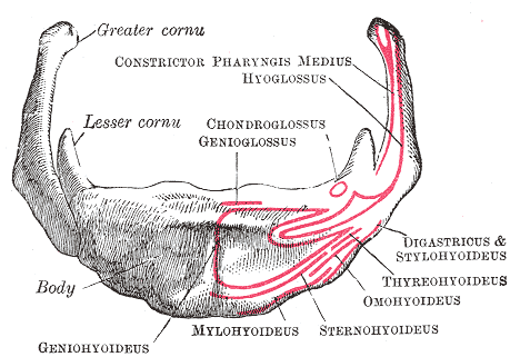
The hyoid bone (lingual bone or tongue-bone) () is a horseshoe-shaped bone situated in the anterior midline of the neck between the chin and the thyroid cartilage. At rest, it lies between the base of the mandible and the third cervical vertebra. Unlike other bones, the hyoid is only distantly articulated to other bones by muscles or ligaments. It is the only bone in the human body that is not connected to any other bones nearby. The hyoid is anchored by muscles from the anterior, posterior and inferior directions, and aids in tongue movement and swallowing. The hyoid bone provides attachment to the muscles of the floor of the mouth and the tongue above, the larynx below, and the epiglottis and pharynx behind. Its name is derived . Structure The hyoid bone is classed as an irregular bone and consists of a central part called the body, and two pairs of horns, the greater and lesser horns. Body The body of the hyoid bone is the central part of the hyoid bone. *At the fron ... [...More Info...] [...Related Items...] OR: [Wikipedia] [Google] [Baidu] |
First Pharyngeal Arch
The pharyngeal arches, also known as visceral arches'','' are structures seen in the embryonic development of vertebrates that are recognisable precursors for many structures. In fish, the arches are known as the branchial arches, or gill arches. In the human embryo, the arches are first seen during the fourth week of development. They appear as a series of outpouchings of mesoderm on both sides of the developing pharynx. The vasculature of the pharyngeal arches is known as the aortic arches. In fish, the branchial arches support the gills. Structure In vertebrates, the pharyngeal arches are derived from all three germ layers (the primary layers of cells that form during embryogenesis). Neural crest cells enter these arches where they contribute to features of the skull and facial skeleton such as bone and cartilage. However, the existence of pharyngeal structures before neural crest cells evolved is indicated by the existence of neural crest-independent mechanisms of pharyn ... [...More Info...] [...Related Items...] OR: [Wikipedia] [Google] [Baidu] |


