|
Extensor Hallucis Brevis
The extensor hallucis brevis is a muscle on the top of the foot that helps to extend the big toe. Structure The extensor hallucis brevis is essentially the medial part of the extensor digitorum brevis muscle. Some anatomists have debated whether these two muscles are distinct entities. The extensor hallucis brevis arises from the calcaneus and inserts on the proximal phalanx of the digit 1 (the big toe). Nerve supply Nerve supplied by lateral terminal branch of Deep Peroneal Nerve (deep fibular nerve) (proximal sciatic branches S1, S2). Same innervation of Extensor Digitorum Brevis Function The extensor hallucis brevis helps to extend the big toe. See also * Extensor digitorum brevis * Extensor hallucis longus The extensor hallucis longus muscle is a thin skeletal muscle, situated between the tibialis anterior and the extensor digitorum longus. It extends the big toe and dorsiflects the foot. It also assists with foot eversion and inversion. Stru ... Additional Im ... [...More Info...] [...Related Items...] OR: [Wikipedia] [Google] [Baidu] |
Calcaneus
In humans and many other primates, the calcaneus (; from the Latin ''calcaneus'' or ''calcaneum'', meaning heel) or heel bone is a bone of the tarsus of the foot which constitutes the heel. In some other animals, it is the point of the hock. Structure In humans, the calcaneus is the largest of the tarsal bones and the largest bone of the foot. Its long axis is pointed forwards and laterally. The talus bone, calcaneus, and navicular bone are considered the proximal row of tarsal bones. In the calcaneus, several important structures can be distinguished:Platzer (2004), p 216 There is a large calcaneal tuberosity located posteriorly on plantar surface with medial and lateral tubercles on its surface. Besides, there is another peroneal tubecle on its lateral surface. On its lower edge on either side are its lateral and medial processes (serving as the origins of the abductor hallucis and abductor digiti minimi). The Achilles tendon is inserted into a roughened area on its sup ... [...More Info...] [...Related Items...] OR: [Wikipedia] [Google] [Baidu] |
Hallux
Toes are the digits (fingers) of the foot of a tetrapod. Animal species such as cats that walk on their toes are described as being ''digitigrade''. Humans, and other animals that walk on the soles of their feet, are described as being ''plantigrade''; ''unguligrade'' animals are those that walk on hooves at the tips of their toes. Structure There are normally five toes present on each human foot. Each toe consists of three phalanx bones, the proximal, middle, and distal, with the exception of the big toe ( la, hallux). For a minority of people, the little toe also is missing a middle bone. The hallux only contains two phalanx bones, the proximal and distal. The joints between each phalanx are the interphalangeal joints. The proximal phalanx bone of each toe articulates with the metatarsal bone of the foot at the metatarsophalangeal joint. Each toe is surrounded by skin, and present on all five toes is a toenail. The toes are, from medial to lateral: * the first t ... [...More Info...] [...Related Items...] OR: [Wikipedia] [Google] [Baidu] |
Dorsalis Pedis Artery
In human anatomy, the dorsalis pedis artery (dorsal artery of foot) is a blood vessel of the lower limb. It arises from the anterior tibial artery, and ends at the first intermetatarsal space (as the first dorsal metatarsal artery and the deep plantar artery). It carries oxygenated blood to the dorsal side of the foot. It is useful for taking a pulse. It is also at risk during anaesthesia of the deep peroneal nerve. Structure The dorsalis pedis artery is located 1/3 from medial malleolus of the ankle. It arises at the anterior aspect of the ankle joint and is a continuation of the anterior tibial artery. It ends at the proximal part of the first intermetatarsal space. Here, it divides into two branches, the first dorsal metatarsal artery, and the deep plantar artery. It is covered by skin and fascia, but is fairly superficial. The dorsalis pedis communicates with the plantar blood supply of the foot through the deep plantar artery. Along its course, it is accompanied by a deep ... [...More Info...] [...Related Items...] OR: [Wikipedia] [Google] [Baidu] |
Deep Fibular Nerve
The deep fibular nerve (also known as deep peroneal nerve) begins at the bifurcation of the common fibular nerve between the fibula and upper part of the fibularis longus, passes infero-medially, deep to the extensor digitorum longus, to the anterior surface of the interosseous membrane, and comes into relation with the anterior tibial artery above the middle of the leg; it then descends with the artery to the front of the ankle-joint, where it divides into a ''lateral'' and a '' medial terminal branch''. Structure Lateral side of the leg The deep fibular nerve is the nerve of the anterior compartment of the leg and the dorsum of the foot. It is one of the terminal branches of the common fibular nerve. It corresponds to the posterior interosseus nerve of the forearm. It begins at the lateral side of the fibula bone, and then enters the anterior compartment by piercing the anterior intermuscular septum. It then pierces the extensor digitorum longus and lies next to the anterior t ... [...More Info...] [...Related Items...] OR: [Wikipedia] [Google] [Baidu] |
Flexor Hallucis Brevis Muscle
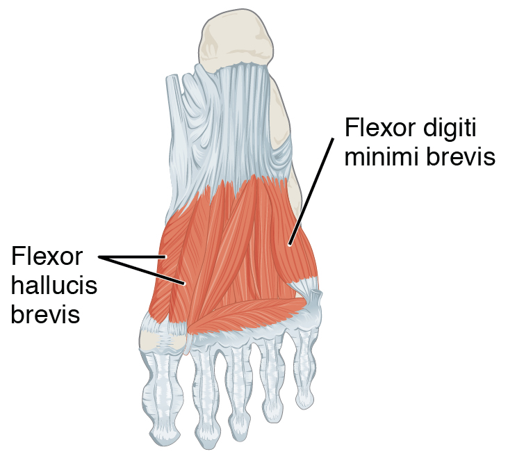
Flexor hallucis brevis muscle is a muscle of the foot that flexes the big toe. Structure Flexor hallucis brevis muscle arises, by a pointed tendinous process, from the medial part of the under surface of the cuboid bone, from the contiguous portion of the third cuneiform, and from the prolongation of the tendon of the tibialis posterior muscle which is attached to that bone. It divides in front into two portions, which are inserted into the medial and lateral sides of the base of the first phalanx of the great toe, a sesamoid bone being present in each tendon at its insertion. The medial portion is blended with the abductor hallucis muscle previous to its insertion; the lateral portion (sometimes described as the first plantar interosseus) with the adductor hallucis muscle. The tendon of the flexor hallucis longus muscle lies in a groove between the two. Its tendon usually contains two sesamoid bones at the point under the first metatarsophalangeal joint. Innervation The medi ... [...More Info...] [...Related Items...] OR: [Wikipedia] [Google] [Baidu] |
Muscle
Skeletal muscles (commonly referred to as muscles) are organs of the vertebrate muscular system and typically are attached by tendons to bones of a skeleton. The muscle cells of skeletal muscles are much longer than in the other types of muscle tissue, and are often known as muscle fibers. The muscle tissue of a skeletal muscle is striated – having a striped appearance due to the arrangement of the sarcomeres. Skeletal muscles are voluntary muscles under the control of the somatic nervous system. The other types of muscle are cardiac muscle which is also striated and smooth muscle which is non-striated; both of these types of muscle tissue are classified as involuntary, or, under the control of the autonomic nervous system. A skeletal muscle contains multiple fascicles – bundles of muscle fibers. Each individual fiber, and each muscle is surrounded by a type of connective tissue layer of fascia. Muscle fibers are formed from the fusion of developmental myoblasts ... [...More Info...] [...Related Items...] OR: [Wikipedia] [Google] [Baidu] |
Big Toe
Toes are the digits (fingers) of the foot of a tetrapod. Animal species such as cats that walk on their toes are described as being ''digitigrade''. Humans, and other animals that walk on the soles of their feet, are described as being ''plantigrade''; ''unguligrade'' animals are those that walk on hooves at the tips of their toes. Structure There are normally five toes present on each human foot. Each toe consists of three phalanx bones, the proximal, middle, and distal, with the exception of the big toe ( la, hallux). For a minority of people, the little toe also is missing a middle bone. The hallux only contains two phalanx bones, the proximal and distal. The joints between each phalanx are the interphalangeal joints. The proximal phalanx bone of each toe articulates with the metatarsal bone of the foot at the metatarsophalangeal joint. Each toe is surrounded by skin, and present on all five toes is a toenail. The toes are, from medial to lateral: * the first toe ... [...More Info...] [...Related Items...] OR: [Wikipedia] [Google] [Baidu] |
Extensor Digitorum Brevis
The extensor digitorum brevis muscle (sometimes EDB) is a muscle on the upper surface of the foot that helps extend digits 2 through 4. Structure The muscle originates from the forepart of the upper and lateral surface of the calcaneus (in front of the groove for the peroneus brevis tendon), from the interosseous talocalcaneal ligament and the stem of the inferior extensor retinaculum. The fibres pass obliquely forwards and medially across the dorsum of the foot and end in four tendons. The medial part of the muscle, also known as extensor hallucis brevis, ends in a tendon which crosses the dorsalis pedis artery and inserts into the dorsal surface of the base of the proximal phalanx of the great toe. The other three tendons insert into the lateral sides of the tendons of extensor digitorum longus for the second, third and fourth toes. Nerve supply Nerve supply: lateral terminal branch of Deep Peroneal Nerve (deep fibular nerve) (proximal sciatic branches L4-L5, but most clinica ... [...More Info...] [...Related Items...] OR: [Wikipedia] [Google] [Baidu] |
Extensor Hallucis Longus
The extensor hallucis longus muscle is a thin skeletal muscle, situated between the tibialis anterior and the extensor digitorum longus. It extends the big toe and dorsiflects the foot. It also assists with foot eversion and inversion. Structure The extensor hallucis longus muscle arises from the anterior surface of the fibula for about the middle two-fourths of its extent, medial to the origin of the extensor digitorum longus muscle. It also arises from the interosseous membrane of the leg to a similar extent. The anterior tibial vessels and deep fibular nerve lie between it and the tibialis anterior. The fibers pass downward, and end in a tendon, which occupies the anterior border of the muscle, passes through a distinct compartment in the cruciate crural ligament, crosses from the lateral to the medial side of the anterior tibial vessels near the bend of the ankle, and is inserted into the base of the distal phalanx of the great toe. Opposite the metatarsophalangeal ar ... [...More Info...] [...Related Items...] OR: [Wikipedia] [Google] [Baidu] |
Calf Muscles
Calf most often refers to: * Calf (animal), the young of domestic cattle. * Calf (leg), in humans (and other primates), the back portion of the lower leg Calf or calves may also refer to: Biology and animal byproducts *Veal, meat from calves *Calfskin, leather *Vellum, calf hide processed as a writing material *Calf-binding, a leather book binding Geography * The Calf, a peak in the Yorkshire Dales, UK *Calf, an island off Newfoundland; see Bull, Cow and Calf *Calf, the product of Ice calving * Calves, Portugal, a hamlet in Póvoa de Varzim, Portugal People *Anthony Calf Other *CALF, the Common Affordable Lightweight Fighter project resulting in the Boeing X-32 *Calf, short for calfdozer, a type of small bulldozer *Calf, part of an early type of internal combustion engine seen in the Ascot (1904 automobile) See also * *List of animal names, for animals whose young are called "calves" * Crus, the entire lower leg * Calve (other) *Calving (other) * Calf I ... [...More Info...] [...Related Items...] OR: [Wikipedia] [Google] [Baidu] |


.jpg)