|
Extraocular Muscles
The extraocular muscles, or extrinsic ocular muscles, are the seven extrinsic muscles of the eye in human eye, humans and other animals. Six of the extraocular muscles, the four recti muscles, and the superior oblique muscle, superior and inferior oblique muscles, control Eye movement, movement of the eye. The other muscle, the Levator palpebrae superioris muscle, levator palpebrae superioris, controls eyelid elevation and depression, elevation. The actions of the six muscles responsible for eye movement depend on the position of the eye at the time of muscle contraction. The ciliary muscle, pupillary sphincter muscle and pupillary dilator muscle sometimes are called intrinsic ocular muscles or intraocular muscles. Structure Since only a small part of the eye called the Fovea centralis, fovea provides sharp vision, the eye must move to follow a target. Eye movements must be precise and fast. This is seen in scenarios like reading, where the reader must shift gaze constantly. Alt ... [...More Info...] [...Related Items...] OR: [Wikipedia] [Google] [Baidu] |
MRI Scan
Magnetic resonance imaging (MRI) is a medical imaging technique used in radiology to generate pictures of the anatomy and the physiological processes inside the body. MRI scanners use strong magnetic fields, magnetic field gradients, and radio waves to form images of the organs in the body. MRI does not involve X-rays or the use of ionizing radiation, which distinguishes it from computed tomography (CT) and positron emission tomography (PET) scans. MRI is a medical application of nuclear magnetic resonance (NMR) which can also be used for imaging in other NMR applications, such as NMR spectroscopy. MRI is widely used in hospitals and clinics for medical diagnosis, staging and follow-up of disease. Compared to CT, MRI provides better contrast in images of soft tissues, e.g. in the brain or abdomen. However, it may be perceived as less comfortable by patients, due to the usually longer and louder measurements with the subject in a long, confining tube, although "ope ... [...More Info...] [...Related Items...] OR: [Wikipedia] [Google] [Baidu] |
Extrinsic Muscles
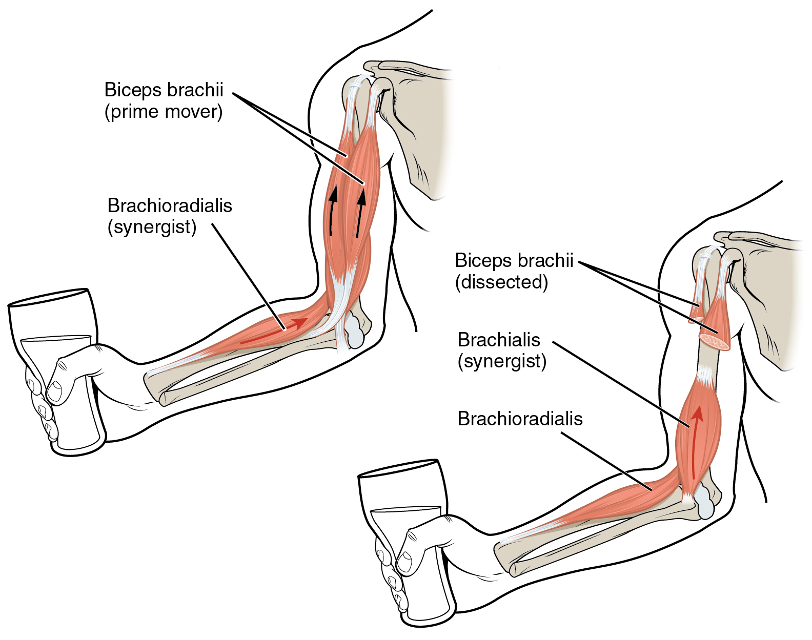
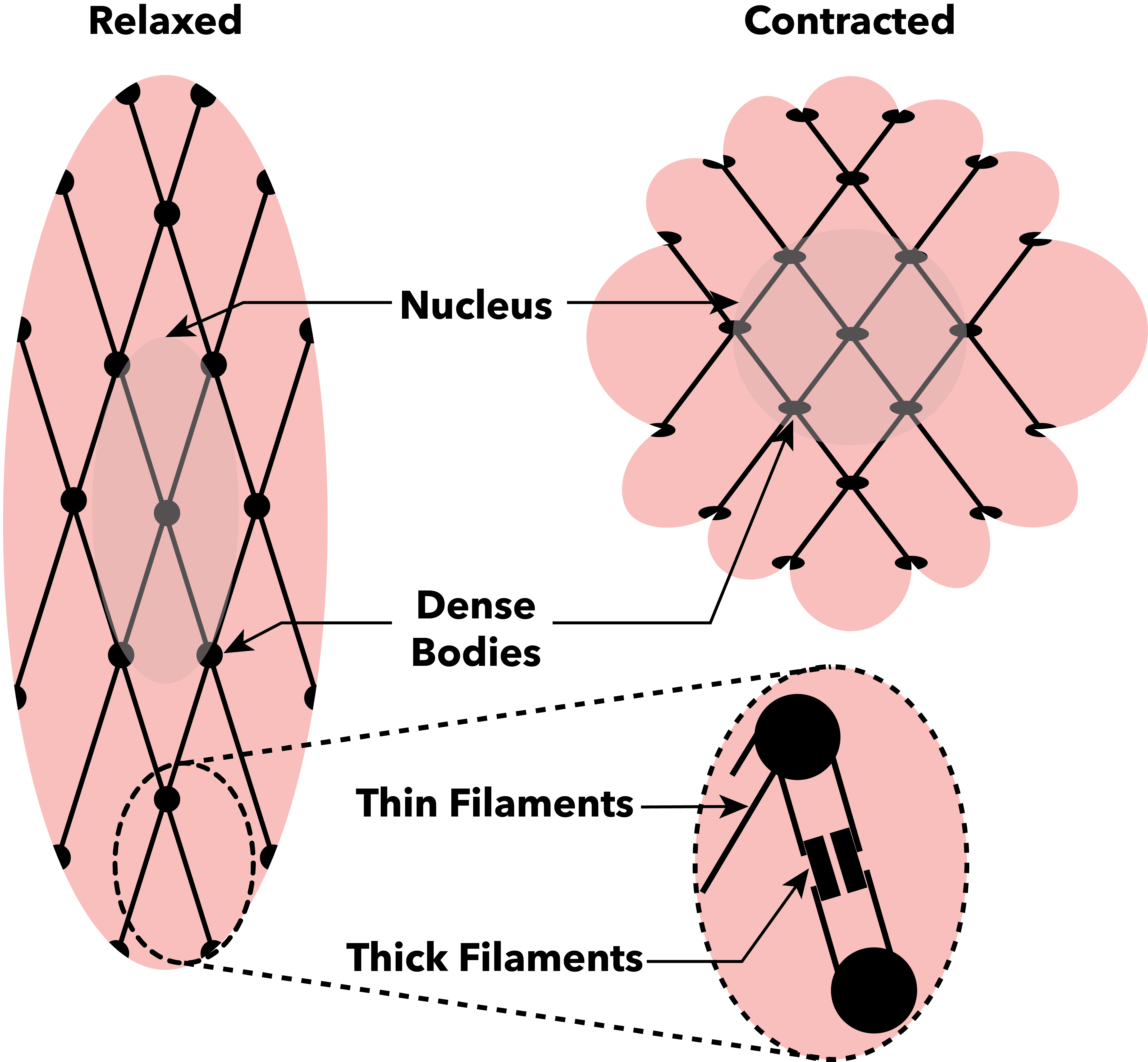
Anatomical terminology is used to uniquely describe aspects of skeletal muscle, cardiac muscle, and smooth muscle such as their actions, structure, size, and location. Types There are three types of muscle tissue in the body: skeletal, smooth, and cardiac. Skeletal muscle Skeletal muscle, or "voluntary muscle", is a striated muscle tissue that primarily joins to bone with tendons. Skeletal muscle enables movement of bones, and maintains posture. The widest part of a muscle that pulls on the tendons is known as the belly. Muscle slip A muscle slip is a slip of muscle that can either be an anatomical variant, or a branching of a muscle as in rib connections of the serratus anterior muscle. Smooth muscle Smooth muscle is involuntary and found in parts of the body where it conveys action without conscious intent. The majority of this type of muscle tissue is found in the digestive and urinary systems where it acts by propelling forward food, chyme, and feces in the former and ur ... [...More Info...] [...Related Items...] OR: [Wikipedia] [Google] [Baidu] |
1412 Extraocular Muscles
141 may refer to: * 141 (number), an integer * AD 141, a year of the Julian calendar * 141 BC, a year of the pre-Julian Roman calendar * 141 Lumen 141 Lumen is a carbonaceous asteroid from the intermediate asteroid belt, approximately 130 kilometers in diameter. It is an identified Eunomia family#Interlopers, Eunomian interloper. Description It was discovered on January 13, 1875, by th ..., a main-belt asteroid * Lockheed C-141 Starlifter, a retired American military aircraft {{numberdis ... [...More Info...] [...Related Items...] OR: [Wikipedia] [Google] [Baidu] |
Intraocular Muscle
Intrinsic ocular muscles or intraocular muscles{{cite book , last1=Ludwig , first1=Parker E. , last2=Aslam , first2=Sanah , last3=Czyz , first3=Craig N. , title=StatPearls , date=2024 , publisher=StatPearls Publishing , chapter-url=https://www.ncbi.nlm.nih.gov/books/NBK470534/ , chapter=Anatomy, Head and Neck: Eye Muscles , pmid=29262013 are muscles of the inside of the eye structure. The intraocular muscles are responsible for adjusting the shape of the lens and the size of the pupil. They're different from the extraocular muscles that are outside of the eye and control the external movement of the eye. There are three intrisic ocular muscles: the ciliary muscle, pupillary sphincter muscle ( sphincter pupillae) and pupillary dilator muscle (dilator pupillae). All of them are smooth muscles. The ciliary muscle is attached to the zonular fibers and the zonular fibers are the suspensory ligaments of the lens. The ciliary muscle controls accommodation by altering the shape of ... [...More Info...] [...Related Items...] OR: [Wikipedia] [Google] [Baidu] |
Pupillary Dilator Muscle
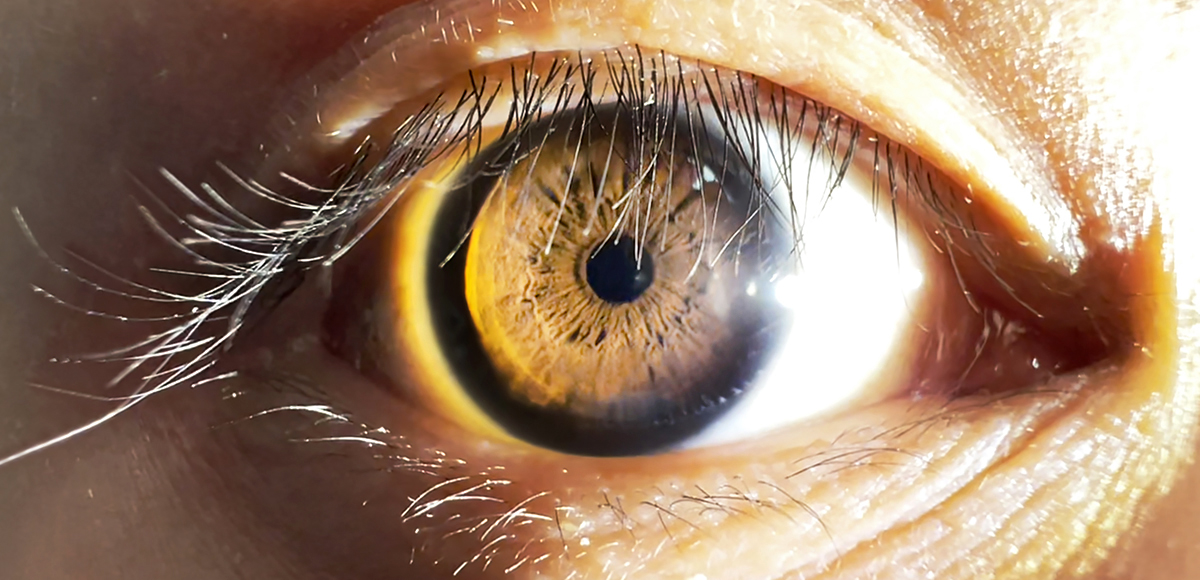
The iris dilator muscle (pupil dilator muscle, pupillary dilator, radial muscle of iris, radiating fibers), is a smooth muscle of the eye, running radially in the iris and therefore fit as a dilator. The pupillary dilator consists of a spokelike arrangement of modified contractile cells called myoepithelial cells. These cells are stimulated by the sympathetic nervous system. When stimulated, the cells contract, widening the pupil and allowing more light to enter the eye. The ciliary muscle, pupillary sphincter muscle and pupillary dilator muscle sometimes are called intrinsic ocular muscles or intraocular muscles. Structure Innervation It is innervated by the sympathetic system, which acts by releasing noradrenaline, which acts on α1-receptors. Thus, when presented with a threatening stimulus that activates the fight-or-flight response, this innervation contracts the muscle and dilates the pupil, thus temporarily letting more light reach the retina. The dilator muscle is inne ... [...More Info...] [...Related Items...] OR: [Wikipedia] [Google] [Baidu] |
Pupillary Sphincter Muscle
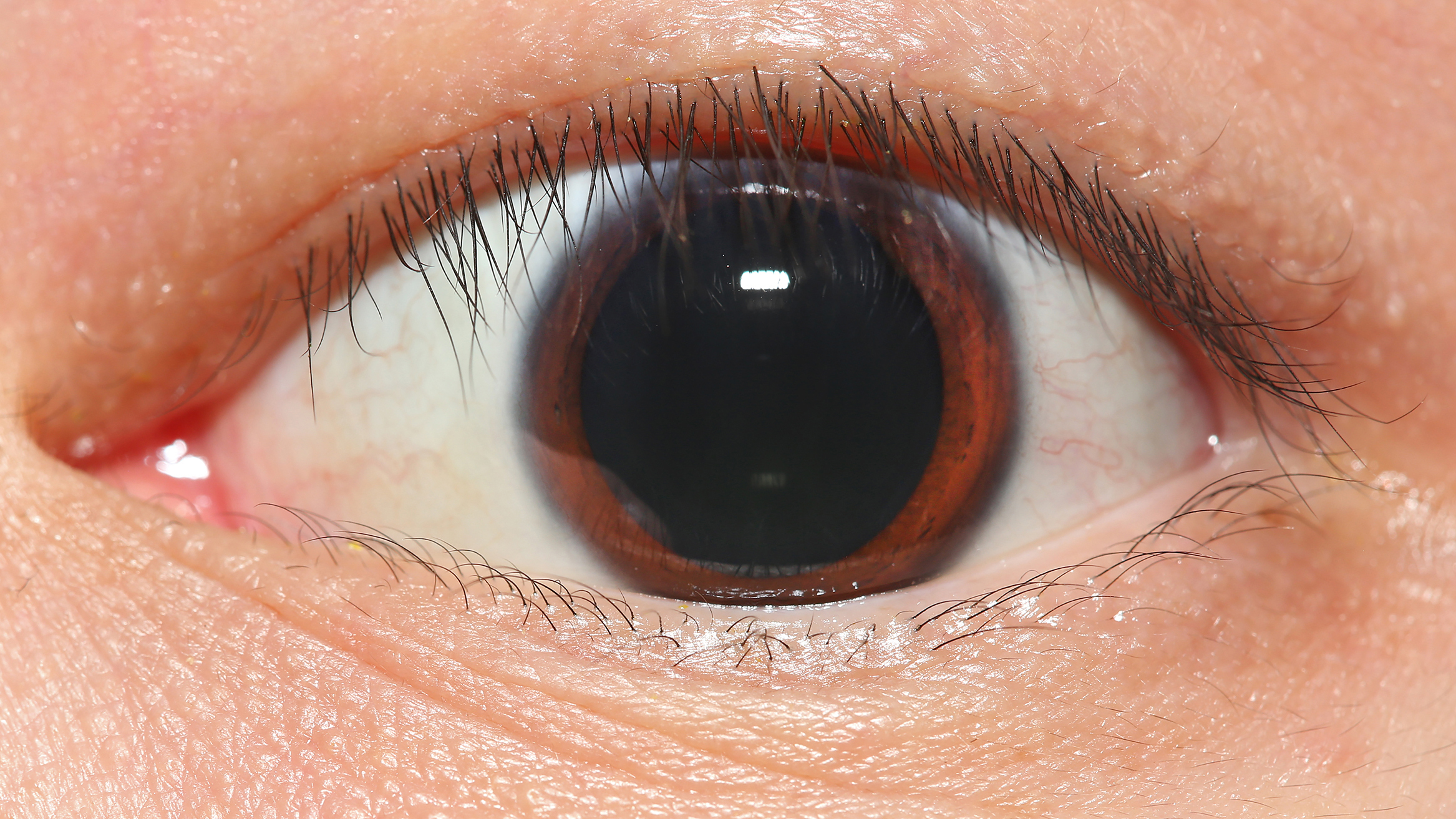
The iris sphincter muscle (pupillary sphincter, pupillary constrictor, circular muscle of iris, circular fibers) is a muscle in the part of the eye called the iris. It encircles the pupil of the iris, appropriate to its function as a constrictor of the pupil. The ciliary muscle, pupillary sphincter muscle and pupillary dilator muscle sometimes are called intrinsic ocular muscles or intraocular muscles. Comparative anatomy This structure is found in vertebrates and in some cephalopods. General structure All the myocytes are of the smooth muscle type. Its dimensions are about 0.75 mm wide by 0.15 mm thick. Mode of action In humans, it functions to constrict the pupil in bright light (pupillary light reflex) or during accommodation. In , the muscle cells themselves are photosensitive causing iris action without brain input. Innervation It is controlled by parasympathetic postganglionic fibers releasing acetylcholine acting primarily on the muscarinic acetylcholine r ... [...More Info...] [...Related Items...] OR: [Wikipedia] [Google] [Baidu] |
Ciliary Muscle
The ciliary muscle is an intrinsic muscle of the eye formed as a ring of smooth muscleSchachar, Ronald A. (2012). "Anatomy and Physiology." (Chapter 4) . in the eye's middle layer, the uvea ( vascular layer). It controls accommodation for viewing objects at varying distances and regulates the flow of aqueous humor into Schlemm's canal. It also changes the shape of the lens within the eye but not the size of the pupil which is carried out by the sphincter pupillae muscle and dilator pupillae. The ciliary muscle, pupillary sphincter muscle and pupillary dilator muscle sometimes are called intrinsic ocular muscles or intraocular muscles. Structure Development The ciliary muscle develops from mesenchyme within the choroid and is considered a cranial neural crest derivative. Nerve supply The ciliary muscle receives parasympathetic fibers from the short ciliary nerves that arise from the ciliary ganglion. The parasympathetic postganglionic fibers are part of cranial n ... [...More Info...] [...Related Items...] OR: [Wikipedia] [Google] [Baidu] |
Muscle Contraction
Muscle contraction is the activation of Tension (physics), tension-generating sites within muscle cells. In physiology, muscle contraction does not necessarily mean muscle shortening because muscle tension can be produced without changes in muscle length, such as when holding something heavy in the same position. The termination of muscle contraction is followed by muscle relaxation, which is a return of the muscle fibers to their low tension-generating state. For the contractions to happen, the muscle cells must rely on the change in action of two types of Myofilament, filaments: thin and thick filaments. The major constituent of thin filaments is a chain formed by helical coiling of two strands of actin, and thick filaments dominantly consist of chains of the Motor protein, motor-protein myosin. Together, these two filaments form myofibrils - the basic functional organelles in the skeletal muscle system. In vertebrates, Muscle cell#Muscle contraction in striated muscle, skele ... [...More Info...] [...Related Items...] OR: [Wikipedia] [Google] [Baidu] |
Elevation And Depression
Motion, the process of movement, is described using specific anatomical terms. Motion includes movement of organs, joints, limbs, and specific sections of the body. The terminology used describes this motion according to its direction relative to the anatomical position of the body parts involved. Anatomists and others use a unified set of terms to describe most of the movements, although other, more specialized terms are necessary for describing unique movements such as those of the hands, feet, and eyes. In general, motion is classified according to the anatomical plane it occurs in. ''Flexion'' and ''extension'' are examples of ''angular'' motions, in which two axes of a joint are brought closer together or moved further apart. ''Rotational'' motion may occur at other joints, for example the shoulder, and are described as ''internal'' or ''external''. Other terms, such as ''elevation'' and ''depression'', describe movement above or below the horizontal plane. Many anatomica ... [...More Info...] [...Related Items...] OR: [Wikipedia] [Google] [Baidu] |
Levator Palpebrae Superioris Muscle
The levator palpebrae superioris () is the muscle in the orbit that elevates the upper eyelid. Structure The levator palpebrae superioris originates from inferior surface of the lesser wing of the sphenoid bone, just above the optic foramen. It broadens and decreases in thickness (becomes thinner) and becomes the levator aponeurosis. This portion inserts on the skin of the upper eyelid, as well as the superior tarsal plate. It is a skeletal muscle. The superior tarsal muscle, a smooth muscle, is attached to the levator palpebrae superioris, and inserts on the superior tarsal plate as well. Blood supply The levator palebrae superioris receives its blood supply from branches of the ophthalmic artery, specifically, muscular branches and the supraorbital artery. Blood is drained into the superior ophthalmic vein. Nerve supply The levator palpebrae superioris receives motor innervation from the superior division of the oculomotor nerve. The smooth muscle that originates from ... [...More Info...] [...Related Items...] OR: [Wikipedia] [Google] [Baidu] |
Eye Movement
Eye movement includes the voluntary or involuntary movement of the eyes. Eye movements are used by a number of organisms (e.g. primates, rodents, flies, birds, fish, cats, crabs, octopus) to fixate, inspect and track visual objects of interests. A special type of eye movement, rapid eye movement, occurs during REM sleep. The eyes are the visual organs of the human body, and move using a system of six muscles. The retina, a specialised type of tissue containing photoreceptors, senses light. These specialised cells convert light into electrochemical signals. These signals travel along the optic nerve fibers to the brain, where they are interpreted as vision in the visual cortex. Primates and many other vertebrates use three types of voluntary eye movement to track objects of interest: smooth pursuit, vergence shifts and saccades. These types of movements appear to be initiated by a small cortical region in the brain's frontal lobe. This is corroborated by removal of the ... [...More Info...] [...Related Items...] OR: [Wikipedia] [Google] [Baidu] |


