|
Embryological Origins Of The Mouth And Anus
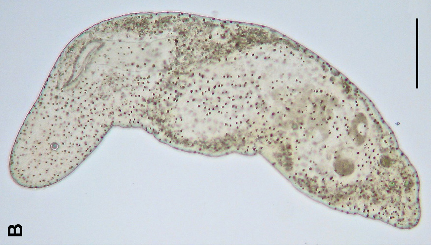
The embryological origin of the mouth and anus is an important characteristic, and forms the morphological basis for separating bilaterian animals into two natural groupings: the protostomes and deuterostomes. In animals at least as complex as an earthworm, a dent forms in one side of the early, spheroidal embryo. This dent, the blastopore, deepens to become the archenteron, the first phase in the growth of the gut. In deuterostomes, the original dent becomes the anus, while the gut eventually tunnels through the embryo until it reaches the other side, forming an opening that becomes the mouth. It was originally thought that the blastopore of the protostomes formed the mouth, and the anus formed second when the gut tunneled through the embryo. More recent research has shown that our understanding of protostome mouth formation is somewhat less secure than we had thought. Acoelomorpha, which form a sister group to the rest of the bilaterian animals, have a single mouth that ... [...More Info...] [...Related Items...] OR: [Wikipedia] [Google] [Baidu] |
Bilateria
Bilateria () is a large clade of animals characterised by bilateral symmetry during embryonic development. This means their body plans are laid around a longitudinal axis with a front (or "head") and a rear (or "tail") end, as well as a left–right–symmetrical belly ( ventral) and back ( dorsal) surface. Nearly all bilaterians maintain a bilaterally symmetrical body as adults; the most notable exception is the echinoderms, which have pentaradial symmetry as adults, but bilateral symmetry as embryos. With few exceptions, bilaterian embryos are triploblastic, having three germ layers: endoderm, mesoderm and ectoderm, and have complete digestive tracts with a separate mouth and anus. Some bilaterians lack body cavities, while others have a primary body cavity derived from the blastocoel, or a secondary cavity, the coelom. Cephalization is a characteristic feature among most bilaterians, where the sense organs and central nerve ganglia become concentrated at th ... [...More Info...] [...Related Items...] OR: [Wikipedia] [Google] [Baidu] |
Embryology Of Digestive System
Embryology (from Ancient Greek, Greek ἔμβρυον, ''embryon'', "the unborn, embryo"; and -λογία, ''-logy, -logia'') is the branch of animal biology that studies the Prenatal development (biology), prenatal development of gametes (sex cells), fertilization, and development of embryos and fetuses. Additionally, embryology encompasses the study of congenital disorders that occur before birth, known as teratology. Early embryology was proposed by Marcello Malpighi, and known as preformationism, the theory that organisms develop from pre-existing miniature versions of themselves. Aristotle proposed the theory that is now accepted, Epigenesis (biology), epigenesis. Epigenesis (biology), Epigenesis is the idea that organisms develop from seed or egg in a sequence of steps. Modern embryology developed from the work of Karl Ernst von Baer, though accurate observations had been made in Italy by anatomists such as Aldrovandi and Leonardo da Vinci in the Renaissance. Comparative ... [...More Info...] [...Related Items...] OR: [Wikipedia] [Google] [Baidu] |
Gastrulation
Gastrulation is the stage in the early embryonic development of most animals, during which the blastula (a single-layered hollow sphere of cells), or in mammals, the blastocyst, is reorganized into a two-layered or three-layered embryo known as the gastrula. Before gastrulation, the embryo is a continuous epithelial sheet of cells; by the end of gastrulation, the embryo has begun differentiation to establish distinct cell lineages, set up the basic axes of the body (e.g. dorsal–ventral, anterior–posterior), and internalized one or more cell types, including the prospective gut. Gastrula layers In triploblastic organisms, the gastrula is trilaminar (three-layered). These three germ layers are the ectoderm (outer layer), mesoderm (middle layer), and endoderm (inner layer).Mundlos 2009p. 422/ref>McGeady, 2004: p. 34 In diploblastic organisms, such as Cnidaria and Ctenophora, the gastrula has only ectoderm and endoderm. The two layers are also sometimes referred to as ... [...More Info...] [...Related Items...] OR: [Wikipedia] [Google] [Baidu] |
Embryology
Embryology (from Ancient Greek, Greek ἔμβρυον, ''embryon'', "the unborn, embryo"; and -λογία, ''-logy, -logia'') is the branch of animal biology that studies the Prenatal development (biology), prenatal development of gametes (sex cells), fertilization, and development of embryos and fetuses. Additionally, embryology encompasses the study of congenital disorders that occur before birth, known as teratology. Early embryology was proposed by Marcello Malpighi, and known as preformationism, the theory that organisms develop from pre-existing miniature versions of themselves. Aristotle proposed the theory that is now accepted, Epigenesis (biology), epigenesis. Epigenesis (biology), Epigenesis is the idea that organisms develop from seed or egg in a sequence of steps. Modern embryology developed from the work of Karl Ernst von Baer, though accurate observations had been made in Italy by anatomists such as Aldrovandi and Leonardo da Vinci in the Renaissance. Comparative ... [...More Info...] [...Related Items...] OR: [Wikipedia] [Google] [Baidu] |
Hindgut
The hindgut (or epigaster) is the posterior ( caudal) part of the alimentary canal. In mammals, it includes the distal one third of the transverse colon and the splenic flexure, the descending colon, sigmoid colon and up to the ano-rectal junction. In zoology, the term ''hindgut'' refers also to the cecum and ascending colon. Structure Blood supply Arterial supply is by the inferior mesenteric artery, and venous drainage is to the portal venous system. Lymphatic drainage is to the chyle cistern. Nerve supply The hindgut is innervated via the inferior mesenteric plexus. Sympathetic innervation is from the lumbar splanchnic nerves (L1-L2), parasympathetic innervation is from S2-S4. Development Additional images File:Gray985.png, Abdominal part of digestive tube and its attachment to the primitive or common mesentery. Human embryo of six weeks. File:Gray1115.png, Tail end of human embryo twenty-five to twenty-nine days old. File:Illacme plenipes female with 170 segments a ... [...More Info...] [...Related Items...] OR: [Wikipedia] [Google] [Baidu] |
Cloacal Membrane
The cloacal membrane is the membrane that covers the embryonic cloaca during the development of the urinary and reproductive organs. It is formed by ectoderm and endoderm coming into contact with each other. As the human embryo grows and caudal folding continues, the urorectal septum divides the cloaca into a ventral urogenital sinus The urogenital sinus is a body part of a human or other Placentalia, placental only present in the development of the urinary system, development of the urinary and development of the reproductive organs, reproductive organs. It is the ventral p ... and dorsal anorectal canal. Before the urorectal septum has an opportunity to fuse with the cloacal membrane, the membrane ruptures, exposing the urogenital sinus and dorsal anorectal canal to the exterior. Later on, an ectodermal plug, the anal membrane, forms to create the lower third of the rectum. It ruptures in the seventh week of gestation. References External links * * Diagram at unsw.e ... [...More Info...] [...Related Items...] OR: [Wikipedia] [Google] [Baidu] |
Foregut
The foregut in humans is the anterior part of the alimentary canal, from the distal esophagus to the first half of the duodenum, at the entrance of the bile duct. Beyond the stomach, the foregut is attached to the abdominal walls by mesentery. The foregut arises from the endoderm, developing from the folding primitive gut, and is developmentally distinct from the midgut and hindgut. Although the term “foregut” is typically used in reference to the anterior section of the primitive gut, components of the adult gut can also be described with this designation. Pain in the epigastric region, just below the intersection of the ribs, typically refers to structures in the adult foregut. Adult foregut Components * Esophagus * Respiratory tract (lower respiratory tract) * Stomach * Duodenum (up to ampulla of vater) * Liver * Gallbladder * Pancreas * Spleen – The spleen arises from the mesodermal dorsal mesentery (the foregut arises from the endoderm not mesoderm). But the sp ... [...More Info...] [...Related Items...] OR: [Wikipedia] [Google] [Baidu] |
Buccopharyngeal Membrane
The region where the crescentic masses of the ectoderm and endoderm come into direct contact with each other constitutes a thin membrane, the buccopharyngeal membrane (or oropharyngeal membrane), which forms a septum between the primitive mouth and pharynx. In front of the buccopharyngeal area, where the lateral crescents of mesoderm fuse in the middle line, the pericardium The pericardium (: pericardia), also called pericardial sac, is a double-walled sac containing the heart and the roots of the great vessels. It has two layers, an outer layer made of strong inelastic connective tissue (fibrous pericardium), ... is afterward developed, and this region is therefore designated the ''pericardial area''. The buccopharyngeal membranes serve as a respiratory surface in a wide variety of amphibians and reptiles. In this type of respiration, membranes in the mouth and throat are permeable to oxygen and carbon dioxide. In some species that remain submerged in water for long period ... [...More Info...] [...Related Items...] OR: [Wikipedia] [Google] [Baidu] |
Urbilaterian
The urbilaterian (from German ur- 'original') is the hypothetical last common ancestor of the bilaterian clade, i.e., all animals having a bilateral symmetry. Appearance Its appearance is a matter of debate, for no representative has been (or may or may not ever be) identified in the fossil record. Two reconstructed urbilaterian morphologies can be considered: first, the less complex ancestral form forming the common ancestor to Xenacoelomorpha and Nephrozoa; and second, the more complex (coelomate) urbilaterian ancestral to both protostomes and deuterostomes, sometimes referred to as the "urnephrozoan". Since most protostomes and deuterostomes share features — e.g. nephridia (and the derived kidneys), through guts, blood vessels and nerve ganglia— that are useful only in relatively large (macroscopic) organisms, their common ancestor ought also to have been macroscopic. However, such large animals should have left traces in the sediment in which they moved, and evidenc ... [...More Info...] [...Related Items...] OR: [Wikipedia] [Google] [Baidu] |
Deuterostomia
Deuterostomes (from Ancient Greek, Greek: ) are bilaterian animals of the superphylum Deuterostomia (), typically characterized by their anus forming before the mouth during embryogenesis, embryonic development. Deuterostomia comprises three Phylum, phyla: chordate, Chordata, Echinodermata, hemichordate, Hemichordata, and the extinct clade Cambroernida. In deuterostomes, the developing embryo's first opening (the blastopore) becomes the anus and cloaca, while the mouth is formed at a different site later on. This was initially the group's distinguishing characteristic, but deuterostomy has since been discovered among protostomes as well. The deuterostomes are also known as enterocoelomates, because their coelom develops through pouching of the gut, enterocoely. Deuterostomia's sister clade is Protostomia, animals that develop mouth first and whose digestive tract development is more varied. Protostomia includes the ecdysozoans and spiralians, as well as the extinct ''Kimberella'' ... [...More Info...] [...Related Items...] OR: [Wikipedia] [Google] [Baidu] |
Protostomia
Protostomia () is the clade of animals once thought to be characterized by the formation of the organism's mouth before its anus during embryonic development. This nature has since been discovered to be extremely variable among Protostomia's members, although the reverse is typically true of its sister clade, Deuterostomia. Well-known examples of protostomes are arthropods, molluscs, annelids, flatworms and nematodes. They are also called schizocoelomates since schizocoely typically occurs in them. Together with the Deuterostomia and Xenacoelomorpha, these form the clade Bilateria, animals with bilateral symmetry, anteroposterior axis and three germ layers. Protostomy In animals at least as complex as earthworms, the first phase in gut development involves the embryo forming a dent on one side (the blastopore) which deepens to become its digestive tube (the archenteron). In the sister-clade, the deuterostomes (), the original dent becomes the anus while the gut event ... [...More Info...] [...Related Items...] OR: [Wikipedia] [Google] [Baidu] |


