|
Combined Pulmonary Fibrosis And Emphysema
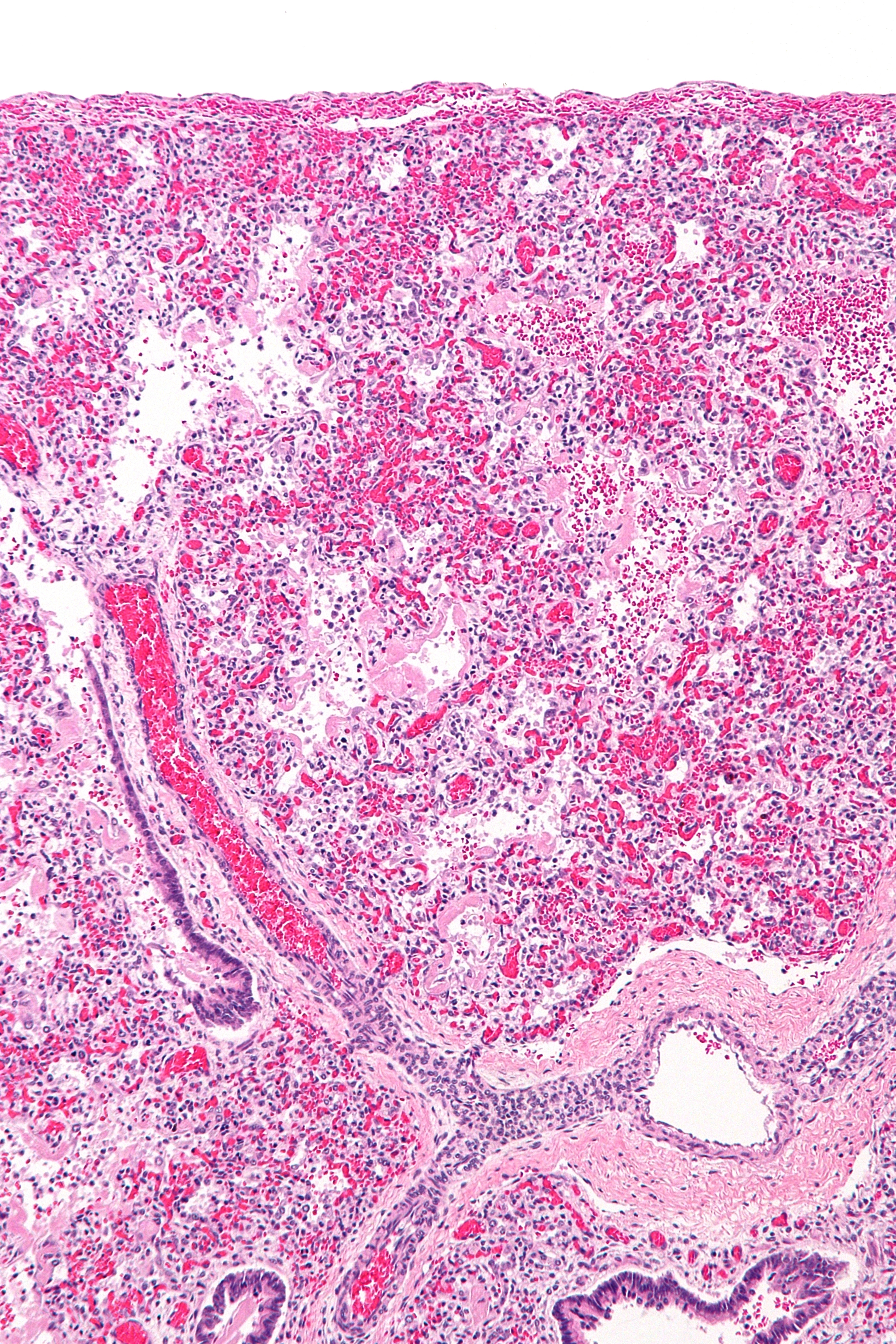
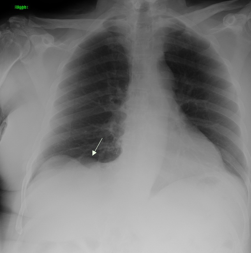
Combined pulmonary fibrosis and emphysema (CPFE), describes a medical syndrome involving both pulmonary fibrosis and emphysema. The combination is most commonly found in male smokers. Pulmonary function tests typically show preserved lung volume with very low transfer factor. __TOC__ Presentation CFPE is characterised by shortness of breath, and reduced oxygen concentration (reflecting gas exchange abnormalities). Imaging shows upper-lobe emphysema, and lower-lobe interstitial fibrosis. CFPE is often complicated by pulmonary hypertension, acute lung injury, lung cancer Lung cancer, also known as lung carcinoma (since about 98–99% of all lung cancers are carcinomas), is a malignant lung tumor characterized by uncontrolled cell growth in tissues of the lung. Lung carcinomas derive from transformed, malign ..., and coronary artery disease. Diagnosis The diagnosis is confirmed with high resolution CT scan. References External links {{Medicine, state=collapsed L ... [...More Info...] [...Related Items...] OR: [Wikipedia] [Google] [Baidu] |
Syndrome
A syndrome is a set of medical signs and symptoms which are correlated with each other and often associated with a particular disease or disorder. The word derives from the Greek language, Greek σύνδρομον, meaning "concurrence". When a syndrome is paired with a definite cause this becomes a disease. In some instances, a syndrome is so closely linked with a pathogenesis or cause that disease#Terminology, the words ''syndrome'', ''disease'', and ''disorder'' end up being used interchangeably for them. This substitution of terminology often confuses the reality and meaning of medical diagnoses. This is especially true of heredity, inherited syndromes. About one third of all phenotypes that are listed in Online Mendelian Inheritance in Man, OMIM are described as dysmorphic, which usually refers to the facial gestalt. For example, Down syndrome, Wolf–Hirschhorn syndrome, and Andersen–Tawil syndrome are disorders with known pathogeneses, so each is more than just a set of sig ... [...More Info...] [...Related Items...] OR: [Wikipedia] [Google] [Baidu] |
Pulmonary Fibrosis
Pulmonary fibrosis is a condition in which the lungs become scarred over time. Symptoms include shortness of breath, a dry cough, feeling tired, weight loss, and nail clubbing. Complications may include pulmonary hypertension, respiratory failure, pneumothorax, and lung cancer. Causes include environmental pollution, certain medications, connective tissue diseases, infections, and interstitial lung diseases. Idiopathic pulmonary fibrosis (IPF), an interstitial lung disease of unknown cause, is most common. Diagnosis may be based on symptoms, medical imaging, lung biopsy, and lung function tests. There is no cure and there are limited treatment options available. Treatment is directed towards efforts to improve symptoms and may include oxygen therapy and pulmonary rehabilitation. Certain medications may be used to try to slow the worsening of scarring. Lung transplantation may occasionally be an option. At least 5 million people are affected globally. Life expectancy is gene ... [...More Info...] [...Related Items...] OR: [Wikipedia] [Google] [Baidu] |
Emphysema
Emphysema, or pulmonary emphysema, is a lower respiratory tract disease, characterised by air-filled spaces ( pneumatoses) in the lungs, that can vary in size and may be very large. The spaces are caused by the breakdown of the walls of the alveoli and they replace the spongy lung parenchyma. This reduces the total alveolar surface available for gas exchange leading to a reduction in oxygen supply for the blood. Emphysema usually affects the middle aged or older population because it takes time to develop with the effects of tobacco smoking, and other risk factors. Alpha-1 antitrypsin deficiency is a genetic risk factor that may lead to the condition presenting earlier. When associated with significant airflow limitation, emphysema is a major subtype of chronic obstructive pulmonary disease ( COPD), a progressive lung disease characterized by long-term breathing problems and poor airflow. Without COPD, the finding of emphysema on a CT lung scan still confers a higher mortal ... [...More Info...] [...Related Items...] OR: [Wikipedia] [Google] [Baidu] |
Dyspnea
Shortness of breath (SOB), also medically known as dyspnea (in AmE) or dyspnoea (in BrE), is an uncomfortable feeling of not being able to breathe well enough. The American Thoracic Society defines it as "a subjective experience of breathing discomfort that consists of qualitatively distinct sensations that vary in intensity", and recommends evaluating dyspnea by assessing the intensity of its distinct sensations, the degree of distress and discomfort involved, and its burden or impact on the patient's activities of daily living. Distinct sensations include effort/work to breathe, chest tightness or pain, and "air hunger" (the feeling of not enough oxygen). The tripod position is often assumed to be a sign. Dyspnea is a normal symptom of heavy physical exertion but becomes pathological if it occurs in unexpected situations, when resting or during light exertion. In 85% of cases it is due to asthma, pneumonia, cardiac ischemia, interstitial lung disease, congestive heart fai ... [...More Info...] [...Related Items...] OR: [Wikipedia] [Google] [Baidu] |
Hypoxemia
Hypoxemia is an abnormally low level of oxygen in the blood. More specifically, it is oxygen deficiency in arterial blood. Hypoxemia has many causes, and often causes hypoxia as the blood is not supplying enough oxygen to the tissues of the body. Definition ''Hypoxemia'' refers to the low level of oxygen in blood, and the more general term ''hypoxia'' is an abnormally low oxygen content in any tissue or organ, or the body as a whole. Hypoxemia can cause hypoxia (hypoxemic hypoxia), but hypoxia can also occur via other mechanisms, such as anemia. Hypoxemia is usually defined in terms of reduced partial pressure of oxygen (mm Hg) in arterial blood, but also in terms of reduced content of oxygen (ml oxygen per dl blood) or percentage saturation of hemoglobin (the oxygen-binding protein within red blood cells) with oxygen, which is either found singly or in combination. While there is general agreement that an arterial blood gas measurement which shows that the partial pressure ... [...More Info...] [...Related Items...] OR: [Wikipedia] [Google] [Baidu] |
Gas Exchange
Gas exchange is the physical process by which gases move passively by diffusion across a surface. For example, this surface might be the air/water interface of a water body, the surface of a gas bubble in a liquid, a gas-permeable membrane, or a biological membrane that forms the boundary between an organism and its extracellular environment. Gases are constantly consumed and produced by cellular and metabolic reactions in most living things, so an efficient system for gas exchange between, ultimately, the interior of the cell(s) and the external environment is required. Small, particularly unicellular organisms, such as bacteria and protozoa, have a high surface-area to volume ratio. In these creatures the gas exchange membrane is typically the cell membrane. Some small multicellular organisms, such as flatworms, are also able to perform sufficient gas exchange across the skin or cuticle that surrounds their bodies. However, in most larger organisms, which have a small surf ... [...More Info...] [...Related Items...] OR: [Wikipedia] [Google] [Baidu] |
Medical Imaging
Medical imaging is the technique and process of imaging the interior of a body for clinical analysis and medical intervention, as well as visual representation of the function of some organs or tissues (physiology). Medical imaging seeks to reveal internal structures hidden by the skin and bones, as well as to diagnose and treat disease. Medical imaging also establishes a database of normal anatomy and physiology to make it possible to identify abnormalities. Although imaging of removed organs and tissues can be performed for medical reasons, such procedures are usually considered part of pathology instead of medical imaging. Measurement and recording techniques that are not primarily designed to produce images, such as electroencephalography (EEG), magnetoencephalography (MEG), electrocardiography (ECG), and others, represent other technologies that produce data susceptible to representation as a parameter graph versus time or maps that contain data about the measureme ... [...More Info...] [...Related Items...] OR: [Wikipedia] [Google] [Baidu] |
Acute Lung Injury
Acute respiratory distress syndrome (ARDS) is a type of respiratory failure characterized by rapid onset of widespread inflammation in the lungs. Symptoms include shortness of breath (dyspnea), rapid breathing (tachypnea), and bluish skin coloration (cyanosis). For those who survive, a decreased quality of life is common. Causes may include sepsis, pancreatitis, trauma, pneumonia, and aspiration. The underlying mechanism involves diffuse injury to cells which form the barrier of the microscopic air sacs of the lungs, surfactant dysfunction, activation of the immune system, and dysfunction of the body's regulation of blood clotting. In effect, ARDS impairs the lungs' ability to exchange oxygen and carbon dioxide. Adult diagnosis is based on a PaO2/FiO2 ratio (ratio of partial pressure arterial oxygen and fraction of inspired oxygen) of less than 300 mm Hg despite a positive end-expiratory pressure (PEEP) of more than 5 cm H2O. Cardiogenic pulmonary edema, as t ... [...More Info...] [...Related Items...] OR: [Wikipedia] [Google] [Baidu] |
Lung Cancer
Lung cancer, also known as lung carcinoma (since about 98–99% of all lung cancers are carcinomas), is a malignant lung tumor characterized by uncontrolled cell growth in tissues of the lung. Lung carcinomas derive from transformed, malignant cells that originate as epithelial cells, or from tissues composed of epithelial cells. Other lung cancers, such as the rare sarcomas of the lung, are generated by the malignant transformation of connective tissues (i.e. nerve, fat, muscle, bone), which arise from mesenchymal cells. Lymphomas and melanomas (from lymphoid and melanocyte cell lineages) can also rarely result in lung cancer. In time, this uncontrolled growth can metastasize (spreading beyond the lung) either by direct extension, by entering the lymphatic circulation, or via hematogenous, bloodborne spread – into nearby tissue or other, more distant parts of the body. Most cancers that originate from within the lungs, known as primary lung cancers, are carcinomas. The t ... [...More Info...] [...Related Items...] OR: [Wikipedia] [Google] [Baidu] |
Coronary Artery Disease
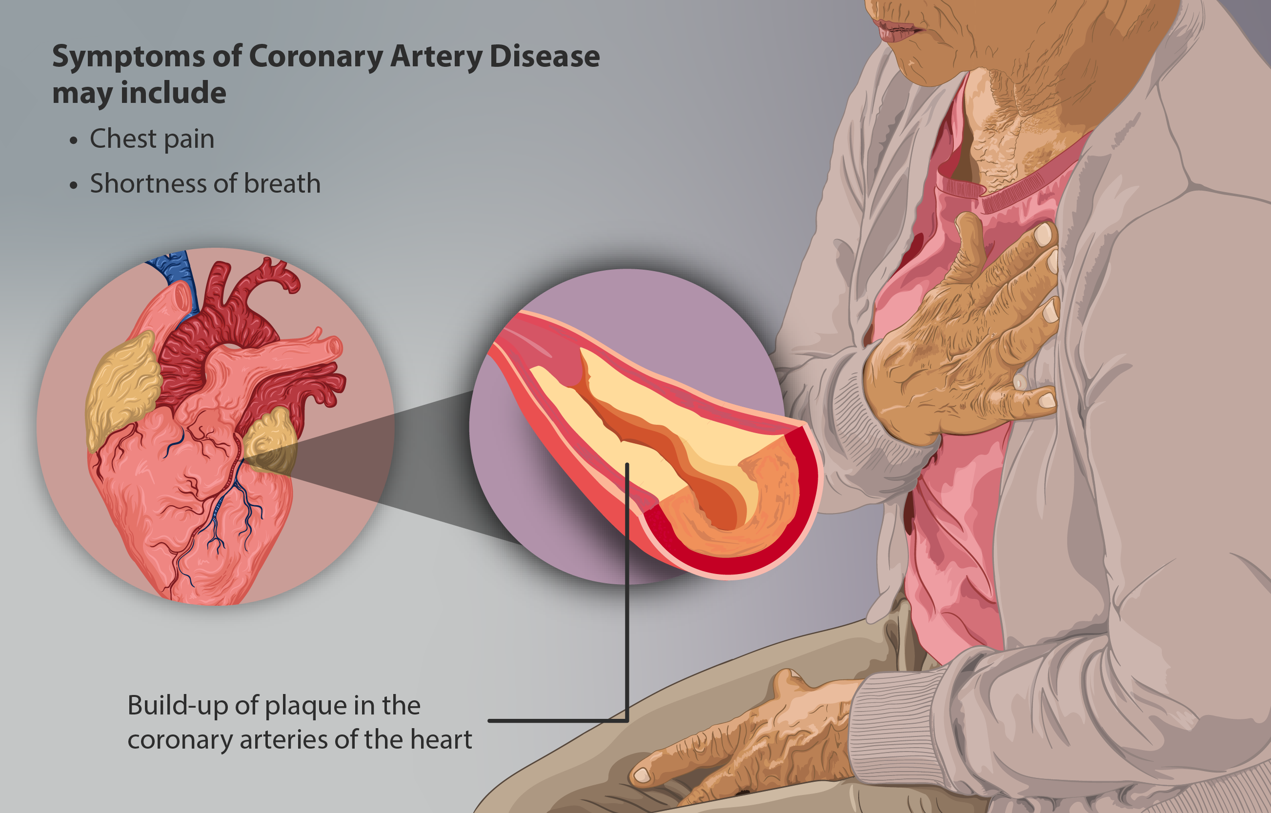
Coronary artery disease (CAD), also called coronary heart disease (CHD), ischemic heart disease (IHD), myocardial ischemia, or simply heart disease, involves the reduction of blood flow to the heart muscle due to build-up of atherosclerotic plaque in the arteries of the heart. It is the most common of the cardiovascular diseases. Types include stable angina, unstable angina, myocardial infarction, and sudden cardiac death. A common symptom is chest pain or discomfort which may travel into the shoulder, arm, back, neck, or jaw. Occasionally it may feel like heartburn. Usually symptoms occur with exercise or emotional stress, last less than a few minutes, and improve with rest. Shortness of breath may also occur and sometimes no symptoms are present. In many cases, the first sign is a heart attack. Other complications include heart failure or an abnormal heartbeat. Risk factors include high blood pressure, smoking, diabetes, lack of exercise, obesity, high blood choles ... [...More Info...] [...Related Items...] OR: [Wikipedia] [Google] [Baidu] |
High-resolution Computed Tomography
High-resolution computed tomography (HRCT) is a type of computed tomography (CT) with specific techniques to enhance image resolution. It is used in the diagnosis of various health problems, though most commonly for lung disease, by assessing the lung parenchyma. On the other hand, HRCT of the temporal bone is used to diagnose various middle ear diseases such as otitis media, cholesteatoma, and evaluations after ear operations. Technique HRCT is performed using a conventional CT scanner. However, imaging parameters are chosen so as to maximize spatial resolution: a narrow slice width is used (usually 1–2 mm), a high spatial resolution image reconstruction algorithm is used, field of view is minimized, so as to minimize the size of each pixel, and other scan factors (e.g. focal spot) may be optimized for resolution at the expense of scan speed. Depending on the suspected diagnosis, the scan may be performed in both inspiration and expiration. In inspiration images are ... [...More Info...] [...Related Items...] OR: [Wikipedia] [Google] [Baidu] |


.jpg)