|
Cat Intelligence
Cat intelligence is the capacity of the domesticated cat to solve problems and adapt to its environment. Researchers have shown feline intelligence to include the ability to acquire new behavior that applies knowledge to new situations, communicating needs and desires within a social group and responding to training cues. The brain Brain size The brain of the domesticated cat is about long and weighs . If a typical cat is taken to be long with a weight of , then the brain would be at 0.91% of its total body mass, compared to 2.33% of total body mass in the average human. Within the encephalization quotient proposed by Jerison in 1973, values above one are classified big-brained, while values lower than one are small-brained. The domestic cat is attributed a value of between 1–1.71; relative to human value, that is 7.44–7.8. The largest brains in the family Felidae are those of the tigers in Java and Bali. It is debated whether there exists a causal relationship be ... [...More Info...] [...Related Items...] OR: [Wikipedia] [Google] [Baidu] |
Visual Cortex
The visual cortex of the brain is the area of the cerebral cortex that processes visual information. It is located in the occipital lobe. Sensory input originating from the eyes travels through the lateral geniculate nucleus in the thalamus and then reaches the visual cortex. The area of the visual cortex that receives the sensory input from the lateral geniculate nucleus is the primary visual cortex, also known as visual area 1 ( V1), Brodmann area 17, or the striate cortex. The extrastriate areas consist of visual areas 2, 3, 4, and 5 (also known as V2, V3, V4, and V5, or Brodmann area 18 and all Brodmann area 19). Both hemispheres of the brain include a visual cortex; the visual cortex in the left hemisphere receives signals from the right visual field, and the visual cortex in the right hemisphere receives signals from the left visual field. Introduction The primary visual cortex (V1) is located in and around the calcarine fissure in the occipital lobe. Each hemisphere's V ... [...More Info...] [...Related Items...] OR: [Wikipedia] [Google] [Baidu] |
Cerebral Cortex (journal)
''Cerebral Cortex'' is a scientific journal in the neuroscience area, focusing on the development, organization, plasticity, and function of the cerebral cortex, including the hippocampus. It is published by Oxford University Press, and had as its founding editor Patricia Goldman-Rakic Patricia Goldman-Rakic ( ; née Shoer, April 22, 1937 – July 31, 2003) was an American professor of neuroscience, neurology, psychiatry and psychology at Yale University School of Medicine. She pioneered multidisciplinary research of t .... References External links Official website {{DEFAULTSORT:Cerebral Cortex (Journal) Neuroscience journals Oxford University Press academic journals Cognitive science journals Publications established in 1991 Monthly journals ... [...More Info...] [...Related Items...] OR: [Wikipedia] [Google] [Baidu] |
Experimental Brain Research
''Experimental Brain Research'' is a peer-reviewed scientific journal covering research in neuroscience. The journal was established in 1966 and is published by Springer Science+Business Media. According to the ''Journal Citation Reports'', the journal has a 2020 5-year impact factor The impact factor (IF) or journal impact factor (JIF) of an academic journal is a scientometric index calculated by Clarivate that reflects the yearly mean number of citations of articles published in the last two years in a given journal, as ... of 2.166. References External links * {{Official website, https://www.springer.com/biomed/neuroscience/journal/221 Neuroscience journals English-language journals Springer Science+Business Media academic journals Publications established in 1966 Monthly journals ... [...More Info...] [...Related Items...] OR: [Wikipedia] [Google] [Baidu] |
Corpus Callosum
The corpus callosum (Latin for "tough body"), also callosal commissure, is a wide, thick nerve tract, consisting of a flat bundle of commissural fibers, beneath the cerebral cortex in the brain. The corpus callosum is only found in placental mammals. It spans part of the longitudinal fissure, connecting the left and right cerebral hemispheres, enabling communication between them. It is the largest white matter structure in the human brain, about in length and consisting of 200–300 million axonal projections. A number of separate nerve tracts, classed as subregions of the corpus callosum, connect different parts of the hemispheres. The main ones are known as the genu, the rostrum, the trunk or body, and the splenium. Structure The corpus callosum forms the floor of the longitudinal fissure that separates the two cerebral hemispheres. Part of the corpus callosum forms the roof of the lateral ventricles. The corpus callosum has four main parts – individual nerve ... [...More Info...] [...Related Items...] OR: [Wikipedia] [Google] [Baidu] |
American Journal Of Psychiatry
''The American Journal of Psychiatry'' is a monthly peer-reviewed medical journal covering all aspects of psychiatry, and is the official journal of the American Psychiatric Association. The first volume was issued in 1844, at which time it was known as the ''American Journal of Insanity''. The title changed to the current form with the July issue of 1921. According to the ''Journal Citation Reports'', the journal has a 2020 impact factor The impact factor (IF) or journal impact factor (JIF) of an academic journal is a scientometric index calculated by Clarivate that reflects the yearly mean number of citations of articles published in the last two years in a given journal, as ... of 18.112. Ethical concerns Several complaints, including legal cases, have charged ''The American Journal of Psychiatry'' with being complicit in pharmaceutical industry corruption of clinical trial results. In a Department of Justice case against Forest Pharmaceuticals, Forest pleaded guilty t ... [...More Info...] [...Related Items...] OR: [Wikipedia] [Google] [Baidu] |
Frontal Lobes
The frontal lobe is the largest of the four major lobes of the brain in mammals, and is located at the front of each cerebral hemisphere (in front of the parietal lobe and the temporal lobe). It is parted from the parietal lobe by a groove between tissues called the central sulcus and from the temporal lobe by a deeper groove called the lateral sulcus (Sylvian fissure). The most anterior rounded part of the frontal lobe (though not well-defined) is known as the frontal pole, one of the three poles of the cerebrum. The frontal lobe is covered by the frontal cortex. The frontal cortex includes the premotor cortex, and the primary motor cortex – parts of the motor cortex. The front part of the frontal cortex is covered by the prefrontal cortex. There are four principal gyri in the frontal lobe. The precentral gyrus is directly anterior to the central sulcus, running parallel to it and contains the primary motor cortex, which controls voluntary movements of specific body part ... [...More Info...] [...Related Items...] OR: [Wikipedia] [Google] [Baidu] |
Amygdala
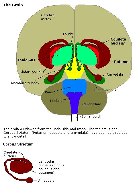
The amygdala (; plural: amygdalae or amygdalas; also '; Latin from Greek, , ', 'almond', 'tonsil') is one of two almond-shaped clusters of nuclei located deep and medially within the temporal lobes of the brain's cerebrum in complex vertebrates, including humans. Shown to perform a primary role in the processing of memory, decision making, and emotional responses (including fear, anxiety, and aggression), the amygdalae are considered part of the limbic system. The term "amygdala" was first introduced by Karl Friedrich Burdach in 1822. Structure The regions described as amygdala nuclei encompass several structures of the cerebrum with distinct connectional and functional characteristics in humans and other animals. Among these nuclei are the basolateral complex, the cortical nucleus, the medial nucleus, the central nucleus, and the intercalated cell clusters. The basolateral complex can be further subdivided into the lateral, the basal, and the accessory basal ... [...More Info...] [...Related Items...] OR: [Wikipedia] [Google] [Baidu] |
Hippocampus
The hippocampus (via Latin from Greek , ' seahorse') is a major component of the brain of humans and other vertebrates. Humans and other mammals have two hippocampi, one in each side of the brain. The hippocampus is part of the limbic system, and plays important roles in the consolidation of information from short-term memory to long-term memory, and in spatial memory that enables navigation. The hippocampus is located in the allocortex, with neural projections into the neocortex in humans, as well as primates. The hippocampus, as the medial pallium, is a structure found in all vertebrates. In humans, it contains two main interlocking parts: the hippocampus proper (also called ''Ammon's horn''), and the dentate gyrus. In Alzheimer's disease (and other forms of dementia), the hippocampus is one of the first regions of the brain to suffer damage; short-term memory loss and disorientation are included among the early symptoms. Damage to the hippocampus can also result f ... [...More Info...] [...Related Items...] OR: [Wikipedia] [Google] [Baidu] |
Lateral Geniculate Nucleus
In neuroanatomy, the lateral geniculate nucleus (LGN; also called the lateral geniculate body or lateral geniculate complex) is a structure in the thalamus and a key component of the mammalian visual pathway. It is a small, ovoid, ventral projection of the thalamus where the thalamus connects with the optic nerve. There are two LGNs, one on the left and another on the right side of the thalamus. In humans, both LGNs have six layers of neurons (grey matter) alternating with optic fibers (white matter). The LGN receives information directly from the ascending retinal ganglion cells via the optic tract and from the reticular activating system. Neurons of the LGN send their axons through the optic radiation, a direct pathway to the primary visual cortex. In addition, the LGN receives many strong feedback connections from the primary visual cortex. In humans as well as other mammals, the two strongest pathways linking the eye to the brain are those projecting to the dorsal part of th ... [...More Info...] [...Related Items...] OR: [Wikipedia] [Google] [Baidu] |
Epithalamus
The epithalamus is a posterior (dorsal) segment of the diencephalon. The epithalamus includes the habenular nuclei and their interconnecting fibers, the habenular commissure, the stria medullaris and the pineal gland. Functions The function of the epithalamus is to connect the limbic system to other parts of the brain. The epithalamus also serves as a connecting point for the dorsal diencephalic conduction system, which is responsible for carrying information from the limbic forebrain to limbic midbrain structures. Some functions of its components include the secretion of melatonin and secretion of hormones from the pituitary gland (by the pineal gland circadian rhythms), regulation of motor pathways and emotions, and how energy is conserved in the body. A study has shown that the lateral habenula, an epithalamic structure, produces spontaneous theta oscillatory activity that was correlated with theta oscillation in the hippocampus. The same study also found that the increase ... [...More Info...] [...Related Items...] OR: [Wikipedia] [Google] [Baidu] |
Hypothalamus
The hypothalamus () is a part of the brain that contains a number of small nuclei with a variety of functions. One of the most important functions is to link the nervous system to the endocrine system via the pituitary gland. The hypothalamus is located below the thalamus and is part of the limbic system. In the terminology of neuroanatomy, it forms the ventral Standard anatomical terms of location are used to unambiguously describe the anatomy of animals, including humans. The terms, typically derived from Latin or Greek roots, describe something in its standard anatomical position. This position prov ... part of the diencephalon. All vertebrate brains contain a hypothalamus. In humans, it is the size of an Almond#Nut, almond. The hypothalamus is responsible for regulating certain Metabolism, metabolic biological process, processes and other activities of the autonomic nervous system. It biosynthesis, synthesizes and secretes certain neurohormones, called releasing hor ... [...More Info...] [...Related Items...] OR: [Wikipedia] [Google] [Baidu] |


