|
Calcifying Odontogenic Cyst
Calcifying odotogenic cyst (COC) is a rare developmental lesion that comes from odontogenic epithelium. It is also known as a calcifying cystic odontogenic tumor, which is a proliferation of odontogenic epithelium and scattered nest of ghost cells and calcifications that may form the lining of a cyst, or present as a solid mass. It can appear in any location in the oral cavity, but more commonly affects the anterior (front) mandible and maxilla. It is most common in individuals in their 20s to 30s, but can be seen at almost any age, regardless of gender. On dental radiographs, the calcifying odontogenic cyst appears as a unilocular (one circle) radiolucency (dark area). In one-third of cases, an impacted tooth is involved. Histologically, cells that are described as " ghost cells", enlarged eosinophilic epithelial cells without nuclei, are present within the epithelial lining and may undergo calcification. Signs and symptoms Most calcifying odontogenic cysts appear asymptom ... [...More Info...] [...Related Items...] OR: [Wikipedia] [Google] [Baidu] |
Dentistry
Dentistry, also known as dental medicine and oral medicine, is the branch of medicine focused on the teeth, gums, and mouth. It consists of the study, diagnosis, prevention, management, and treatment of diseases, disorders, and conditions of the mouth, most commonly focused on dentition (the development and arrangement of teeth) as well as the oral mucosa. Dentistry may also encompass other aspects of the craniofacial complex including the temporomandibular joint. The practitioner is called a dentist. The history of dentistry is almost as ancient as the history of humanity and civilization with the earliest evidence dating from 7000 BC to 5500 BC. Dentistry is thought to have been the first specialization in medicine which have gone on to develop its own accredited degree with its own specializations. Dentistry is often also understood to subsume the now largely defunct medical specialty of stomatology (the study of the mouth and its disorders and diseases) for which reas ... [...More Info...] [...Related Items...] OR: [Wikipedia] [Google] [Baidu] |
Asymptomatic
In medicine, any disease is classified asymptomatic if a patient tests as carrier for a disease or infection but experiences no symptoms. Whenever a medical condition fails to show noticeable symptoms after a diagnosis it might be considered asymptomatic. Infections of this kind are usually called subclinical infections. Diseases such as mental illnesses or psychosomatic conditions are considered subclinical if they present some individual symptoms but not all those normally required for a clinical diagnosis. The term clinically silent is also found. Producing only a few, mild symptoms, disease is paucisymptomatic. Symptoms appearing later, after an asymptomatic incubation period, mean a pre-symptomatic period has existed. Importance Knowing that a condition is asymptomatic is important because: * It may develop symptoms later and only then require treatment. * It may resolve itself or become benign. * It may be contagious, and the contribution of asymptomatic and pre-symptom ... [...More Info...] [...Related Items...] OR: [Wikipedia] [Google] [Baidu] |
Dystrophic Calcification
Dystrophic calcification (DC) is the calcification occurring in degenerated or necrotic tissue, as in hyalinized scars, degenerated foci in leiomyomas, and caseous nodules. This occurs as a reaction to tissue damage, including as a consequence of medical device implantation. Dystrophic calcification can occur even if the amount of calcium in the blood is not elevated (a systemic mineral imbalance would elevate calcium levels in the blood and all tissues) and cause metastatic calcification. Basophilic calcium salt deposits aggregate, first in the mitochondria, then progressively throughout the cell. These calcifications are an indication of previous microscopic cell injury, occurring in areas of cell necrosis when activated phosphatases bind calcium ions to phospholipids in the membrane. Calcification in dead tissue #Caseous necrosis in T.B. is most common site of dystrophic calcification. #Liquefactive necrosis in chronic abscesses may get calcified. #Fat necrosis following ... [...More Info...] [...Related Items...] OR: [Wikipedia] [Google] [Baidu] |
Dentin
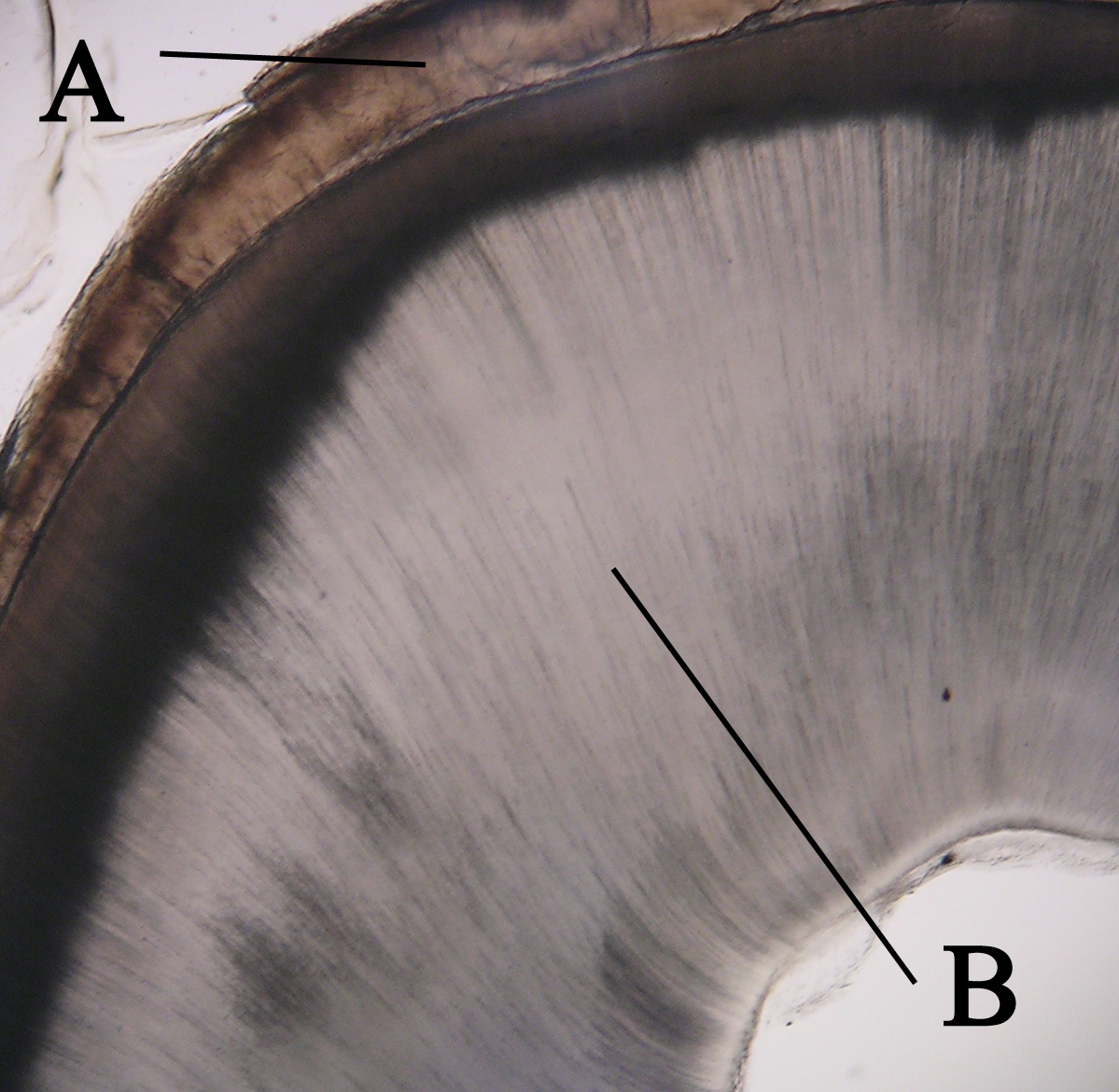
Dentin () (American English) or dentine ( or ) (British English) ( la, substantia eburnea) is a calcified tissue of the body and, along with enamel, cementum, and pulp, is one of the four major components of teeth. It is usually covered by enamel on the crown and cementum on the root and surrounds the entire pulp. By volume, 45% of dentin consists of the mineral hydroxyapatite, 33% is organic material, and 22% is water. Yellow in appearance, it greatly affects the color of a tooth due to the translucency of enamel. Dentin, which is less mineralized and less brittle than enamel, is necessary for the support of enamel. Dentin rates approximately 3 on the Mohs scale of mineral hardness. There are two main characteristics which distinguish dentin from enamel: firstly, dentin forms throughout life; secondly, dentin is sensitive and can become hypersensitive to changes in temperature due to the sensory function of odontoblasts, especially when enamel recedes and dentin channels bec ... [...More Info...] [...Related Items...] OR: [Wikipedia] [Google] [Baidu] |
Basophilic
Basophilic is a technical term used by pathologists. It describes the appearance of cells, tissues and cellular structures as seen through the microscope after a histological section has been stained with a basic dye. The most common such dye is haematoxylin. The name basophilic refers to the characteristic of these structures to be stained very well by basic dyes. This can be explained by their charges. Basic dyes are cationic, i.e. contain positive charges, and thus they stain anionic structures (i.e. structures containing negative charges), such as the phosphate backbone of DNA in the cell nucleus and ribosomes. "Basophils" are cells that "love" (from greek "-phil") basic dyes, for example haematoxylin, azure and methylene blue. Specifically, this term refers to: * basophil granulocytes * anterior pituitary basophils An abnormal increase in basophil granulocytes is therefore also described as basophilia.https://www.collinsdictionary.com/de/worterbuch/englisch/basophi ... [...More Info...] [...Related Items...] OR: [Wikipedia] [Google] [Baidu] |
Acellular
Non-cellular life, or acellular life is life that exists without a cellular structure for at least part of its life cycle. Historically, most (descriptive) definitions of life postulated that an organism must be composed of one or more cells, but this is no longer considered necessary, and modern criteria allow for forms of life based on other structural arrangements. The primary candidates for non-cellular life are viruses. Some biologists consider viruses to be organisms, but others do not. Their primary objection is that no known viruses are capable of autonomous reproduction: they must rely on cells to copy them. Engineers sometimes use the term "artificial life" to refer to software and robots inspired by biological processes, but these do not satisfy any biological definition of life. Viruses as non-cellular life The nature of viruses was unclear for many years following their discovery as pathogens. They were described as poisons or toxins at first, then as "infect ... [...More Info...] [...Related Items...] OR: [Wikipedia] [Google] [Baidu] |
Keratinization
Keratin () is one of a family of structural fibrous proteins also known as ''scleroproteins''. Alpha-keratin (α-keratin) is a type of keratin found in vertebrates. It is the key structural material making up scales, hair, nails, feathers, horns, claws, hooves, and the outer layer of skin among vertebrates. Keratin also protects epithelial cells from damage or stress. Keratin is extremely insoluble in water and organic solvents. Keratin monomers assemble into bundles to form intermediate filaments, which are tough and form strong unmineralized epidermal appendages found in reptiles, birds, amphibians, and mammals. Excessive keratinization participate in fortification of certain tissues such as in horns of cattle and rhinos, and armadillos' osteoderm. The only other biological matter known to approximate the toughness of keratinized tissue is chitin. Keratin comes in two types, the primitive, softer forms found in all vertebrates and harder, derived forms found only am ... [...More Info...] [...Related Items...] OR: [Wikipedia] [Google] [Baidu] |
Enamel Organ
The enamel organ, also known as the dental organ, is a cellular aggregation seen in a developing tooth and it lies above the dental papilla. The enamel organ which is differentiated from the primitive oral epithelium lining the stomodeum.The enamel organ is responsible for the formation of enamel, initiation of dentine formation, establishment of the shape of a tooth's crown, and establishment of the dentoenamel junction. The enamel organ has four layers; the inner enamel epithelium, outer enamel epithelium, stratum intermedium, and the stellate reticulum. The dental papilla, the differentiated ectomesenchyme deep to the enamel organ, will produce dentin and the dental pulp. The surrounding ectomesenchyme tissue, the dental follicle, is the primitive cementum, periodontal ligament and alveolar bone beneath the tooth root. The site where the internal enamel epithelium and external enamel epithelium coalesce is the cervical root, important in proliferation of the dental root ... [...More Info...] [...Related Items...] OR: [Wikipedia] [Google] [Baidu] |
Infarction
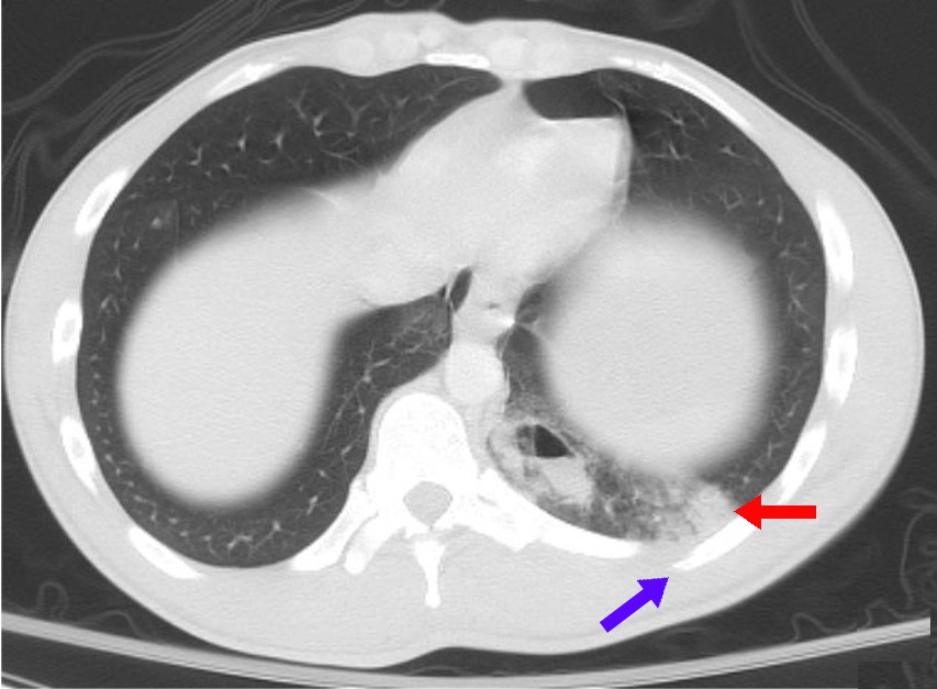
Infarction is tissue death ( necrosis) due to inadequate blood supply to the affected area. It may be caused by artery blockages, rupture, mechanical compression, or vasoconstriction. The resulting lesion is referred to as an infarct (from the Latin ''infarctus'', "stuffed into"). Causes Infarction occurs as a result of prolonged ischemia, which is the insufficient supply of oxygen and nutrition to an area of tissue due to a disruption in blood supply. The blood vessel supplying the affected area of tissue may be blocked due to an obstruction in the vessel (e.g., an arterial embolus, thrombus, or atherosclerotic plaque), compressed by something outside of the vessel causing it to narrow (e.g., tumor, volvulus, or hernia), ruptured by trauma causing a loss of blood pressure downstream of the rupture, or vasoconstricted, which is the narrowing of the blood vessel by contraction of the muscle wall rather than an external force (e.g., cocaine vasoconstriction le ... [...More Info...] [...Related Items...] OR: [Wikipedia] [Google] [Baidu] |
Ischemia
Ischemia or ischaemia is a restriction in blood supply to any tissue, muscle group, or organ of the body, causing a shortage of oxygen that is needed for cellular metabolism (to keep tissue alive). Ischemia is generally caused by problems with blood vessels, with resultant damage to or dysfunction of tissue i.e. hypoxia and microvascular dysfunction. It also implies local hypoxia in a part of a body resulting from constriction (such as vasoconstriction, thrombosis, or embolism). Ischemia causes not only insufficiency of oxygen, but also reduced availability of nutrients and inadequate removal of metabolic wastes. Ischemia can be partial (poor perfusion) or total blockage. The inadequate delivery of oxygenated blood to the organs must be resolved either by treating the cause of the inadequate delivery or reducing the oxygen demand of the system that needs it. For example, patients with myocardial ischemia have a decreased blood flow to the heart and are prescribed with ... [...More Info...] [...Related Items...] OR: [Wikipedia] [Google] [Baidu] |
Coagulative Necrosis
Coagulative necrosis is a type of accidental cell death typically caused by ischemia or infarction. In coagulative necrosis, the architectures of dead tissue are preserved for at least a couple of days. It is believed that the injury denatures structural proteins as well as lysosomal enzymes, thus blocking the proteolysis of the damaged cells. The lack of lysosomal enzymes allows it to maintain a "coagulated" morphology for some time. Like most types of necrosis, if enough viable cells are present around the affected area, regeneration will usually occur. Coagulative necrosis occurs in most bodily organs, excluding the brain. Different diseases are associated with coagulative necrosis, including acute tubular necrosis and acute myocardial infarction. Coagulative necrosis can also be induced by high local temperature; it is a desired effect of treatments such as high intensity focused ultrasound applied to cancerous cells. Causes Coagulative necrosis is most commonly caused by ... [...More Info...] [...Related Items...] OR: [Wikipedia] [Google] [Baidu] |
Tooth Development
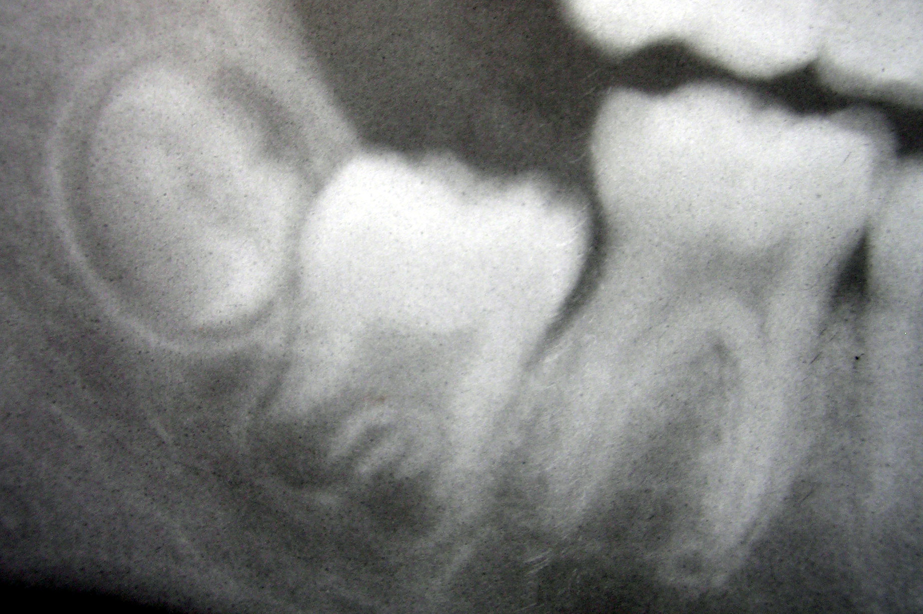
Tooth development or odontogenesis is the complex process by which teeth form from embryonic cells, grow, and erupt into the mouth. For human teeth to have a healthy oral environment, all parts of the tooth must develop during appropriate stages of fetal development. Primary (baby) teeth start to form between the sixth and eighth week of prenatal development, and permanent teeth begin to form in the twentieth week.Ten Cate's Oral Histology, Nanci, Elsevier, 2013, pages 70-94 If teeth do not start to develop at or near these times, they will not develop at all, resulting in hypodontia or anodontia. A significant amount of research has focused on determining the processes that initiate tooth development. It is widely accepted that there is a factor within the tissues of the first pharyngeal arch that is necessary for the development of teeth. Overview The tooth germ is an aggregation of cells that eventually forms a tooth.University of Texas Medical Branch. These cells ar ... [...More Info...] [...Related Items...] OR: [Wikipedia] [Google] [Baidu] |


