|
Craniofacial Abnormality
Craniofacial abnormalities are congenital musculoskeletal disorders which primarily affect the cranium and facial bones. They are associated with the development of the pharyngeal arches. Approximately, 5% of the UK or USA population present with dentofacial deformities requiring Orthognathic surgery, jaw surgery, and Orthodontics, brace therapy, as a part of their definitive treatment. Notable conditions * Platybasia * Arrhinia - absence of the nose * Craniosynostosis - premature fusion of the cranial sutures * Cyclopia Cyclopia (named after the Greek mythology characters cyclopes), also known as alobar holoprosencephaly, is the most extreme form of holoprosencephaly and is a congenital disorder (birth defect) characterized by the failure of the embryonic prosen ... - one eye * Mobius syndrome - paralysis of the facial muscles References External links Congenital disorders of musculoskeletal system {{musculoskeletal-stub ... [...More Info...] [...Related Items...] OR: [Wikipedia] [Google] [Baidu] |
Human Skull
The skull, or cranium, is typically a bony enclosure around the brain of a vertebrate. In some fish, and amphibians, the skull is of cartilage. The skull is at the head end of the vertebrate. In the human, the skull comprises two prominent parts: the neurocranium and the facial skeleton, which evolved from the first pharyngeal arch. The skull forms the frontmost portion of the axial skeleton and is a product of cephalization and vesicular enlargement of the brain, with several special senses structures such as the eyes, ears, nose, tongue and, in fish, specialized tactile organs such as barbels near the mouth. The skull is composed of three types of bone: cranial bones, facial bones and ossicles, which is made up of a number of fused flat and irregular bones. The cranial bones are joined at firm fibrous junctions called sutures and contains many foramina, fossae, processes, and sinuses. In zoology, the openings in the skull are called fenestrae, the ... [...More Info...] [...Related Items...] OR: [Wikipedia] [Google] [Baidu] |
Musculoskeletal System
The human musculoskeletal system (also known as the human locomotor system, and previously the activity system) is an organ system that gives humans the ability to move using their Muscular system, muscular and Human skeleton, skeletal systems. The musculoskeletal system provides form, support, stability, and movement to the body. The human musculoskeletal system is made up of the bones of the human skeleton, skeleton, muscles, cartilage, tendons, ligaments, joints, and other connective tissue that supports and binds tissues and organs together. The musculoskeletal system's primary functions include supporting the body, allowing motion, and protecting vital organs. The skeletal portion of the system serves as the main storage system for calcium and phosphorus and contains critical components of the hematopoietic system. This system describes how bones are connected to other bones and muscle fibers via connective tissue such as tendons and ligaments. The bones provide stability t ... [...More Info...] [...Related Items...] OR: [Wikipedia] [Google] [Baidu] |
Human Skull
The skull, or cranium, is typically a bony enclosure around the brain of a vertebrate. In some fish, and amphibians, the skull is of cartilage. The skull is at the head end of the vertebrate. In the human, the skull comprises two prominent parts: the neurocranium and the facial skeleton, which evolved from the first pharyngeal arch. The skull forms the frontmost portion of the axial skeleton and is a product of cephalization and vesicular enlargement of the brain, with several special senses structures such as the eyes, ears, nose, tongue and, in fish, specialized tactile organs such as barbels near the mouth. The skull is composed of three types of bone: cranial bones, facial bones and ossicles, which is made up of a number of fused flat and irregular bones. The cranial bones are joined at firm fibrous junctions called sutures and contains many foramina, fossae, processes, and sinuses. In zoology, the openings in the skull are called fenestrae, the ... [...More Info...] [...Related Items...] OR: [Wikipedia] [Google] [Baidu] |
Facial Bone
The facial skeleton comprises the ''facial bones'' that may attach to build a portion of the skull. The remainder of the skull is the neurocranium. In human anatomy and development, the facial skeleton is sometimes called the ''membranous viscerocranium'', which comprises the mandible and dermatocranial elements that are not part of the braincase. Structure In the human skull, the facial skeleton consists of fourteen bones in the face: * Inferior turbinal (2) * Lacrimal bones (2) * Mandible * Maxilla (2) * Nasal bones (2) * Palatine bones (2) * Vomer * Zygomatic bones (2) Variations Elements of the ''cartilaginous viscerocranium'' (i.e., splanchnocranial elements), such as the hyoid bone, are sometimes considered part of the facial skeleton. The ethmoid bone (or a part of it) and also the sphenoid bone are sometimes included, but otherwise considered part of the neurocranium. Because the maxillary bones are fused, they are often collectively listed as only one bone. The mandi ... [...More Info...] [...Related Items...] OR: [Wikipedia] [Google] [Baidu] |
Pharyngeal Arch
The pharyngeal arches, also known as visceral arches'','' are transient structures seen in the Animal embryonic development, embryonic development of humans and other vertebrates, that are recognisable precursors for many structures. In fish, the arches support the Fish gill, gills and are known as the branchial arches, or gill arches. In the human embryo, the arches are first seen during the fourth week of human embryonic development, development. They appear as a series of outpouchings of mesoderm on both sides of the developing pharynx. The vasculature of the pharyngeal arches are the aortic arches that arise from the aortic sac. Structure In humans and other vertebrates, the pharyngeal arches are derived from all three germ layers (the primary layers of cells that form during embryonic development). Neural crest cells enter these arches where they contribute to features of the skull and facial skeleton such as bone and cartilage. However, the existence of pharyngeal structu ... [...More Info...] [...Related Items...] OR: [Wikipedia] [Google] [Baidu] |
Orthognathic Surgery
Orthognathic surgery (), also known as corrective jaw surgery or simply jaw surgery, is surgery designed to correct conditions of the jaw and lower face related to structure, growth, airway issues including sleep apnea, TMJ disorders, malocclusion problems primarily arising from skeletal disharmonies, and other orthodontic dental bite problems that cannot be treated easily with braces, as well as the broad range of facial imbalances, disharmonies, asymmetries, and malproportions where correction may be considered to improve facial aesthetics and self-esteem. The origins of orthognathic surgery belong in oral surgery, and the basic operations related to the surgical removal of impacted or displaced teeth – especially where indicated by orthodontics to enhance dental treatments of malocclusion and dental crowding. One of the first published cases of orthognathic surgery was the one from Dr. Simon P. Hullihen in 1849. Originally coined by Harold Hargis, it was more widely popul ... [...More Info...] [...Related Items...] OR: [Wikipedia] [Google] [Baidu] |
Orthodontics
Orthodontics (also referred to as orthodontia) is a dentistry specialty that addresses the diagnosis, prevention, management, and correction of mal-positioned teeth and jaws, as well as misaligned bite patterns. It may also address the modification of facial growth, known as dentofacial orthopedics. Abnormal alignment of the teeth and jaws is very common. The approximate worldwide prevalence of malocclusion was as high as 56%. However, conclusive evidence-based medicine, scientific evidence for the Health benefit (medicine), health benefits of orthodontic treatment is lacking, although patients with completed treatment have reported a higher quality of life than that of untreated patients undergoing orthodontic treatment. The main reason for the prevalence of these malocclusions is diets with less fresh fruit and vegetables and overall softer foods in childhood, causing smaller jaws with less room for the teeth to erupt. Treatment may require several months to a few years and enta ... [...More Info...] [...Related Items...] OR: [Wikipedia] [Google] [Baidu] |
Elsevier
Elsevier ( ) is a Dutch academic publishing company specializing in scientific, technical, and medical content. Its products include journals such as ''The Lancet'', ''Cell (journal), Cell'', the ScienceDirect collection of electronic journals, ''Trends (journals), Trends'', the ''Current Opinion (Elsevier), Current Opinion'' series, the online citation database Scopus, the SciVal tool for measuring research performance, the ClinicalKey search engine for clinicians, and the ClinicalPath evidence-based cancer care service. Elsevier's products and services include digital tools for Data management platform, data management, instruction, research analytics, and assessment. Elsevier is part of the RELX Group, known until 2015 as Reed Elsevier, a publicly traded company. According to RELX reports, in 2022 Elsevier published more than 600,000 articles annually in over 2,800 journals. As of 2018, its archives contained over 17 million documents and 40,000 Ebook, e-books, with over one b ... [...More Info...] [...Related Items...] OR: [Wikipedia] [Google] [Baidu] |
Platybasia
Platybasia is a spinal disease of a malformed relationship between the occipital bone and cervical spine In tetrapods, cervical vertebrae (: vertebra) are the vertebrae of the neck, immediately below the skull. Truncal vertebrae (divided into thoracic and lumbar vertebrae in mammals) lie caudal (toward the tail) of cervical vertebrae. In sauro .... It may be caused by Paget's disease. Platybasia is also a feature of Gorlin-Goltz syndrome, commonly known as basal cell nevus syndrome. It may be developmental in origin or due to softening of the skull base bone, allowing it to be pushed upward. The developmental variant is the result of a wider angle between the skull base of the frontal or anterior fossa and the clivus, which defines the anterior wall of the posterior fossa. It is sometimes associated with other congenital abnormalities of the bone structure, such as fusion of the first cervical vertebrae to the skull atlas assimilation. Further reading * * Virchow R. ... [...More Info...] [...Related Items...] OR: [Wikipedia] [Google] [Baidu] |
Arrhinia
Arrhinia (alternatively spelled "arhinia") is the congenital partial or complete absence of the nose at birth. It is an extremely rare condition, with few reported cases in the history of modern medicine. It is generally classified as a craniofacial abnormality. The cause of arrhinia is not known. One study of the literature found that all cases had presented a normal antenatal history. Conditions Arrhinia is associated with the following conditions and syndromes: * Arrhinia with choanal atresia and microphthalmia syndrome (Bosma arhinia microphthalmia syndrome) * Holoprosencephaly 1 * Holoprosencephaly 13, X-linked * Holoprosencephaly-radial heart renal anomalies syndrome History The phenomenon of congenital arrhinia, which refers to the complete absence of the nose from birth, was initially documented in French literature during the 1800s. Over time, a few additional cases of individuals born with congenital arrhinia, some with accompanying eye defects and others without ... [...More Info...] [...Related Items...] OR: [Wikipedia] [Google] [Baidu] |
Craniosynostosis
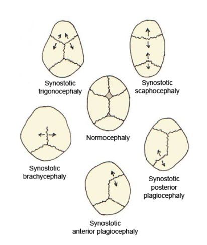
Craniosynostosis is a condition in which one or more of the fibrous sutures in a young infant's skull prematurely fuses by turning into bone (ossification), thereby changing the growth pattern of the skull. Because the skull cannot expand perpendicular to the fused suture, it compensates by growing more in the direction parallel to the closed sutures. Sometimes the resulting growth pattern provides the necessary space for the growing brain, but results in an abnormal head shape and abnormal facial features. In cases in which the compensation does not effectively provide enough space for the growing brain, craniosynostosis results in increased intracranial pressure leading possibly to visual impairment, sleeping impairment, eating difficulties, or an impairment of mental development combined with a significant reduction in IQ. Craniosynostosis occurs in one in 2000 births. Craniosynostosis is part of a syndrome in 15% to 40% of affected patients, but it usually occurs as an isol ... [...More Info...] [...Related Items...] OR: [Wikipedia] [Google] [Baidu] |
Cyclopia
Cyclopia (named after the Greek mythology characters cyclopes), also known as alobar holoprosencephaly, is the most extreme form of holoprosencephaly and is a congenital disorder (birth defect) characterized by the failure of the embryonic prosencephalon to properly divide the Orbit (anatomy), orbits of the eye into two cavities. Its incidence is 1 in 16,000 in born animals and 1 in 200 in miscarriage, miscarried fetuses. Signs and symptoms Typically, the nose is either missing or not functional. This deformity (called Proboscis (anomaly), proboscis) forms above the center eye and is characteristic of a form of cyclopia called rhinencephaly or rhinocephaly. Most such embryos are either naturally miscarriage, miscarried or are stillbirth, stillborn upon childbirth, delivery. Although cyclopia is rare, several cyclopic human babies are preserved in medical museums (e.g. The University of Amsterdam, Vrolik Museum, Amsterdam, Trivandrum Medical College). Some extreme cases of cycl ... [...More Info...] [...Related Items...] OR: [Wikipedia] [Google] [Baidu] |


