|
Cervical Lymph Node
Cervical lymph nodes are lymph nodes found in the neck. Of the 800 lymph nodes in the human body, 300 are in the neck. Cervical lymph nodes are subject to a number of different pathological conditions including neoplasm, tumours, infection and inflammation. Classification There are approximately 300 lymph nodes in the neck, and they can be classified in a number of different ways. History The classification of the cervical lymph nodes is generally attributed to Henri Rouvière in his 1932 publication "Anatomie des Lymphatiques de l'Homme" Rouviere described the cervical lymph nodes as a collar which surrounded the aerodigestive tract, upper aerodigestive tract, consisting of submental, facial, submandibular, parotid, mastoid, occipital and retropharyngeal nodes, together with two chains that run in the long axis of the neck, the anterior cervical and postero-lateral cervical groups. However, this system was based upon anatomical landmarks found in dissection, making it ... [...More Info...] [...Related Items...] OR: [Wikipedia] [Google] [Baidu] |
Cervix
The cervix (: cervices) or cervix uteri is a dynamic fibromuscular sexual organ of the female reproductive system that connects the vagina with the uterine cavity. The human female cervix has been documented anatomically since at least the time of Hippocrates, over 2,000 years ago. The cervix is approximately 4 cm long with a diameter of approximately 3 cm and tends to be described as a cylindrical shape, although the front and back walls of the cervix are contiguous. The size of the cervix changes throughout a woman's life cycle. For example, women in the fertile years of their reproductive cycle tend to have larger cervixes than postmenopausal women; likewise, women who have produced offspring have a larger cervix than those who have not. In relation to the vagina, the part of the cervix that opens to the uterus is called the ''internal os'' and the opening of the cervix in the vagina is called the ''external os''. Between them is a conduit commonly called the cervic ... [...More Info...] [...Related Items...] OR: [Wikipedia] [Google] [Baidu] |
Hyoid
The hyoid-bone (lingual-bone or tongue-bone) () is a horseshoe-shaped bone situated in the anterior midline of the neck between the chin and the thyroid-cartilage. At rest, it lies between the base of the mandible and the third cervical vertebra. Unlike other bones, the hyoid is only distantly articulated to other bones by muscles or ligaments. It is the only bone in the human body that is not connected to any other bones. The hyoid is anchored by muscles from the anterior, posterior and inferior directions, and aids in tongue movement and swallowing. The hyoid bone provides attachment to the muscles of the floor of the mouth and the tongue above, the larynx below, and the epiglottis and pharynx behind. Its name is derived . Structure The hyoid bone is classed as an irregular bone and consists of a central part called the body, and two pairs of horns, the greater and lesser horns. Body The body of the hyoid bone is the central part of the hyoid bone. *At the front ... [...More Info...] [...Related Items...] OR: [Wikipedia] [Google] [Baidu] |
Inferior (anatomy)
Standard anatomical terms of location are used to describe unambiguously the anatomy of humans and other animals. The terms, typically derived from Latin or Greek roots, describe something in its standard anatomical position. This position provides a definition of what is at the front ("anterior"), behind ("posterior") and so on. As part of defining and describing terms, the body is described through the use of anatomical planes and axes. The meaning of terms that are used can change depending on whether a vertebrate is a biped or a quadruped, due to the difference in the neuraxis, or if an invertebrate is a non-bilaterian. A non-bilaterian has no anterior or posterior surface for example but can still have a descriptor used such as proximal or distal in relation to a body part that is nearest to, or furthest from its middle. International organisations have determined vocabularies that are often used as standards for subdisciplines of anatomy. For example, '' Terminol ... [...More Info...] [...Related Items...] OR: [Wikipedia] [Google] [Baidu] |
Posterior (anatomy)
Standard anatomical terms of location are used to describe unambiguously the anatomy of humans and other animals. The terms, typically derived from Latin or Greek language, Greek roots, describe something in its standard anatomical position. This position provides a definition of what is at the front ("anterior"), behind ("posterior") and so on. As part of defining and describing terms, the body is described through the use of anatomical planes and anatomical axes, axes. The meaning of terms that are used can change depending on whether a vertebrate is a biped or a quadruped, due to the difference in the neuraxis, or if an invertebrate is a non-bilaterian. A non-bilaterian has no anterior or posterior surface for example but can still have a descriptor used such as proximal or distal in relation to a body part that is nearest to, or furthest from its middle. International organisations have determined vocabularies that are often used as standards for subdisciplines of anatomy. ... [...More Info...] [...Related Items...] OR: [Wikipedia] [Google] [Baidu] |
Lateral Mass Of Atlas
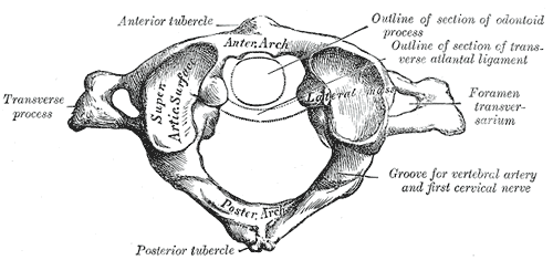
In anatomy, the atlas (C1) is the most superior (first) cervical vertebra of the spine and is located in the neck. The bone is named for Atlas of Greek mythology, just as Atlas bore the weight of the heavens, the first cervical vertebra supports the head. However, the term atlas was first used by the ancient Romans for the seventh cervical vertebra (C7) due to its suitability for supporting burdens. In Greek mythology, Atlas was condemned to bear the weight of the heavens as punishment for rebelling against Zeus. Ancient depictions of Atlas show the globe of the heavens resting at the base of his neck, on C7. Sometime around 1522, anatomists decided to call the first cervical vertebra the atlas. Scholars believe that by switching the designation atlas from the seventh to the first cervical vertebra Renaissance anatomists were commenting that the point of man's burden had shifted from his shoulders to his head—that man's true burden was not a physical load, but rather, his min ... [...More Info...] [...Related Items...] OR: [Wikipedia] [Google] [Baidu] |
CT Scan
A computed tomography scan (CT scan), formerly called computed axial tomography scan (CAT scan), is a medical imaging technique used to obtain detailed internal images of the body. The personnel that perform CT scans are called radiographers or radiology technologists. CT scanners use a rotating X-ray tube and a row of detectors placed in a gantry (medical), gantry to measure X-ray Attenuation#Radiography, attenuations by different tissues inside the body. The multiple X-ray measurements taken from different angles are then processed on a computer using tomographic reconstruction algorithms to produce Tomography, tomographic (cross-sectional) images (virtual "slices") of a body. CT scans can be used in patients with metallic implants or pacemakers, for whom magnetic resonance imaging (MRI) is Contraindication, contraindicated. Since its development in the 1970s, CT scanning has proven to be a versatile imaging technique. While CT is most prominently used in medical diagnosis, i ... [...More Info...] [...Related Items...] OR: [Wikipedia] [Google] [Baidu] |
Sternocleidomastoid Muscle
The sternocleidomastoid muscle is one of the largest and most superficial cervical muscles. The primary actions of the muscle are rotation of the head to the opposite side and Anatomical terms of motion#Flexion and extension, flexion of the neck. The sternocleidomastoid is innervated by the accessory nerve. Etymology and location It is given the name ''sternocleidomastoid'' because it originates at the manubrium of the Human sternum, sternum (''sterno-'') and the clavicle (''cleido-'') and has an Insertion (anatomy), insertion at the mastoid process of the temporal bone of the human skull, skull. Structure The sternocleidomastoid muscle originates from two locations: the manubrium of the Human sternum, sternum and the clavicle, hence it is said to have two heads: sternal head and clavicular head. It travels obliquely across the side of the neck and inserts at the mastoid process of the temporal bone of the human skull, skull by a thin aponeurosis. The sternocleidomastoid is thick ... [...More Info...] [...Related Items...] OR: [Wikipedia] [Google] [Baidu] |
Base Of The Skull
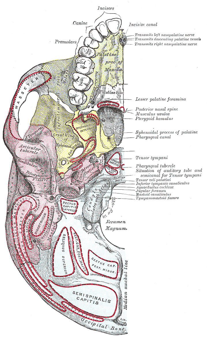
The base of skull, also known as the cranial base or the cranial floor, is the most inferior area of the skull. It is composed of the endocranium and the lower parts of the calvaria. Structure Structures found at the base of the skull are for example: Bones There are five bones that make up the base of the skull: *Ethmoid bone *Sphenoid bone *Occipital bone *Frontal bone *Temporal bone Sinuses * Occipital sinus * Superior sagittal sinus * Superior petrosal sinus Foramina of the skull * Foramen cecum *Optic foramen * Foramen lacerum * Foramen rotundum * Foramen magnum * Foramen ovale *Jugular foramen * Internal auditory meatus * Mastoid foramen * Sphenoidal emissary foramen * Foramen spinosum Sutures * Frontoethmoidal suture * Sphenofrontal suture * Sphenopetrosal suture * Sphenoethmoidal suture * Petrosquamous suture * Sphenosquamosal suture Other * Sphenoidal lingula * Subarcuate fossa * Dorsum sellae * Jugular process * Petro-occipital fissure * Condylar canal * J ... [...More Info...] [...Related Items...] OR: [Wikipedia] [Google] [Baidu] |
Accessory Nerve
The accessory nerve, also known as the eleventh cranial nerve, cranial nerve XI, or simply CN XI, is a cranial nerve that supplies the sternocleidomastoid and trapezius muscles. It is classified as the eleventh of twelve pairs of cranial nerves because part of it was formerly believed to originate in the brain. The sternocleidomastoid muscle tilts and rotates the head, whereas the trapezius muscle, connecting to the scapula, acts to shrug the shoulder. Traditional descriptions of the accessory nerve divide it into a spinal part and a cranial part. The cranial component rapidly joins the vagus nerve, and there is ongoing debate about whether the cranial part should be considered part of the accessory nerve proper. Consequently, the term "accessory nerve" usually refers only to nerve supplying the sternocleidomastoid and trapezius muscles, also called the spinal accessory nerve. Strength testing of these muscles can be measured during a neurological examination to assess func ... [...More Info...] [...Related Items...] OR: [Wikipedia] [Google] [Baidu] |
Internal Jugular Vein
The internal jugular vein is a paired jugular vein that collects blood from the brain and the superficial parts of the face and neck. This vein runs in the carotid sheath with the common carotid artery and vagus nerve. It begins in the posterior compartment of the jugular foramen, at the base of the skull. It is somewhat dilated at its origin, which is called the ''superior bulb''. This vein also has a common trunk into which drains the anterior branch of the retromandibular vein, the facial vein, and the lingual vein. It runs down the side of the neck in a vertical direction, being at one end lateral to the internal carotid artery, and then lateral to the common carotid artery, and at the root of the neck, it unites with the subclavian vein to form the brachiocephalic vein (innominate vein); a little above its termination is a second dilation, the ''inferior bulb''. Above, it lies upon the rectus capitis lateralis, behind the internal carotid artery and the nerves pa ... [...More Info...] [...Related Items...] OR: [Wikipedia] [Google] [Baidu] |
Upper Jugular Nodes
The deep cervical lymph nodes are a group of cervical lymph nodes in the neck that form a chain along the internal jugular vein within the carotid sheath. Structure Classification The deep cervical lymph nodes are subdivided into a superior group and an inferior group. Alternatively, they can be divided into deep anterior cervical lymph nodes and deep lateral cervical lymph nodes. They can also be divided into three groups: "superior deep jugular", "middle deep jugular", and "inferior deep jugular". Relations The deep cervical lymph nodes are contained in the carotid sheath in the neck, close to the internal jugular vein. They connect to the meningeal lymphatic vessels superiorly. Afferents All lymphatic vessels of the head and neck ultimately drain to the deep cervical lymph nodes - either by way of other lymph nodes or directly from tissues. CNS lymphatic vessels have been found to drain to the deep cervical lymph nodes in a 2016 animal study. Efferents Effe ... [...More Info...] [...Related Items...] OR: [Wikipedia] [Google] [Baidu] |
Mandible
In jawed vertebrates, the mandible (from the Latin ''mandibula'', 'for chewing'), lower jaw, or jawbone is a bone that makes up the lowerand typically more mobilecomponent of the mouth (the upper jaw being known as the maxilla). The jawbone is the skull's only movable, posable bone, sharing Temporomandibular joint, joints with the cranium's temporal bones. The mandible hosts the lower Human tooth, teeth (their depth delineated by the alveolar process). Many muscles attach to the bone, which also hosts nerves (some connecting to the teeth) and blood vessels. Amongst other functions, the jawbone is essential for chewing food. Owing to the Neolithic Revolution, Neolithic advent of agriculture (), human jaws evolved to be Human jaw shrinkage, smaller. Although it is the strongest bone of the facial skeleton, the mandible tends to deform in old age; it is also subject to Mandibular fracture, fracturing. Surgery allows for the removal of jawbone fragments (or its entirety) as well a ... [...More Info...] [...Related Items...] OR: [Wikipedia] [Google] [Baidu] |


