|
Cardiac MRI
Cardiac magnetic resonance imaging (cardiac MRI, CMR), also known as cardiovascular MRI, is a magnetic resonance imaging (MRI) technology used for non-invasive assessment of the function and structure of the cardiovascular system. Conditions in which it is performed include congenital heart disease, cardiomyopathies and valvular heart disease, diseases of the aorta such as dissection, aneurysm and coarctation, coronary heart disease. It can also be used to look at pulmonary veins. It is contraindicated if there are some implanted metal or electronic devices such as some intracerebral clips or claustrophobia. Conventional MRI sequences are adapted for cardiac imaging by using ECG gating and high temporal resolution protocols. The development of cardiac MRI is an active field of research and continues to see a rapid expansion of new and emerging techniques. Uses Cardiovascular MRI is complementary to other imaging techniques, such as echocardiography, cardiac CT, and nuclear med ... [...More Info...] [...Related Items...] OR: [Wikipedia] [Google] [Baidu] |
MRI Sequence
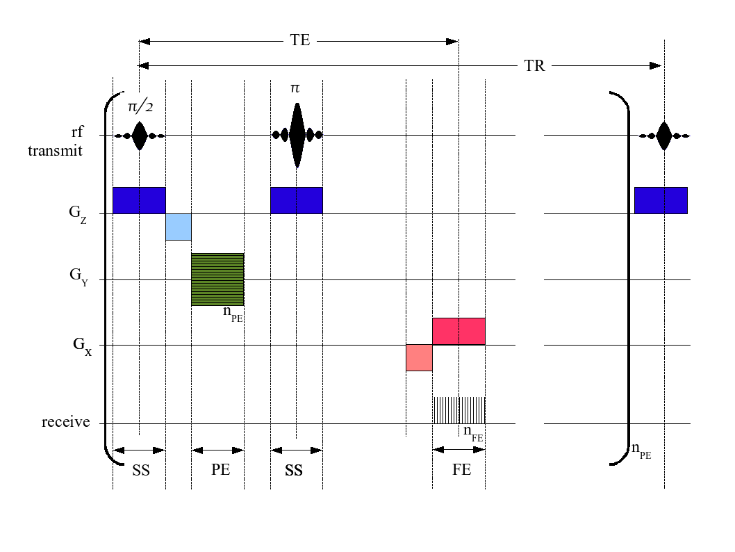
An MRI pulse sequence in magnetic resonance imaging (MRI) is a particular setting of pulse sequences and pulsed field gradients, resulting in a particular image appearance. A multiparametric MRI is a combination of two or more sequences, and/or including Magnetic resonance imaging#Other specialized configurations, other specialized MRI configurations such as In vivo magnetic resonance spectroscopy, spectroscopy. Spin echo T1 and T2 Each tissue returns to its equilibrium state after excitation by the independent relaxation processes of T1 (Spin–lattice relaxation, spin-lattice; that is, magnetization in the same direction as the static magnetic field) and T2 (Spin-spin relaxation time, spin-spin; transverse to the static magnetic field). To create a T1-weighted image, magnetization is allowed to recover before measuring the MR signal by changing the repetition time (TR). This image weighting is useful for assessing the cerebral cortex, identifying fatty tissue, characteriz ... [...More Info...] [...Related Items...] OR: [Wikipedia] [Google] [Baidu] |
Clipping (medicine)
Clipping is a surgical procedure performed to treat an aneurysm. If the aneurysm is intracranial, a craniotomy is performed, and afterwards an Elgiloy (Phynox) or titanium Sugita clip is affixed around the aneurysm's neck. Surgical clipping was introduced by Walter Dandy of the Johns Hopkins Hospital in 1937. It consists of performing a craniotomy A craniotomy is a surgery, surgical operation in which a bone flap is temporarily removed from the Human skull, skull to access the Human brain, brain. Craniotomies are often critical operations, performed on patients who are suffering from brain ..., exposing the aneurysm, and closing the base of the aneurysm with a clip chosen specifically for the site. The surgical technique has been modified and improved over the years. Surgical clipping has a lower rate of aneurysm recurrence after treatment. Titanium Aneurysm Clips are being used to clip aneurysms and the procedure is known as aneurysm clipping. References Neurosurgery [...More Info...] [...Related Items...] OR: [Wikipedia] [Google] [Baidu] |
Microvasculature
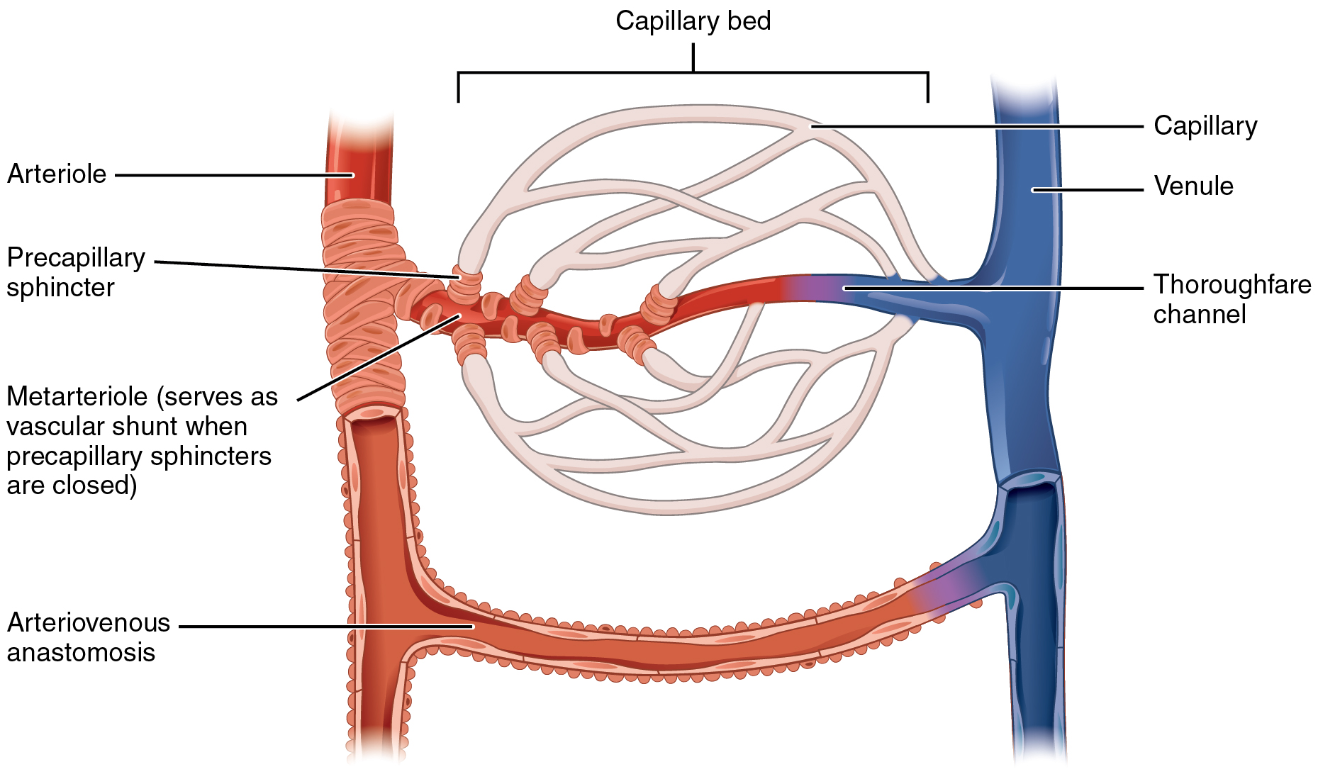
The microcirculation is the circulation of the blood in the smallest blood vessels, the microvessels of the microvasculature present within organ tissues. The microvessels include terminal arterioles, metarterioles, capillaries, and venules. Arterioles carry oxygenated blood to the capillaries, and blood flows out of the capillaries through venules into veins. In addition to these blood vessels, the microcirculation also includes lymphatic capillaries and collecting ducts. The main functions of the microcirculation are the delivery of oxygen and nutrients and the removal of carbon dioxide (CO2). It also serves to regulate blood flow and tissue perfusion, thereby affecting blood pressure and responses to inflammation which can include edema (swelling). Most vessels of the microcirculation are lined by flattened cells of the endothelium and many of them are surrounded by contractile cells called pericytes. The endothelium provides a smooth surface for the flow of blood and regu ... [...More Info...] [...Related Items...] OR: [Wikipedia] [Google] [Baidu] |
Epicardial Coronary Arteries
The pericardium (: pericardia), also called pericardial sac, is a double-walled sac containing the heart and the roots of the great vessels. It has two layers, an outer layer made of strong inelastic connective tissue (fibrous pericardium), and an inner layer made of serous membrane (serous pericardium). It encloses the pericardial cavity, which contains pericardial fluid, and defines the middle mediastinum. It separates the heart from interference of other structures, protects it against infection and blunt trauma, and lubricates the heart's movements. The English name originates from the Ancient Greek prefix ''peri-'' (περί) 'around' and the suffix ''-cardion'' (κάρδιον) 'heart'. Anatomy The pericardium is a tough fibroelastic sac which covers the heart from all sides except at the cardiac root (where the great vessels join the heart) and the bottom (where only the serous pericardium exists to cover the upper surface of the central tendon of diaphragm). The ... [...More Info...] [...Related Items...] OR: [Wikipedia] [Google] [Baidu] |
Vasodilation
Vasodilation, also known as vasorelaxation, is the widening of blood vessels. It results from relaxation of smooth muscle cells within the vessel walls, in particular in the large veins, large arteries, and smaller arterioles. Blood vessel walls are composed of endothelial tissue and a basal membrane lining the lumen of the vessel, concentric smooth muscle layers on top of endothelial tissue, and an adventitia over the smooth muscle layers. Relaxation of the smooth muscle layer allows the blood vessel to dilate, as it is held in a semi-constricted state by sympathetic nervous system activity. Vasodilation is the opposite of vasoconstriction, which is the narrowing of blood vessels. When blood vessels dilate, the flow of blood is increased due to a decrease in vascular resistance and increase in cardiac output. Vascular resistance is the amount of force circulating blood must overcome in order to allow perfusion of body tissues. Narrow vessels create more vascular resista ... [...More Info...] [...Related Items...] OR: [Wikipedia] [Google] [Baidu] |
Congenital Heart Defect
A congenital heart defect (CHD), also known as a congenital heart anomaly, congenital cardiovascular malformation, and congenital heart disease, is a defect in the structure of the heart or great vessels that is present at birth. A congenital heart defect is classed as a cardiovascular disease. Signs and symptoms depend on the specific type of defect. Symptoms can vary from none to life-threatening. When present, symptoms are variable and may include rapid breathing, bluish skin (cyanosis), poor weight gain, and feeling tired. CHD does not cause chest pain. Most congenital heart defects are not associated with other diseases. A complication of CHD is heart failure. Congenital heart defects are the most common birth defect. In 2015, they were present in 48.9 million people globally. They affect between 4 and 75 per 1,000 live births, depending upon how they are diagnosed. In about 6 to 19 per 1,000 they cause a moderate to severe degree of problems. Congenital heart defects are t ... [...More Info...] [...Related Items...] OR: [Wikipedia] [Google] [Baidu] |
Iron Overload
Iron overload is the abnormal and increased accumulation of total iron in the body, leading to organ damage. The primary mechanism of organ damage is oxidative stress, as elevated intracellular iron levels increase free radical formation via the Fenton reaction. Iron overload is often ''primary'' (i.e hereditary haemochromatosis, aceruloplasminemia) but may also be ''secondary'' to other causes (i.e. transfusional iron overload). Iron deposition most commonly occurs in the liver, pancreas, skin, heart, and joints. People with iron overload classically present with the triad of liver cirrhosis, secondary diabetes mellitus, and bronze skin. However, due to earlier detection nowadays, symptoms are often limited to general chronic malaise, arthralgia, and hepatomegaly. Signs and symptoms Organs most commonly affected by hemochromatosis include the liver, heart, and endocrine glands. Hemochromatosis may present with the following clinical syndromes: * liver: chronic liver di ... [...More Info...] [...Related Items...] OR: [Wikipedia] [Google] [Baidu] |
Myocarditis
Myocarditis is inflammation of the cardiac muscle. Myocarditis can progress to inflammatory cardiomyopathy when there is associated ventricular remodeling and cardiac dysfunction due to chronic inflammation. Symptoms can include shortness of breath, chest pain, Exercise intolerance, decreased ability to exercise, and an irregular heartbeat. The duration of problems can vary from hours to months. Complications may include heart failure, due to dilated cardiomyopathy or cardiac arrest. Myocarditis is most often due to a viral infection. Other causes include bacterial infections, certain medications, toxins and autoimmune disorders. A diagnosis may be supported by an electrocardiogram (ECG), increased troponin, cardiac magnetic resonance imaging, heart MRI, and occasionally a heart biopsy. An echocardiogram, ultrasound of the heart is important to rule out other potential causes, such as valvular heart disease, heart valve problems. Treatment depends on both the severity and the c ... [...More Info...] [...Related Items...] OR: [Wikipedia] [Google] [Baidu] |
Cardiomyopathy
Cardiomyopathy is a group of primary diseases of the heart muscle. Early on there may be few or no symptoms. As the disease worsens, shortness of breath, feeling tired, and swelling of the legs may occur, due to the onset of heart failure. An irregular heart beat and fainting may occur. Those affected are at an increased risk of sudden cardiac death. As of 2013, cardiomyopathies are defined as "disorders characterized by morphologically and functionally abnormal myocardium in the absence of any other disease that is sufficient, by itself, to cause the observed phenotype." Types of cardiomyopathy include hypertrophic cardiomyopathy, dilated cardiomyopathy, restrictive cardiomyopathy, arrhythmogenic right ventricular dysplasia, and Takotsubo cardiomyopathy (broken heart syndrome). In hypertrophic cardiomyopathy the heart muscle enlarges and thickens. In dilated cardiomyopathy the ventricles enlarge and weaken. In restrictive cardiomyopathy the ventricle stiffens. In ... [...More Info...] [...Related Items...] OR: [Wikipedia] [Google] [Baidu] |
Coronary Artery Disease
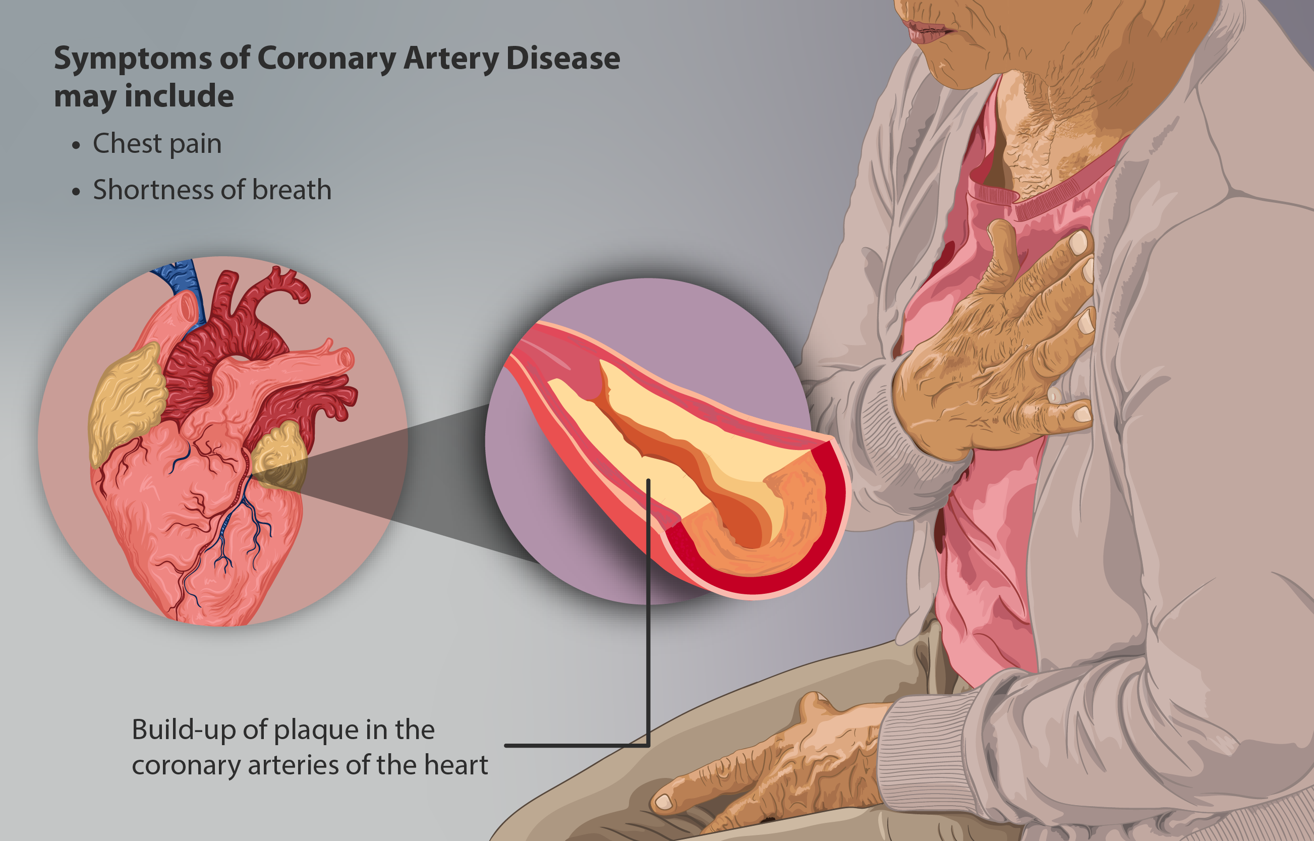
Coronary artery disease (CAD), also called coronary heart disease (CHD), or ischemic heart disease (IHD), is a type of cardiovascular disease, heart disease involving Ischemia, the reduction of blood flow to the cardiac muscle due to a build-up of atheromatous plaque in the Coronary arteries, arteries of the heart. It is the most common of the cardiovascular diseases. CAD can cause stable angina, unstable angina, myocardial ischemia, and myocardial infarction. A common symptom is angina, which is chest pain or discomfort that may travel into the shoulder, arm, back, neck, or jaw. Occasionally it may feel like heartburn. In stable angina, symptoms occur with exercise or emotional Psychological stress, stress, last less than a few minutes, and improve with rest. Shortness of breath may also occur and sometimes no symptoms are present. In many cases, the first sign is a Myocardial infarction, heart attack. Other complications include heart failure or an Heart arrhythmia, abnormal h ... [...More Info...] [...Related Items...] OR: [Wikipedia] [Google] [Baidu] |
Nuclear Medicine
Nuclear medicine (nuclear radiology, nucleology), is a medical specialty involving the application of radioactivity, radioactive substances in the diagnosis and treatment of disease. Nuclear imaging is, in a sense, ''radiology done inside out'', because it records radiation radiant exitance, emitted from within the body rather than radiation that is transmittance, transmitted through the body from external sources like X-ray generators. In addition, nuclear medicine scans differ from radiology, as the emphasis is not on imaging anatomy, but on the function. For such reason, it is called a Functional imaging, physiological imaging modality. Single photon emission computed tomography (SPECT) and positron emission tomography (PET) scans are the two most common imaging modalities in nuclear medicine. Diagnostic medical imaging Diagnostic In nuclear medicine imaging, radiopharmaceuticals are taken internally, for example, through inhalation, intravenously, or orally. Then, externa ... [...More Info...] [...Related Items...] OR: [Wikipedia] [Google] [Baidu] |
Computed Tomography Of The Heart
A computed tomography scan (CT scan), formerly called computed axial tomography scan (CAT scan), is a medical imaging technique used to obtain detailed internal images of the body. The personnel that perform CT scans are called radiographers or radiology technologists. CT scanners use a rotating X-ray tube and a row of detectors placed in a gantry to measure X-ray attenuations by different tissues inside the body. The multiple X-ray measurements taken from different angles are then processed on a computer using tomographic reconstruction algorithms to produce tomographic (cross-sectional) images (virtual "slices") of a body. CT scans can be used in patients with metallic implants or pacemakers, for whom magnetic resonance imaging (MRI) is contraindicated. Since its development in the 1970s, CT scanning has proven to be a versatile imaging technique. While CT is most prominently used in medical diagnosis, it can also be used to form images of non-living objects. The 1979 Nobel ... [...More Info...] [...Related Items...] OR: [Wikipedia] [Google] [Baidu] |


