|
Birth Trauma (physical)
Birth trauma refers to damage of the tissues and organs of a newly delivered child, often as a result of physical pressure or trauma during childbirth. The term also encompasses the long term consequences, often of cognitive nature, of damage to the brain or cranium. Medical study of birth trauma dates to the 16th century, and the morphological consequences of mishandled delivery are described in Renaissance-era medical literature. Birth injury occupies a unique area of concern and study in the medical canon. In ICD-10 "birth trauma" occupied 49 individual codes (P10-Р15). However, there are often clear distinctions to be made between brain damage caused by birth trauma and that induced by intrauterine asphyxia. It is also crucial to distinguish between "birth trauma" and "birth injury". Birth injuries encompass any systemic damages incurred during delivery ( hypoxic, toxic, biochemical, infection factors, etc.), but "birth trauma" focuses largely on mechanical damage. Caput su ... [...More Info...] [...Related Items...] OR: [Wikipedia] [Google] [Baidu] |
Obstetrics
Obstetrics is the field of study concentrated on pregnancy, childbirth and the postpartum period. As a medical specialty, obstetrics is combined with gynecology under the discipline known as obstetrics and gynecology (OB/GYN), which is a surgical field. Main areas Prenatal care Prenatal care is important in screening for various complications of pregnancy. This includes routine office visits with physical exams and routine lab tests along with telehealth care for women with low-risk pregnancies: Image:Ultrasound_image_of_a_fetus.jpg, 3D ultrasound of fetus (about 14 weeks gestational age) Image:Sucking his thumb and waving.jpg, Fetus at 17 weeks Image:3dultrasound 20 weeks.jpg, Fetus at 20 weeks First trimester Routine tests in the first trimester of pregnancy generally include: * Complete blood count * Blood type ** Rh-negative antenatal patients should receive RhoGAM at 28 weeks to prevent Rh disease. * Indirect Coombs test (AGT) to assess risk of hemoly ... [...More Info...] [...Related Items...] OR: [Wikipedia] [Google] [Baidu] |
Pathogenesis
Pathogenesis is the process by which a disease or disorder develops. It can include factors which contribute not only to the onset of the disease or disorder, but also to its progression and maintenance. The word comes from Greek πάθος ''pathos'' 'suffering, disease' and γένεσις ''genesis'' 'creation'. Description Types of pathogenesis include microbial infection, inflammation, malignancy and tissue breakdown. For example, bacterial pathogenesis is the process by which bacteria cause infectious illness. Most diseases are caused by multiple processes. For example, certain cancers arise from dysfunction of the immune system (skin tumors and lymphoma after a renal transplant, which requires immunosuppression), Streptococcus pneumoniae is spread through contact with respiratory secretions, such as saliva, mucus, or cough droplets from an infected person and colonizes the upper respiratory tract and begins to multiply. The pathogenic mechanisms of a disease (or cond ... [...More Info...] [...Related Items...] OR: [Wikipedia] [Google] [Baidu] |
Birth Asphyxia And Birth Trauma World Map - DALY - WHO2002
Birth is the act or process of bearing or bringing forth offspring, also referred to in technical contexts as parturition. In mammals, the process is initiated by hormones which cause the muscular walls of the uterus to contract, expelling the fetus at a developmental stage when it is ready to feed and breathe. In some species the offspring is precocial and can move around almost immediately after birth but in others it is altricial and completely dependent on parenting. In marsupials, the fetus is born at a very immature stage after a short gestation and develops further in its mother's womb pouch. It is not only mammals that give birth. Some reptiles, amphibians, fish and invertebrates carry their developing young inside them. Some of these are ovoviviparous, with the eggs being hatched inside the mother's body, and others are viviparous, with the embryo developing inside her body, as in the case of mammals. Mammals Large mammals, such as primates, cattle, horses, some an ... [...More Info...] [...Related Items...] OR: [Wikipedia] [Google] [Baidu] |
Clavicle
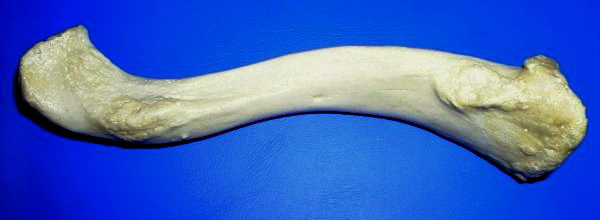
The clavicle, or collarbone, is a slender, S-shaped long bone approximately 6 inches (15 cm) long that serves as a strut between the shoulder blade and the sternum (breastbone). There are two clavicles, one on the left and one on the right. The clavicle is the only long bone in the body that lies horizontally. Together with the shoulder blade, it makes up the shoulder girdle. It is a palpable bone and, in people who have less fat in this region, the location of the bone is clearly visible. It receives its name from the Latin ''clavicula'' ("little key"), because the bone rotates along its axis like a key when the shoulder is abducted. The clavicle is the most commonly fractured bone. It can easily be fractured by impacts to the shoulder from the force of falling on outstretched arms or by a direct hit. Structure The collarbone is a thin doubly curved long bone that connects the arm to the trunk of the body. Located directly above the first rib, it acts as a strut t ... [...More Info...] [...Related Items...] OR: [Wikipedia] [Google] [Baidu] |
Intraventricular Hemorrhage
Intraventricular hemorrhage (IVH), also known as intraventricular bleeding, is a bleeding into the brain's ventricular system, where the cerebrospinal fluid is produced and circulates through towards the subarachnoid space. It can result from physical trauma or from hemorrhagic stroke. 30% of intraventricular hemorrhage (IVH) are primary, confined to the ventricular system and typically caused by intraventricular trauma, aneurysm, vascular malformations, or tumors, particularly of the choroid plexus. However 70% of IVH are secondary in nature, resulting from an expansion of an existing intraparenchymal or subarachnoid hemorrhage. Intraventricular hemorrhage has been found to occur in 35% of moderate to severe traumatic brain injuries. Thus the hemorrhage usually does not occur without extensive associated damage, and so the outcome is rarely good.Dawodu S. 2007"Traumatic Brain Injury: Definition, Epidemiology, Pathophysiology"Emedicine.com. Retrieved on June 19, 2007.Vinas FC and ... [...More Info...] [...Related Items...] OR: [Wikipedia] [Google] [Baidu] |
Epidural Hemorrhage
Epidural hematoma is when bleeding occurs between the tough outer membrane covering the brain (dura mater) and the skull. Often there is loss of consciousness following a head injury, a brief regaining of consciousness, and then loss of consciousness again. Other symptoms may include headache, confusion, vomiting, and an inability to move parts of the body. Complications may include seizures. The cause is typically head injury that results in a break of the temporal bone and bleeding from the middle meningeal artery. Occasionally it can occur as a result of a bleeding disorder or blood vessel malformation. Diagnosis is typically by a CT scan or MRI. When this condition occurs in the spine it is known as a spinal epidural hematoma. Treatment is generally by urgent surgery in the form of a craniotomy or burr hole. Without treatment, death typically results. The condition occurs in one to four percent of head injuries. Typically it occurs in young adults. Males are more often ... [...More Info...] [...Related Items...] OR: [Wikipedia] [Google] [Baidu] |
Subarachnoid Hemorrhage
Subarachnoid hemorrhage (SAH) is bleeding into the subarachnoid space—the area between the arachnoid membrane and the pia mater surrounding the brain. Symptoms may include a severe headache of rapid onset, vomiting, decreased level of consciousness, fever, and sometimes seizures. Neck stiffness or neck pain are also relatively common. In about a quarter of people a small bleed with resolving symptoms occurs within a month of a larger bleed. SAH may occur as a result of a head injury or spontaneously, usually from a ruptured cerebral aneurysm. Risk factors for spontaneous cases include high blood pressure, smoking, family history, alcoholism, and cocaine use. Generally, the diagnosis can be determined by a CT scan of the head if done within six hours of symptom onset. Occasionally, a lumbar puncture is also required. After confirmation further tests are usually performed to determine the underlying cause. Treatment is by prompt neurosurgery or endovascular coiling. Me ... [...More Info...] [...Related Items...] OR: [Wikipedia] [Google] [Baidu] |
Subdural Hemorrhage
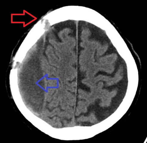
A subdural hematoma (SDH) is a type of bleeding in which a collection of blood—usually but not always associated with a traumatic brain injury—gathers between the inner layer of the dura mater and the arachnoid mater of the meninges surrounding the brain. It usually results from tears in bridging veins that cross the subdural space. Subdural hematomas may cause an increase in the pressure inside the skull, which in turn can cause compression of and damage to delicate brain tissue. Acute subdural hematomas are often life-threatening. Chronic subdural hematomas have a better prognosis if properly managed. In contrast, epidural hematomas are usually caused by tears in arteries, resulting in a build-up of blood between the dura mater and the skull. The third type of brain hemorrhage, known as a subarachnoid hemorrhage, causes bleeding into the subarachnoid space between the arachnoid mater and the pia mater. __TOC__ Signs and symptoms The symptoms of a subdural hematoma ha ... [...More Info...] [...Related Items...] OR: [Wikipedia] [Google] [Baidu] |
Subgaleal Hemorrhage
Subgaleal hemorrhage, also known as subgaleal hematoma, is bleeding in the potential space between the skull periosteum and the scalp galea aponeurosis. Symptoms The diagnosis is generally clinical, with a fluctuant boggy mass developing over the scalp (especially over the occiput) with superficial skin bruising. The swelling develops gradually 12–72 hours after delivery, although it may be noted immediately after delivery in severe cases. Subgaleal hematoma growth is insidious, as it spreads across the whole calvaria and may not be recognized for hours to days. If enough blood accumulates, a visible fluid wave may be seen. Patients may develop periorbital ecchymosis ("raccoon eyes"). Patients with subgaleal hematoma may present with hemorrhagic shock given the volume of blood that can be lost into the potential space between the skull periosteum and the scalp galea aponeurosis, which has been found to be as high as 20-40% of the neonatal blood volume in some studies. The swe ... [...More Info...] [...Related Items...] OR: [Wikipedia] [Google] [Baidu] |
Cephalohematoma
A cephalohaematoma is a hemorrhage of blood between the skull and the periosteum of any age human, including a newborn baby secondary to rupture of blood vessels crossing the periosteum. Because the swelling is subperiosteal, its boundaries are limited by the individual bones, in contrast to a caput succedaneum. Symptoms and signs Swelling appears after 2-3 days after birth. If severe the child may develop jaundice, anemia or hypotension. In some cases it may be an indication of a linear skull fracture or be at risk of an infection leading to osteomyelitis or meningitis. The swelling of a cephalohematoma takes weeks to resolve as the blood clot is slowly absorbed from the periphery towards the centre. In time the swelling hardens (calcification) leaving a relatively softer centre so that it appears as a 'depressed fracture'. Cephalohematoma should be distinguished from another scalp bleeding called subgaleal hemorrhage (also called subaponeurotic hemorrhage), which is blood bet ... [...More Info...] [...Related Items...] OR: [Wikipedia] [Google] [Baidu] |
Caput Succedaneum
Caput succedaneum is a neonatal condition involving a serosanguinous, subcutaneous, extraperiosteal fluid collection with poorly defined margins caused by the pressure of the presenting part of the scalp against the dilating cervix (tourniquet effect of the cervix) during delivery. It involves bleeding below the scalp and above the periosteum. See also * Cephalohematoma * Cephalic * Chignon (medical term) * Hematoma * Subgaleal hemorrhage Subgaleal hemorrhage, also known as subgaleal hematoma, is bleeding in the potential space between the skull periosteum and the scalp galea aponeurosis. Symptoms The diagnosis is generally clinical, with a fluctuant boggy mass developing over the ... References External links {{DEFAULTSORT:Caput Succedaneum Birth trauma Vascular-related cutaneous conditions ... [...More Info...] [...Related Items...] OR: [Wikipedia] [Google] [Baidu] |
Brachial Plexus Injury
A brachial plexus injury (BPI), also known as brachial plexus lesion, is an injury to the brachial plexus, the network of nerves that conducts signals from the spinal cord to the shoulder, arm and hand. These nerves originate in the fifth, sixth, seventh and eighth cervical (C5–C8), and first thoracic (T1) spinal nerves, and innervate the muscles and skin of the chest, shoulder, arm and hand. Brachial plexus injuries can occur as a result of shoulder trauma, tumours, or inflammation, or obstetric. Obstetric injuries may occur from mechanical injury involving shoulder dystocia during difficult childbirth, with a prevalence of 1 in 1000 births. "The brachial plexus may be injured by falls from a height on to the side of the head and shoulder, whereby the nerves of the plexus are violently stretched. The brachial plexus may also be injured by direct violence or gunshot wounds, by violent traction on the arm, or by efforts at reducing a dislocation of the shoulder joint". The rare P ... [...More Info...] [...Related Items...] OR: [Wikipedia] [Google] [Baidu] |

.jpg)


