|
Bcl-2-associated Death Promoter
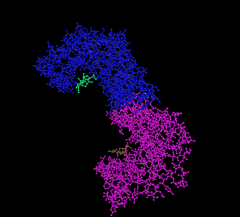
The BCL2 associated agonist of cell death (BAD) protein is a pro-apoptotic member of the Bcl-2 gene family which is involved in initiating apoptosis. BAD is a member of the BH3-only family, a subfamily of the Bcl-2 family. It does not contain a C-terminal transmembrane domain for outer mitochondrial membrane and nuclear envelope targeting, unlike most other members of the Bcl-2 family. After activation, it is able to form a heterodimer with anti-apoptotic proteins and prevent them from stopping apoptosis. Mechanism of action Bax/ Bak are believed to initiate apoptosis by forming a pore in the mitochondrial outer membrane that allows cytochrome c to escape into the cytoplasm and activate the pro-apoptotic caspase cascade. The anti-apoptotic Bcl-2 and Bcl-xL proteins inhibit cytochrome c release through the mitochondrial pore and also inhibit activation of the cytoplasmic caspase cascade by cytochrome c. Dephosphorylated BAD forms a heterodimer with Bcl-2 and Bcl-xL, inactivati ... [...More Info...] [...Related Items...] OR: [Wikipedia] [Google] [Baidu] |
Protein
Proteins are large biomolecules and macromolecules that comprise one or more long chains of amino acid residues. Proteins perform a vast array of functions within organisms, including catalysing metabolic reactions, DNA replication, responding to stimuli, providing structure to cells and organisms, and transporting molecules from one location to another. Proteins differ from one another primarily in their sequence of amino acids, which is dictated by the nucleotide sequence of their genes, and which usually results in protein folding into a specific 3D structure that determines its activity. A linear chain of amino acid residues is called a polypeptide. A protein contains at least one long polypeptide. Short polypeptides, containing less than 20–30 residues, are rarely considered to be proteins and are commonly called peptides. The individual amino acid residues are bonded together by peptide bonds and adjacent amino acid residues. The sequence of amino acid ... [...More Info...] [...Related Items...] OR: [Wikipedia] [Google] [Baidu] |
14-3-3
14-3-3 proteins are a family of conserved regulatory molecules that are expressed in all eukaryotic cells. 14-3-3 proteins have the ability to bind a multitude of functionally diverse signaling proteins, including kinases, phosphatases, and transmembrane receptors. More than 200 signaling proteins have been reported as 14-3-3 ligands. Elevated amounts of 14-3-3 proteins in cerebrospinal fluid may be a sign of Creutzfeldt–Jakob disease. Properties Seven genes encode seven distinct 14-3-3 proteins in most mammals (See ''Human genes'' below) and 13-15 genes in many higher plants, though typically in fungi they are present only in pairs. Protists have at least one. Eukaryotes can tolerate the loss of a single 14-3-3 gene if multiple genes are expressed, but deletion of all 14-3-3s (as experimentally determined in yeast) results in death. 14-3-3 proteins are structurally similar to the Tetratrico Peptide Repeat (TPR) superfamily, which generally have 9 or 10 alpha helices, an ... [...More Info...] [...Related Items...] OR: [Wikipedia] [Google] [Baidu] |
YWHAZ
14-3-3 protein zeta/delta (14-3-3ζ) is a protein that in humans is encoded by the ''YWHAZ'' gene on chromosome 8. The protein encoded by this gene is a member of the 14-3-3 protein family and a central hub protein for many signal transduction pathways. 14-3-3ζ is a major regulator of apoptotic pathways critical to cell survival and plays a key role in a number of cancers and neurodegenerative diseases. Structure 14-3-3 proteins generally form ~30 kDa-long homo- or heterodimers. Each of the monomers are composed of 9 antiparallel alpha helices. Four alpha-helices (αC, αE, αG, and αI) form an amphipathic groove that serves as the ligand binding site, which can recognize three types of consensus binding motifs: RXX(pS/pT)XP, RXXX(pS/pT)XP, and (pS/pT)X1-2-COOH (where pS/pT represents phosphorylated serine/threonine). In addition to these primary interactions, the target protein can also bind outside the groove via secondary interactions. In particular, the crystallized st ... [...More Info...] [...Related Items...] OR: [Wikipedia] [Google] [Baidu] |
YWHAQ
14-3-3 protein theta is a protein that in humans is encoded by the ''YWHAQ'' gene. Function This gene product belongs to the 14-3-3 family of proteins that mediate signal transduction by binding to phosphoserine-containing proteins. This highly conserved protein family is found in both plants and mammals, and this protein is 99% identical to the mouse and rat orthologs. This gene is upregulated in patients with amyotrophic lateral sclerosis. It contains in its 5' UTR a 6 bp tandem repeat sequence that is polymorphic; however, there is no correlation between the repeat number and the disease. Interactions YWHAQ has been shown to interact with: * BAX, * BAD, * C-Raf, * CRTC2, * CBL * HDAC5, * MEF2D, * NRIP1, * PFKFB2, * PRKD1, * PRKCZ, * TERT Telomerase reverse transcriptase (abbreviated to TERT, or hTERT in humans) is a catalytic subunit of the enzyme telomerase, which, together with the telomerase RNA component (TERC), comprises the most important unit o ... [...More Info...] [...Related Items...] OR: [Wikipedia] [Google] [Baidu] |
S100A10
S100 calcium-binding protein A10 (S100A10), also known as p11, is a protein that is encoded by the ''S100A10'' gene in humans and the ''S100a10'' gene in other species. S100A10 is a member of the S100 family of proteins containing two EF-hand calcium-binding motifs. S100 proteins are localized in the cytoplasm and/or nucleus of a wide range of cells. They regulate a number of cellular processes such as cell cycle progression and differentiation. The S100 protein is implicated in exocytosis and endocytosis by reorganization of F-actin. The p11 protein is linked with the transport of neurotransmitters. Found in the brain of humans and other mammals, it has been implicated in the regulation of mood. In addition, due to its interaction with serotonin-signaling proteins and its correlation with symptoms of mood disorders, p11 is a new potential target for drug therapy. Gene The S100 gene family, localized in the cytoplasm and nucleus of cells, includes at least 13 members that ... [...More Info...] [...Related Items...] OR: [Wikipedia] [Google] [Baidu] |
MCL1
Induced myeloid leukemia cell differentiation protein Mcl-1 is a protein that in humans is encoded by the ''MCL1'' gene. Function The protein encoded by this gene belongs to the Bcl-2 family. Alternative splicing occurs at this locus and two transcript variants encoding distinct isoforms have been identified. The longer gene product (isoform 1) enhances cell survival by inhibiting apoptosis while the alternatively spliced shorter gene product (isoform 2) promotes apoptosis and is death-inducing. The protein MCL1 has a very short biological half-life of only 20–30 minutes. The loss of MCL1 has a more dramatic impact than the loss of any other anti-apoptotic member of the Bcl-2 family. Loss of the ''Mcl-1'' gene results in embryo death when the embryo is only around 3.5 days old, before it has even implanted. Conditional deletion of ''Mcl-1'' depletes a wide variety of cells, including hematopoietic stem cells, B cell–committed progenitors, T cell–committed progenitors, ... [...More Info...] [...Related Items...] OR: [Wikipedia] [Google] [Baidu] |
BCL2L2
Bcl-2-like protein 2 is a 193-amino acid protein that in humans is encoded by the ''BCL2L2'' gene on chromosome 14 ( band q11.2-q12). It was originally discovered by Leonie Gibson, Suzanne Cory and colleagues at the Walter and Eliza Hall Institute of Medical Research, who called it Bcl-w. Function This gene encodes a pro-survival (anti-apoptotic) member of the bcl-2 protein family, and is most similar to Bcl-xL. The proteins of this family form hetero- or homodimers and act as anti- and pro-apoptotic regulators. Expression of this gene in cells has been shown to contribute to reduced cell apoptosis under cytotoxic conditions. Studies of the related gene in mice indicated a role in the survival of NGF- and BDNF-dependent neurons. Mutation and knockout studies of the mouse gene demonstrated an essential role in adult spermatogenesis. Clinical significance High levels of Bcl-w are seen in many cancers, including glioblastoma, colorectal cancer, non-small-cell lung carcino ... [...More Info...] [...Related Items...] OR: [Wikipedia] [Google] [Baidu] |
BCL2-related Protein A1
Bcl-2-related protein A1 is a protein that in humans is encoded by the ''BCL2A1'' gene. Function This gene encodes a member of the bcl2 protein family. The proteins of this family form hetero- or homodimers and act as anti- and pro-apoptotic regulators that are involved in a wide variety of cellular activities such as embryonic development, homeostasis and tumorigenesis. The protein encoded by this gene is able to reduce the release of pro-apoptotic cytochrome c from mitochondria and block caspase activation. This gene is a direct transcription target of NF-kappa B in response to inflammatory mediators, and has been shown to be up-regulated by different extracellular signals, such as granulocyte-macrophage colony-stimulating factor (GM-CSF), CD40, phorbol ester and inflammatory cytokine TNF and IL-1, which suggests a cytoprotective function that is essential for lymphocyte activation as well as cell survival. In melanocytic cells BCL2A1 gene expression may be regulated by ... [...More Info...] [...Related Items...] OR: [Wikipedia] [Google] [Baidu] |
BCL2-like 1 (gene)
Bcl-2-like protein 1 is a protein encoded in humans by the ''BCL2L1'' gene. Through alternative splicing, the gene encodes both of the human proteins Bcl-xL and Bcl-xS. Function The protein encoded by this gene belongs to the Bcl-2 protein family. Bcl-2 family members form hetero- or homodimers and act as anti- or pro- apoptotic regulators that are involved in a wide variety of cellular activities. The proteins encoded by this gene are located at the outer mitochondrial membrane, and have been shown to regulate outer mitochondrial membrane channel ( voltage-dependent anion channels (VDACs) opening. VDACs regulate mitochondrial membrane potential, and thus controls the production of reactive oxygen species and release of cytochrome C by mitochondria, both of which are the potent inducers of cell apoptosis. Two alternatively spliced transcript variants, which encode distinct isoforms, have been reported. The longer isoform ( Bcl-xL) acts as an apoptotic inhibitor and the shor ... [...More Info...] [...Related Items...] OR: [Wikipedia] [Google] [Baidu] |
Signal Transduction Pathways
Signal transduction is the process by which a chemical or physical signal is transmitted through a cell as a series of molecular events, most commonly protein phosphorylation catalyzed by protein kinases, which ultimately results in a cellular response. Proteins responsible for detecting stimuli are generally termed receptors, although in some cases the term sensor is used. The changes elicited by ligand binding (or signal sensing) in a receptor give rise to a biochemical cascade, which is a chain of biochemical events known as a signaling pathway. When signaling pathways interact with one another they form networks, which allow cellular responses to be coordinated, often by combinatorial signaling events. At the molecular level, such responses include changes in the transcription or translation of genes, and post-translational and conformational changes in proteins, as well as changes in their location. These molecular events are the basic mechanisms controlling cell gro ... [...More Info...] [...Related Items...] OR: [Wikipedia] [Google] [Baidu] |
Calcineurin
Calcineurin (CaN) is a calcium and calmodulin dependent serine/threonine protein phosphatase (also known as protein phosphatase 3, and calcium-dependent serine-threonine phosphatase). It activates the T cells of the immune system and can be blocked by drugs. Calcineurin activates nuclear factor of activated T cell cytoplasmic ( NFATc), a transcription factor, by dephosphorylating it. The activated NFATc is then translocated into the nucleus, where it upregulates the expression of interleukin 2 (IL-2), which, in turn, stimulates the growth and differentiation of the T cell response. Calcineurin is the target of a class of drugs called calcineurin inhibitors, which include ciclosporin, voclosporin, pimecrolimus and tacrolimus. Structure Calcineurin is a heterodimer of a 61-kD calmodulin-binding catalytic subunit, calcineurin A and a 19-kD Ca2+-binding regulatory subunit, calcineurin B. There are three isozymes of the catalytic subunit, each encoded by a separate gene ( PP ... [...More Info...] [...Related Items...] OR: [Wikipedia] [Google] [Baidu] |


