|
Axillary Nerve
The axillary nerve or the circumflex nerve is a nerve of the human body, that originates from the brachial plexus ( upper trunk, posterior division, posterior cord) at the level of the axilla (armpit) and carries nerve fibers from C5 and C6. The axillary nerve travels through the quadrangular space with the posterior circumflex humeral artery and vein to innervate the deltoid and teres minor. Structure The nerve lies at first behind the axillary artery, and in front of the subscapularis, and passes downward to the lower border of that muscle. It then winds from anterior to posterior around the neck of the humerus, in company with the posterior humeral circumflex artery, through the quadrangular space (bounded above by the teres minor, below by the teres major, medially by the long head of the triceps brachii, and laterally by the surgical neck of the humerus), and divides into an anterior, a posterior, and a collateral branch to the long head of the triceps brachii bra ... [...More Info...] [...Related Items...] OR: [Wikipedia] [Google] [Baidu] |
Suprascapular Nerve
The suprascapular nerve is a nerve that branches from the upper trunk of the brachial plexus. It is responsible for the innervation of two of the muscles that originate from the scapula, namely the supraspinatus and infraspinatus muscles. Structure The suprascapular nerve arises from the upper trunk of the brachial plexus which is formed by the union of the ventral rami of the fifth and sixth cervical nerves. After branching from the upper trunk, the nerve passes across the posterior triangle of the neck parallel to the inferior belly of the omohyoid muscle and deep to the trapezius muscle. It then runs along the superior border of the scapula through the suprascapular canal, in which it enters via the suprascapular notch inferior to the superior transverse scapular ligament and enters the supraspinous fossa. It then passes beneath the supraspinatus, and curves around the lateral border of the spine of the scapula through spinogleniod notch to the infraspinous fossa. Funct ... [...More Info...] [...Related Items...] OR: [Wikipedia] [Google] [Baidu] |
Axillary Artery
In human anatomy, the axillary artery is a large blood vessel that conveys oxygenated blood to the lateral aspect of the thorax, the axilla (armpit) and the upper limb. Its origin is at the lateral margin of the first rib, before which it is called the subclavian artery. After passing the lower margin of teres major it becomes the brachial artery. Structure The axillary artery is often referred to as having three parts, with these divisions based on its location relative to the Pectoralis minor muscle, which is superficial to the artery. * First part – the part of the artery superior to the pectoralis minor * Second part – the part of the artery posterior to the pectoralis minor * Third part – the part of the artery inferior to the pectoralis minor. Relations The axillary artery is accompanied by the axillary vein, which lies medial to the artery, along its length. In the axilla, the axillary artery is surrounded by the brachial plexus. The second part of the axill ... [...More Info...] [...Related Items...] OR: [Wikipedia] [Google] [Baidu] |
Dislocated Shoulder
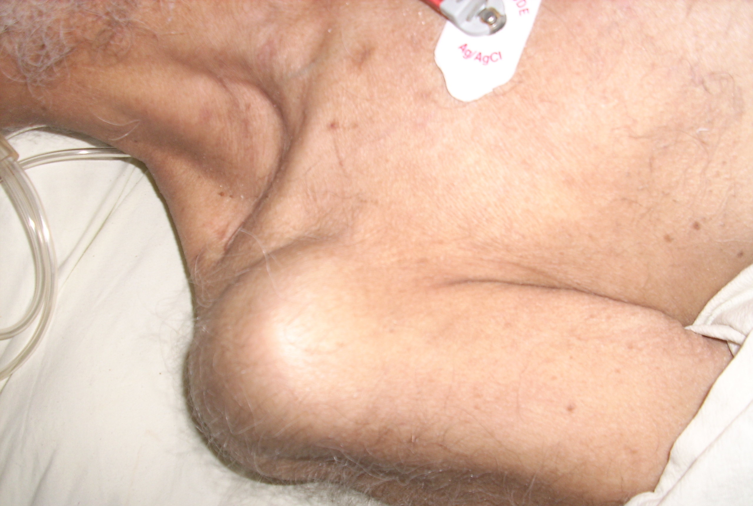
A dislocated shoulder is a condition in which the head of the humerus is detached from the shoulder joint. Symptoms include shoulder pain and instability. Complications may include a Bankart lesion, Hill-Sachs lesion, rotator cuff tear, or injury to the axillary nerve. A shoulder dislocation often occurs as a result of a fall onto an outstretched arm or onto the shoulder. Diagnosis is typically based on symptoms and confirmed by X-rays. They are classified as anterior, posterior, inferior, and superior with most being anterior. Treatment is by shoulder reduction which may be accomplished by a number of techniques. These include traction-countertraction, external rotation, scapular manipulation, and the Stimson technique. After reduction X-rays are recommended for verification. The arm may then be placed in a sling for a few weeks. Surgery may be recommended in those with recurrent dislocations. Not all patients require surgery following a shoulder dislocation. There is mo ... [...More Info...] [...Related Items...] OR: [Wikipedia] [Google] [Baidu] |
Rotator Cuff
The rotator cuff is a group of muscles and their tendons that act to stabilize the human shoulder and allow for its extensive range of motion. Of the seven scapulohumeral muscles, four make up the rotator cuff. The four muscles are the supraspinatus muscle, the infraspinatus muscle, teres minor muscle, and the subscapularis muscle. Structure Muscles composing rotator cuff The supraspinatus muscle spreads out in a horizontal band to insert on the superior facet of the greater tubercle of the humerus. The greater tubercle projects as the most lateral structure of the humeral head. Medial to this, in turn, is the lesser tubercle of the humeral head. The subscapularis muscle origin is divided from the remainder of the rotator cuff origins as it is deep to the scapula. The four tendons of these muscles converge to form the rotator cuff tendon. These tendinous insertions along with the articular capsule, the coracohumeral ligament, and the glenohumeral ligament complex, ... [...More Info...] [...Related Items...] OR: [Wikipedia] [Google] [Baidu] |
Muscle
Skeletal muscles (commonly referred to as muscles) are organs of the vertebrate muscular system and typically are attached by tendons to bones of a skeleton. The muscle cells of skeletal muscles are much longer than in the other types of muscle tissue, and are often known as muscle fibers. The muscle tissue of a skeletal muscle is striated – having a striped appearance due to the arrangement of the sarcomeres. Skeletal muscles are voluntary muscles under the control of the somatic nervous system. The other types of muscle are cardiac muscle which is also striated and smooth muscle which is non-striated; both of these types of muscle tissue are classified as involuntary, or, under the control of the autonomic nervous system. A skeletal muscle contains multiple fascicles – bundles of muscle fibers. Each individual fiber, and each muscle is surrounded by a type of connective tissue layer of fascia. Muscle fibers are formed from the fusion of developmental myoblasts ... [...More Info...] [...Related Items...] OR: [Wikipedia] [Google] [Baidu] |
Cadaver
A cadaver or corpse is a dead human body that is used by medical students, physicians and other scientists to study anatomy, identify disease sites, determine causes of death, and provide tissue to repair a defect in a living human being. Students in medical school study and dissect cadavers as a part of their education. Others who study cadavers include archaeologists and arts students. The term ''cadaver'' is used in courts of law (and, to a lesser extent, also by media outlets such as newspapers) to refer to a dead body, as well as by recovery teams searching for bodies in natural disasters. The word comes from the Latin word ''cadere'' ("to fall"). Related terms include ''cadaverous'' (resembling a cadaver) and ''cadaveric spasm'' (a muscle spasm causing a dead body to twitch or jerk). A cadaver graft (also called “postmortem graft”) is the grafting of tissue from a dead body onto a living human to repair a defect or disfigurement. Cadavers can be observed for their sta ... [...More Info...] [...Related Items...] OR: [Wikipedia] [Google] [Baidu] |
Shoulder Joint
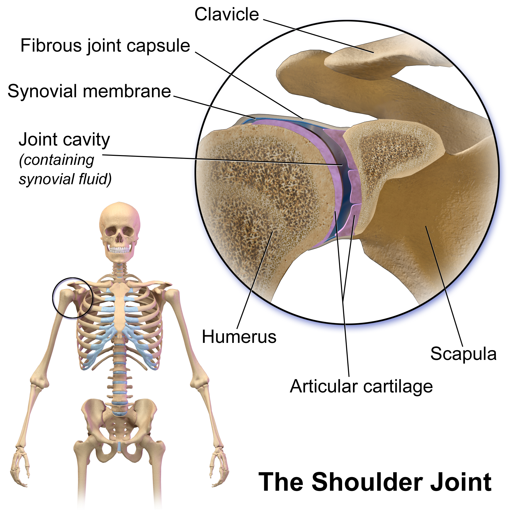
The shoulder joint (or glenohumeral joint from Greek ''glene'', eyeball, + -''oid'', 'form of', + Latin ''humerus'', shoulder) is structurally classified as a synovial ball-and-socket joint and functionally as a diarthrosis and multiaxial joint. It involves an articulation between the glenoid fossa of the scapula (shoulder blade) and the head of the humerus (upper arm bone). Due to the very loose joint capsule that gives a limited interface of the humerus and scapula, it is the most mobile joint of the human body. Structure The shoulder joint is a ball-and-socket joint between the scapula and the humerus. The socket of the glenoid fossa of the scapula is itself quite shallow, but it is made deeper by the addition of the glenoid labrum. The glenoid labrum is a ring of cartilaginous fibre attached to the circumference of the cavity. This ring is continuous with the tendon of the biceps brachii above. Spaces Significant joint spaces are: * The normal glenohumeral space is 4� ... [...More Info...] [...Related Items...] OR: [Wikipedia] [Google] [Baidu] |
Superior Lateral Cutaneous Nerve Of Arm
The superior lateral cutaneous nerve of arm (or superior lateral brachial cutaneous nerve) is the continuation of the posterior branch of the axillary nerve, after it pierces the deep fascia. It contains axons from C5-C6 ventral rami. Structure It sweeps around the posterior border of the deltoideus and supplies the skin over the lower two-thirds of the posterior part of this muscle, as well as that covering the long head of the triceps brachii. See also * Posterior cutaneous nerve of arm (Posterior brachial) * Medial cutaneous nerve of arm (Medial brachial) * Lateral cutaneous nerve of forearm The lateral antebrachial cutaneous nerve (or lateral cutaneous nerve of forearm) (branch of musculocutaneous nerve, also sometimes spelled "antebrachial") passes behind the cephalic vein, and divides, opposite the elbow-joint, into a volar and ... (Lateral antebrachial) Additional images File:Gray811and813.PNG, Cutaneous nerves of right upper extremity. File:Gray812and814.PNG, ... [...More Info...] [...Related Items...] OR: [Wikipedia] [Google] [Baidu] |
Surgical Neck Of The Humerus
The surgical neck of the humerus is a bony constriction at the proximal end of shaft of humerus. It is situated distal to the greater tubercle and lesser tubercle, and proximal to the deltoid tuberosity. Clinical significance The surgical neck is much more frequently fractured than the anatomical neck of the humerus. This type of fracture takes place when the humerus is forced in one direction while the joint capsule and the rotator cuff muscles remain intact. A fracture in this area is most likely to cause damage to the axillary nerve and posterior circumflex humeral artery. Damage to the axillary nerve affects function of the teres minor and deltoid muscles, resulting in loss of abduction of arm (from 15-90 degrees), weak flexion, extension, and rotation of shoulder as well as loss of sensation of the skin over a small part of the lateral shoulder. Additional images File:Neck-of-Humerus.jpg, The difference between anatomical neck and surgical neck of the humerus File:Ill ... [...More Info...] [...Related Items...] OR: [Wikipedia] [Google] [Baidu] |
Surgical Neck
The surgical neck of the humerus is a bony constriction at the proximal end of shaft of humerus. It is situated distal to the greater tubercle and lesser tubercle, and proximal to the deltoid tuberosity. Clinical significance The surgical neck is much more frequently fractured than the anatomical neck of the humerus. This type of fracture takes place when the humerus is forced in one direction while the joint capsule and the rotator cuff muscles remain intact. A fracture in this area is most likely to cause damage to the axillary nerve and posterior circumflex humeral artery. Damage to the axillary nerve affects function of the teres minor and deltoid muscles, resulting in loss of abduction of arm (from 15-90 degrees), weak flexion, extension, and rotation of shoulder as well as loss of sensation of the skin over a small part of the lateral shoulder. Additional images File:Neck-of-Humerus.jpg, The difference between anatomical neck and surgical neck of the humerus File:Il ... [...More Info...] [...Related Items...] OR: [Wikipedia] [Google] [Baidu] |
Triceps Brachii
The triceps, or triceps brachii (Latin for "three-headed muscle of the arm"), is a large muscle on the back of the upper limb of many vertebrates. It consists of 3 parts: the medial, lateral, and long head. It is the muscle principally responsible for extension of the elbow joint (straightening of the arm). Structure The long head arises from the infraglenoid tubercle of the scapula. It extends distally anterior to the teres minor and posterior to the teres major. The medial head arises proximally in the humerus, just inferior to the groove of the radial nerve; from the dorsal (back) surface of the humerus; from the medial intermuscular septum; and its distal part also arises from the lateral intermuscular septum. The medial head is mostly covered by the lateral and long heads, and is only visible distally on the humerus. The lateral head arises from the dorsal surface of the humerus, lateral and proximal to the groove of the radial nerve, from the greater tuber ... [...More Info...] [...Related Items...] OR: [Wikipedia] [Google] [Baidu] |
Teres Major
The teres major muscle is a muscle of the upper limb. It attaches to the scapula and the humerus and is one of the seven scapulohumeral muscles. It is a thick but somewhat flattened muscle. The teres major muscle (from Latin ''teres'', meaning "rounded") is positioned above the latissimus dorsi muscle and assists in the extension and medial rotation of the humerus. This muscle is commonly confused as a rotator cuff muscle, but it is not because it does not attach to the capsule of the shoulder joint, unlike the teres minor muscle for example. Structure The teres major muscle originates on the dorsal surface of the inferior angle and the lower part of the lateral border of the scapula. The fibers of teres major insert into the medial lip of the intertubercular sulcus of the humerus. Relations The tendon, at its insertion, lies behind that of the latissimus dorsi, from which it is separated by a bursa, the two tendons being, however, united along their lower borders for a sh ... [...More Info...] [...Related Items...] OR: [Wikipedia] [Google] [Baidu] |


