|
Axillary Artery
In human anatomy, the axillary artery is a large blood vessel that conveys oxygenated blood to the lateral aspect of the thorax, the axilla (armpit) and the upper limb. Its origin is at the lateral margin of the first rib, before which it is called the subclavian artery. After passing the lower margin of teres major it becomes the brachial artery. Structure The axillary artery is often referred to as having three parts, with these divisions based on its location relative to the Pectoralis minor muscle, which is superficial to the artery. * First part – the part of the artery superior to the pectoralis minor * Second part – the part of the artery posterior to the pectoralis minor * Third part – the part of the artery inferior to the pectoralis minor. Relations The axillary artery is accompanied by the axillary vein, which lies medial to the artery, along its length. In the axilla, the axillary artery is surrounded by the brachial plexus. The second part of the axill ... [...More Info...] [...Related Items...] OR: [Wikipedia] [Google] [Baidu] |
Pectoralis Minor Muscle
Pectoralis minor muscle () is a thin, triangular muscle, situated at the upper part of the chest, beneath the pectoralis major in the human body. Structure Attachments Pectoralis minor muscle arises from the upper margins and outer surfaces of the third, fourth, and fifth ribs, near their costal cartilages and from the aponeuroses covering the intercostalis. The fibers pass superior and lateral and converge to form a flat tendon. This tendon inserts onto the medial border and upper surface of the coracoid process of the scapula. Relations Pectoralis minor muscle forms part of the anterior wall of the axilla. It is covered anteriorly (superficially) by the clavipectoral fascia. The medial pectoral nerve pierces the pectoralis minor and the clavipectoral fascia. In attaching to the coracoid process, the pectoralis minor forms a 'bridge' - structures passing into the upper limb from the thorax will pass directly underneath.http://www.teachmeanatomy.com/muscles-of-the ... [...More Info...] [...Related Items...] OR: [Wikipedia] [Google] [Baidu] |
Pectoralis Minor
Pectoralis minor muscle () is a thin, triangular muscle, situated at the upper part of the chest, beneath the pectoralis major in the human body. Structure Attachments Pectoralis minor muscle arises from the upper margins and outer surfaces of the third, fourth, and fifth ribs, near their costal cartilages and from the aponeuroses covering the intercostalis. The fibers pass superior and lateral and converge to form a flat tendon. This tendon inserts onto the medial border and upper surface of the coracoid process of the scapula. Relations Pectoralis minor muscle forms part of the anterior wall of the axilla. It is covered anteriorly (superficially) by the clavipectoral fascia. The medial pectoral nerve pierces the pectoralis minor and the clavipectoral fascia. In attaching to the coracoid process, the pectoralis minor forms a 'bridge' - structures passing into the upper limb from the thorax will pass directly underneath.http://www.teachmeanatomy.com/muscles-of-the-pecto ... [...More Info...] [...Related Items...] OR: [Wikipedia] [Google] [Baidu] |
Ascending Aorta
The ascending aorta (AAo) is a portion of the aorta commencing at the upper part of the base of the left ventricle, on a level with the lower border of the third costal cartilage behind the left half of the sternum. Structure It passes obliquely upward, forward, and to the right, in the direction of the heart's axis, as high as the upper border of the second right costal cartilage, describing a slight curve in its course, and being situated, about behind the posterior surface of the sternum. The total length is about . Components The aortic root is the portion of the aorta beginning at the aortic annulus and extending to the sinotubular junction. It is sometimes regarded as a part of the ascending aorta, and sometimes regarded as a separate entity from the rest of the ascending aorta. Between each commissure of the aortic valve and opposite the cusps of the aortic valve, three small dilatations called the aortic sinuses. The sinotubular junction is the point in the ascend ... [...More Info...] [...Related Items...] OR: [Wikipedia] [Google] [Baidu] |
Aortic Dissection
Aortic dissection (AD) occurs when an injury to the innermost layer of the aorta allows blood to flow between the layers of the aortic wall, forcing the layers apart. In most cases, this is associated with a sudden onset of severe chest or back pain, often described as "tearing" in character. Also, vomiting, sweating, and lightheadedness may occur. Other symptoms may result from decreased blood supply to other organs, such as stroke, lower extremity ischemia, or mesenteric ischemia. Aortic dissection can quickly lead to death from insufficient blood flow to the heart or complete rupture of the aorta. AD is more common in those with a history of high blood pressure; a number of connective tissue diseases that affect blood vessel wall strength including Marfan syndrome and Ehlers–Danlos syndrome; a bicuspid aortic valve; and previous heart surgery. Major trauma, smoking, cocaine use, pregnancy, a thoracic aortic aneurysm, inflammation of arteries, and abnorma ... [...More Info...] [...Related Items...] OR: [Wikipedia] [Google] [Baidu] |
Cardiac Surgery
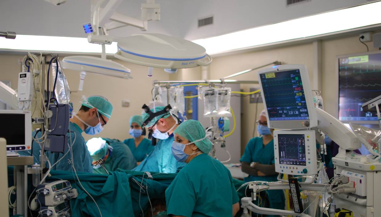
Cardiac surgery, or cardiovascular surgery, is surgery on the heart or great vessels performed by cardiac surgeons. It is often used to treat complications of ischemic heart disease (for example, with coronary artery bypass grafting); to correct congenital heart disease; or to treat valvular heart disease from various causes, including endocarditis, rheumatic heart disease, and atherosclerosis. It also includes heart transplantation. History 19th century The earliest operations on the pericardium (the sac that surrounds the heart) took place in the 19th century and were performed by Francisco Romero (1801) in the city of Almería (Spain), Dominique Jean Larrey (1810), Henry Dalton (1891), and Daniel Hale Williams (1893). The first surgery on the heart itself was performed by Axel Cappelen on 4 September 1895 at Rikshospitalet in Kristiania, now Oslo. Cappelen ligated a bleeding coronary artery in a 24-year-old man who had been stabbed in the left axilla and was in ... [...More Info...] [...Related Items...] OR: [Wikipedia] [Google] [Baidu] |
Cannula
A cannula (; Latin meaning 'little reed'; plural or ) is a tube that can be inserted into the body, often for the delivery or removal of fluid or for the gathering of samples. In simple terms, a cannula can surround the inner or outer surfaces of a trocar needle thus extending the effective needle length by at least half the length of the original needle. Its size mainly ranges from 14 to 24 gauge. Different-sized cannula have different colours as coded. Decannulation is the permanent removal of a cannula ( extubation), especially of a tracheostomy cannula, once a physician determines it is no longer needed for breathing. Medicine Cannulas normally come with a trocar inside. The trocar is a needle, which punctures the body in order to get into the intended space. Many types of cannulas exist: Intravenous cannulas are the most common in hospital use. A variety of cannulas are used to establish cardiopulmonary bypass in cardiac surgery. A nasal cannula is a piece of plastic ... [...More Info...] [...Related Items...] OR: [Wikipedia] [Google] [Baidu] |
Suprascapular Artery
The suprascapular artery is a branch of the thyrocervical trunk on the neck. Structure At first, it passes downward and laterally across the scalenus anterior and phrenic nerve, being covered by the sternocleidomastoid muscle; it then crosses the subclavian artery and the brachial plexus, running behind and parallel with the clavicle and subclavius muscle and beneath the inferior belly of the omohyoid to the superior border of the scapula. It passes over the superior transverse scapular ligament in most of the cases while below it through the suprascapular notch in some cases. The artery then enters the supraspinous fossa of the scapula. It travels close to the bone, running through the suprascapular canal underneath the supraspinatus muscle, to which it supplies branches. It then descends behind the neck of the scapula, through the great scapular notch and under cover of the inferior transverse ligament, to reach the infraspinatous fossa, where it supplies infraspinatu ... [...More Info...] [...Related Items...] OR: [Wikipedia] [Google] [Baidu] |
Dorsal Scapular Artery
The transverse cervical artery (transverse artery of neck or transversa colli artery) is an artery in the neck and a branch of the thyrocervical trunk, running at a higher level than the suprascapular artery. Structure It passes transversely below the inferior belly of the omohyoid muscle to the anterior margin of the trapezius, beneath which it divides into a superficial and a deep branch. It crosses in front of the phrenic nerve and the scalene muscles, and in front of or between the divisions of the brachial plexus, and is covered by the platysma and sternocleidomastoid muscles, and crossed by the omohyoid and trapezius. The transverse cervical artery originates from the thyrocervical trunk, it passes through the posterior triangle of the neck to the anterior border of the levator scapulae muscle, where it divides into deep and superficial branches. * Superficial branch ** Ascending branch ** Descending branch (also known as superficial cervical artery, which supplies th ... [...More Info...] [...Related Items...] OR: [Wikipedia] [Google] [Baidu] |
Teres Major
The teres major muscle is a muscle of the upper limb. It attaches to the scapula and the humerus and is one of the seven scapulohumeral muscles. It is a thick but somewhat flattened muscle. The teres major muscle (from Latin ''teres'', meaning "rounded") is positioned above the latissimus dorsi muscle and assists in the extension and medial rotation of the humerus. This muscle is commonly confused as a rotator cuff muscle, but it is not because it does not attach to the capsule of the shoulder joint, unlike the teres minor muscle for example. Structure The teres major muscle originates on the dorsal surface of the inferior angle and the lower part of the lateral border of the scapula. The fibers of teres major insert into the medial lip of the intertubercular sulcus of the humerus. Relations The tendon, at its insertion, lies behind that of the latissimus dorsi, from which it is separated by a bursa, the two tendons being, however, united along their lower borders for a sh ... [...More Info...] [...Related Items...] OR: [Wikipedia] [Google] [Baidu] |
Posterior Humeral Circumflex Artery
The posterior humeral circumflex artery (posterior circumflex artery, posterior circumflex humeral artery) arises from the third part of axillary artery at the lower border of the subscapularis, and runs posteriorly with the axillary nerve through the quadrangular space. It winds around the surgical neck of the humerus and is distributed to the deltoid muscle and shoulder-joint, anastomosing with the anterior humeral circumflex and deep artery of the arm. It supplies the teres major, teres minor, deltoid, and (long head only) triceps muscles. Additional images File:Gray810.png, Suprascapular and axillary nerves of right side, seen from behind. File:Slide4SSS.JPG, Posterior humeral circumflex artery See also * Anterior humeral circumflex artery The anterior humeral circumflex artery (anterior circumflex artery, anterior circumflex humeral artery) is an artery in the arm. It is one of two circumflexing arteries that branch from the axillary artery, the other being the po ... [...More Info...] [...Related Items...] OR: [Wikipedia] [Google] [Baidu] |
Thoraco-acromial Artery
The thoracoacromial artery (acromiothoracic artery; thoracic axis) is a short trunk that arises from the second part of the axillary artery, its origin being generally overlapped by the upper edge of the pectoralis minor. Structure Projecting forward to the upper border of the Pectoralis minor, it pierces the coracoclavicular fascia The clavipectoral fascia (costocoracoid membrane; coracoclavicular fascia) is a strong fascia situated under cover of the clavicular portion of the pectoralis major. It occupies the interval between the pectoralis minor and subclavius, and pro ... and divides into four branches—pectoral, acromial, clavicular, and deltoid. Additional images File:Gray523.png, The axillary artery and its branches. References External links * * * - "Pectoral Region: Thoracoacromial Artery and its Branches" * - "The axillary artery and its major branches shown in relation to major landmarks." {{Authority control Arteries of the upper limb ... [...More Info...] [...Related Items...] OR: [Wikipedia] [Google] [Baidu] |
Mnemonics
A mnemonic ( ) device, or memory device, is any learning technique that aids information retention or retrieval (remembering) in the human memory for better understanding. Mnemonics make use of elaborative encoding, retrieval cues, and imagery as specific tools to encode information in a way that allows for efficient storage and retrieval. Mnemonics aid original information in becoming associated with something more accessible or meaningful—which, in turn, provides better retention of the information. Commonly encountered mnemonics are often used for lists and in auditory form, such as short poems, acronyms, initialisms, or memorable phrases, but mnemonics can also be used for other types of information and in visual or kinesthetic forms. Their use is based on the observation that the human mind more easily remembers spatial, personal, surprising, physical, sexual, humorous, or otherwise "relatable" information, rather than more abstract or impersonal forms of information. ... [...More Info...] [...Related Items...] OR: [Wikipedia] [Google] [Baidu] |


