|
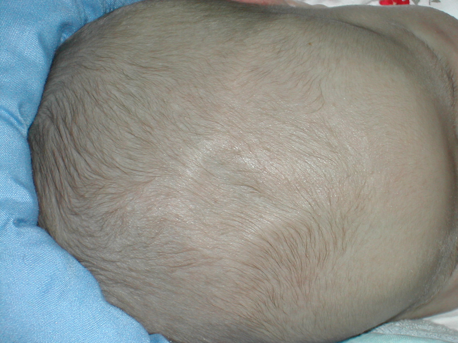
Anterior Fontanelle
The anterior fontanelle (bregmatic fontanelle, frontal fontanelle) is the largest fontanelle, and is placed at the junction of the sagittal suture, coronal suture, and frontal suture; it is lozenge-shaped, and measures about 4 cm in its antero-posterior and 2.5 cm in its transverse diameter. The fontanelle allows the skull to deform during birth to ease its passage through the birth canal and for expansion of the brain after birth. The anterior fontanelle typically closes between the ages of 12 and 18 months. Clinical significance The anterior fontanelle is useful clinically. Examination of an infant includes palpating the anterior fontanelle. A sunken fontanelle indicates dehydration whereas a very tense or bulging anterior fontanelle indicates raised intracranial pressure Intracranial pressure (ICP) is the pressure exerted by fluids such as cerebrospinal fluid (CSF) inside the skull and on the brain tissue. ICP is measured in millimeters of mercury ( mmHg) and at ... [...More Info...] [...Related Items...] OR: [Wikipedia] [Google] [Baidu] |
Fontanelle
A fontanelle (or fontanel) (colloquially, soft spot) is an anatomical feature of the infant human skull comprising soft membranous gaps ( sutures) between the cranial bones that make up the calvaria of a fetus or an infant. Fontanelles allow for stretching and deformation of the neurocranium both during birth and later as the brain expands faster than the surrounding bone can grow. Premature complete ossification of the sutures is called craniosynostosis. After infancy, the anterior fontanelle is known as the bregma. Structure An infant's skull consists of five main bones: two frontal bones, two parietal bones, and one occipital bone. These are joined by fibrous sutures, which allow movement that facilitates childbirth and brain growth. * Posterior fontanelle is triangle-shaped. It lies at the junction between the sagittal suture and lambdoid suture. At birth, the skull features a small posterior fontanelle with an open area covered by a tough membrane, where the two ... [...More Info...] [...Related Items...] OR: [Wikipedia] [Google] [Baidu] |
Sagittal Suture
The sagittal suture, also known as the interparietal suture and the ''sutura interparietalis'', is a dense, fibrous connective tissue joint between the two parietal bones of the skull. The term is derived from the Latin word ''sagitta'', meaning arrow. Structure The sagittal suture is formed from the fibrous connective tissue joint between the two parietal bones of the skull. It has a varied and irregular shape which arises during development. The pattern is different between the inside and the outside. Two anatomical landmarks are found on the sagittal suture: the bregma, and the vertex of the skull. The bregma is formed by the intersection of the sagittal and coronal sutures. The vertex is the highest point on the skull and is often near the midpoint of the sagittal suture. Development At birth, the bones of the skull do not meet. The gap that remains, which is approximately 5 mm wide, allows for the brain to continue to grow normally after birth. The inner parts ... [...More Info...] [...Related Items...] OR: [Wikipedia] [Google] [Baidu] |
Coronal Suture
The coronal suture is a dense, fibrous connective tissue joint that separates the two parietal bones from the frontal bone of the skull. Structure The coronal suture lies between the paired parietal bones and the frontal bone of the skull. It runs from the pterion on each side. Nerve supply The coronal suture is likely supplied by a branch of the trigeminal nerve. Development The coronal suture is derived from the paraxial mesoderm. Clinical significance If certain bones of the skull grow too fast then premature fusion of the sutures may occur. This can result in skull deformities. There are two possible deformities that can be caused by the premature closure of the coronal suture: * a high, tower-like skull called "oxycephaly" or "turret skull". * a twisted and asymmetrical skull called "plagiocephaly". References * "Sagittal suture." ''Stedman's Medical Dictionary, 27th ed.'' (2000). * Moore, Keith L., and T.V.N. Persaud. ''The Developing Human: Clinically Orie ... [...More Info...] [...Related Items...] OR: [Wikipedia] [Google] [Baidu] |
Frontal Suture
The frontal suture is a fibrous joint that divides the two halves of the frontal bone of the skull in infants and children. Typically, it completely fuses between three and nine months of age, with the two halves of the frontal bone being fused together. It is also called the metopic suture, although this term may also refer specifically to a ''persistent frontal suture''. If the suture is not present at birth because both frontal bones have fused (craniosynostosis), it will cause a keel-shaped deformity of the skull called trigonocephaly. Its presence in a fetal skull, along with other cranial sutures and fontanelles, provides a malleability to the skull that can facilitate movement of the head through the cervical canal and vagina during delivery. The dense connective tissue found between the frontal bones is replaced with bone tissue as the child grows older. Persistent frontal suture In some individuals, the suture can persist (totally or partly) into adulthood, and is ref ... [...More Info...] [...Related Items...] OR: [Wikipedia] [Google] [Baidu] |
Lozenge (shape)
A lozenge ( ; symbol: ), often referred to as a diamond, is a form of rhombus. The definition of ''lozenge'' is not strictly fixed, and the word is sometimes used simply as a synonym () for ''rhombus''. Most often, though, lozenge refers to a thin rhombus—a rhombus with two acute and two obtuse angles, especially one with acute angles of 45°. The lozenge shape is often used in parquetry (with acute angles that are 360°/''n'' with ''n'' being an integer higher than 4, because they can be used to form a set of tiles of the same shape and size, reusable to cover the plane in various geometric patterns as the result of a tiling process called tessellation in mathematics) and as decoration on ceramics, silverware and textiles. It also features in heraldry and playing cards. Symbolism The lozenge motif dates from the Neolithic and Paleolithic period in Eastern Europe and represents a sown field and female fertility. The ancient lozenge pattern often shows up in Diamond vault ... [...More Info...] [...Related Items...] OR: [Wikipedia] [Google] [Baidu] |
Dehydration
In physiology, dehydration is a lack of total body water, with an accompanying disruption of metabolic processes. It occurs when free water loss exceeds free water intake, usually due to exercise, disease, or high environmental temperature. Mild dehydration can also be caused by immersion diuresis, which may increase risk of decompression sickness in divers. Most people can tolerate a 3-4% decrease in total body water without difficulty or adverse health effects. A 5-8% decrease can cause fatigue and dizziness. Loss of over ten percent of total body water can cause physical and mental deterioration, accompanied by severe thirst. Death occurs at a loss of between fifteen and twenty-five percent of the body water.Ashcroft F, Life Without Water in Life at the Extremes. Berkeley and Los Angeles, 2000, 134-138. Mild dehydration is characterized by thirst and general discomfort and is usually resolved with oral rehydration. Dehydration can cause hypernatremia (high levels of ... [...More Info...] [...Related Items...] OR: [Wikipedia] [Google] [Baidu] |
Intracranial Pressure
Intracranial pressure (ICP) is the pressure exerted by fluids such as cerebrospinal fluid (CSF) inside the skull and on the brain tissue. ICP is measured in millimeters of mercury ( mmHg) and at rest, is normally 7–15 mmHg for a supine adult. The body has various mechanisms by which it keeps the ICP stable, with CSF pressures varying by about 1 mmHg in normal adults through shifts in production and absorption of CSF. Changes in ICP are attributed to volume changes in one or more of the constituents contained in the cranium. CSF pressure has been shown to be influenced by abrupt changes in intrathoracic pressure during coughing (which is induced by contraction of the diaphragm and abdominal wall muscles, the latter of which also increases intra-abdominal pressure), the valsalva maneuver, and communication with the vasculature (venous and arterial systems). Intracranial hypertension (IH), also called increased ICP (IICP) or raised intracranial pressure (RICP), is elevation o ... [...More Info...] [...Related Items...] OR: [Wikipedia] [Google] [Baidu] |
Neonatal Meningitis
Neonatal meningitis is a serious medical condition in infants that is rapidly fatal if untreated. Meningitis is an inflammation of the meninges, the protective membranes of the central nervous system, is more common in the neonatal period (infants less than 44 days old) than any other time in life, and is an important cause of morbidity and mortality globally. Mortality is roughly half in developing countries and ranges from 8%-12.5% in developed countries. Symptoms seen with neonatal meningitis are often unspecific and may point to several conditions, such as sepsis (whole body inflammation). These can include fever, irritability, and dyspnea. The only method to determine if meningitis is the cause of these symptoms is lumbar puncture (an examination of the cerebrospinal fluid). The most common cause of neonatal meningitis is bacterial infection of blood, known as bacteremia. Organisms responsible are different; most commonly group B streptococci (i.e. '' Streptococcus agalactiae' ... [...More Info...] [...Related Items...] OR: [Wikipedia] [Google] [Baidu] |
Bregma
The bregma is the anatomical point on the skull at which the coronal suture is intersected perpendicularly by the sagittal suture. Structure The bregma is located at the intersection of the coronal suture and the sagittal suture on the superior middle portion of the calvaria. It is the point where the frontal bone and the two parietal bones meet. Development The bregma is known as the anterior fontanelle during infancy. The anterior fontanelle is membranous and closes in the first 18-36 months of life. Clinical significance Cleidocranial dysostosis In the birth defect cleidocranial dysostosis, the anterior fontanelle never closes to form the bregma. Surgical landmark The bregma is often used as a reference point for stereotactic surgery of the brain. It may be identified by blunt scraping of the surface of the skull and washing to make the meeting point of the sutures clearer. Neonatal examination Examination of an infant includes palpating the anterior fontanel ... [...More Info...] [...Related Items...] OR: [Wikipedia] [Google] [Baidu] |


