|
Aβ Fibers
Group Aβ of the type II sensory fiber is a type of sensory fiber, the second of the two main groups of touch receptors. The responses of different type Aβ fibers to these stimuli can be subdivided based on their adaptation properties, traditionally into rapidly adapting (RA) or slowly adapting (SA) neurons. Type II sensory fibers are slowly-adapting (SA), meaning that even when there is no change in touch, they keep respond to stimuli and fire action potentials. In the body, Type II sensory fibers belong to pseudounipolar neurons. The most notable example are neurons with Merkel cell-neurite complexes on their dendrites (sense static touch) and Ruffini endings (sense stretch on the skin and over-extension inside joints). Under pathological conditions they may become hyper-excitable leading to stimuli that would usually elicit sensations of tactile touch causing pain. These changes are in part induced by PGE2 which is produced by COX1, and type II fibers with free nerve en ... [...More Info...] [...Related Items...] OR: [Wikipedia] [Google] [Baidu] |
Type Of Sensory Fiber
An axon (from Greek ἄξων ''áxōn'', axis) or nerve fiber (or nerve fibre: see American and British English spelling differences#-re, -er, spelling differences) is a long, slender cellular extensions, projection of a nerve cell, or neuron, in Vertebrate, vertebrates, that typically conducts electrical impulses known as action potentials away from the Soma (biology), nerve cell body. The function of the axon is to transmit information to different neurons, muscles, and glands. In certain sensory neurons (pseudounipolar neurons), such as those for touch and warmth, the axons are called afferent nerve fibers and the electrical impulse travels along these from the peripheral nervous system, periphery to the cell body and from the cell body to the spinal cord along another branch of the same axon. Axon dysfunction can be the cause of many inherited and acquired neurological disorders that affect both the Peripheral nervous system, peripheral and Central nervous system, central ne ... [...More Info...] [...Related Items...] OR: [Wikipedia] [Google] [Baidu] |
Mechanoreceptors
A mechanoreceptor, also called mechanoceptor, is a sensory receptor that responds to mechanical pressure or distortion. Mechanoreceptors are located on sensory neurons that convert mechanical pressure into electrical signals that, in animals, are sent to the central nervous system. Vertebrate mechanoreceptors Cutaneous mechanoreceptors Cutaneous mechanoreceptors respond to mechanical stimuli that result from physical interaction, including pressure and vibration. They are located in the skin, like other cutaneous receptors. They are all innervated by Aβ fibers, except the mechanorecepting free nerve endings, which are innervated by Aδ fibers. Cutaneous mechanoreceptors can be categorized by what kind of sensation they perceive, by the rate of adaptation, and by morphology. Furthermore, each has a different receptive field. By sensation * The Slowly Adapting type 1 (SA1) mechanoreceptor, with the Merkel corpuscle end-organ (also known as Merkel discs) detect sustaine ... [...More Info...] [...Related Items...] OR: [Wikipedia] [Google] [Baidu] |
Pseudounipolar Neuron
A pseudounipolar neuron is a type of neuron which has one extension from its cell body. This type of neuron contains an axon that has split into two branches. They develop embryologically as bipolar in shape, and are thus termed pseudounipolar instead of unipolar. Structure A pseudounipolar neuron has one axon that projects from the cell body for relatively a very short distance, before splitting into two branches. Pseudounipolar neurons are sensory neurons that have no dendrites, the branched axon serving both functions. The peripheral branch extends from the cell body to organs in the periphery including skin, joints and muscles, and the central branch extends from the cell body to the spinal cord. In the dorsal root ganglia The cell body of a pseudounipolar neuron is located within a dorsal root ganglion. The axon leaves the cell body (and out of the dorsal root ganglion) into the dorsal root, where it splits into two branches. The central branch goes to the dorsal colum ... [...More Info...] [...Related Items...] OR: [Wikipedia] [Google] [Baidu] |
Merkel Cell
Merkel cells, also known as Merkel–Ranvier cells or tactile epithelial cells, are oval-shaped mechanoreceptors essential for light touch sensation and found in the skin of vertebrates. They are abundant in highly sensitive skin like that of the fingertips in humans, and make synaptic contacts with somatosensory afferent nerve fibers. It has been reported that Merkel cells are derived from neural crest cells, though more recent experiments in mammals have indicated that they are epithelial in origin. Merkel cells functionally resemble the enterochromaffin cell, the mechanosensory cell of the gastrointestinal epithelium.Chang W, Kanda H, Ikeda R, Ling J, DeBerry JJ, Gu JG. Merkel disc is a serotonergic synapse in the epidermis for transmitting tactile signals in mammals. Proc Natl Acad Sci U S A. 2016 Sep 13;113(37): E5491-500. doi: 10.1073/pnas.1610176113. Structure Merkel cells are found in the skin and some parts of the mucosa of all vertebrates. In mammalian skin, they ar ... [...More Info...] [...Related Items...] OR: [Wikipedia] [Google] [Baidu] |
Bulbous Corpuscle
The bulbous corpuscle, Ruffini ending or Ruffini corpuscle is a slowly adapting mechanoreceptor A mechanoreceptor, also called mechanoceptor, is a sensory receptor that responds to mechanical pressure or distortion. Mechanoreceptors are located on sensory neurons that convert mechanical pressure into action potential, electrical signals tha ... located in the cutaneous tissue between the dermal papillae and the hypodermis. It is named after Angelo Ruffini. Structure Ruffini corpuscles are enlarged dendritic endings with elongated capsules. Function This spindle-shaped receptor is sensitive to skin stretch, and contributes to the kinesthetic sense of and control of finger position and movement. They are at the highest density around the fingernails where they act in monitoring slippage of objects along the surface of the skin, allowing modulation of grip on an object. Ruffini corpuscles respond to sustained pressure and show very little adaptation. Ruffinian endings are ... [...More Info...] [...Related Items...] OR: [Wikipedia] [Google] [Baidu] |
Prostaglandin E2
Prostaglandin E2 (PGE2), also known as dinoprostone, is a naturally occurring prostaglandin with oxytocic properties that is used as a medication. Dinoprostone is used in labor induction, bleeding after delivery, termination of pregnancy, and in newborn babies to keep the ductus arteriosus open. In babies it is used in those with congenital heart defects until surgery can be carried out. It is also used to manage gestational trophoblastic disease. It may be used within the vagina or by injection into a vein. PGE2 synthesis within the body begins with the activation of arachidonic acid (AA) by the enzyme phospholipase A2. Once activated, AA is oxygenated by cyclooxygenase (COX) enzymes to form prostaglandin endoperoxides. Specifically, prostaglandin G2 (PGG2) is modified by the peroxidase moiety of the COX enzyme to produce prostaglandin H2 (PGH2) which is then converted to PGE2. Common side effects of PGE2 include nausea, vomiting, diarrhea, fever, and excessi ... [...More Info...] [...Related Items...] OR: [Wikipedia] [Google] [Baidu] |
Cytochrome C Oxidase Subunit I
Cytochrome c oxidase I (COX1) also known as mitochondrially encoded cytochrome c oxidase I (MT-CO1) is a protein that is encoded by the ''MT-CO1'' gene in eukaryotes. The gene is also called ''COX1'', ''CO1'', or ''COI''. Cytochrome c oxidase I is the main subunit of the cytochrome c oxidase complex. In humans, mutations in MT-CO1 have been associated with Leber's hereditary optic neuropathy (LHON), acquired Idiopathic disease, idiopathic sideroblastic anemia, Cytochrome c oxidase, Complex IV deficiency, colorectal cancer, sensorineural deafness, and recurrent myoglobinuria. Structure In humans, the MT-CO1 gene is located from nucleotide pairs 5904 to 7444 on the guanine-rich heavy (H) section of Mitochondrial DNA, mtDNA. The gene product is a 57 kDa protein composed of 513 amino acids. Function Cytochrome c oxidase subunit I (CO1 or MT-CO1) is one of three mitochondrial DNA (mtDNA) encoded subunits (MT-CO1, MT-CO2, MT-CO3) of cytochrome c oxidase, also known as Electron t ... [...More Info...] [...Related Items...] OR: [Wikipedia] [Google] [Baidu] |
Free Nerve Ending
A free nerve ending (FNE) or bare nerve ending, is an unspecialized, afferent nerve fiber sending its signal to a sensory neuron. ''Afferent'' in this case means bringing information from the body's periphery toward the brain. They function as cutaneous nociceptors and are essentially used by vertebrates to detect noxious stimuli that often result in pain. Structure Free nerve endings are unencapsulated and have no complex sensory structures. They are the most common type of nerve ending, and are most frequently found in the skin. They penetrate the dermis and end in the stratum granulosum. FNEs infiltrate the middle layers of the dermis and surround hair follicles. Types Free nerve endings have different rates of adaptation, stimulus modalities, and fiber types. Rate of adaptation Different types of FNE can be rapidly adapting, intermediate adapting, or slowly adapting. A delta type II fibers are fast-adapting while A delta type I and C fibers are slowly adapting. Modali ... [...More Info...] [...Related Items...] OR: [Wikipedia] [Google] [Baidu] |
Proprioception
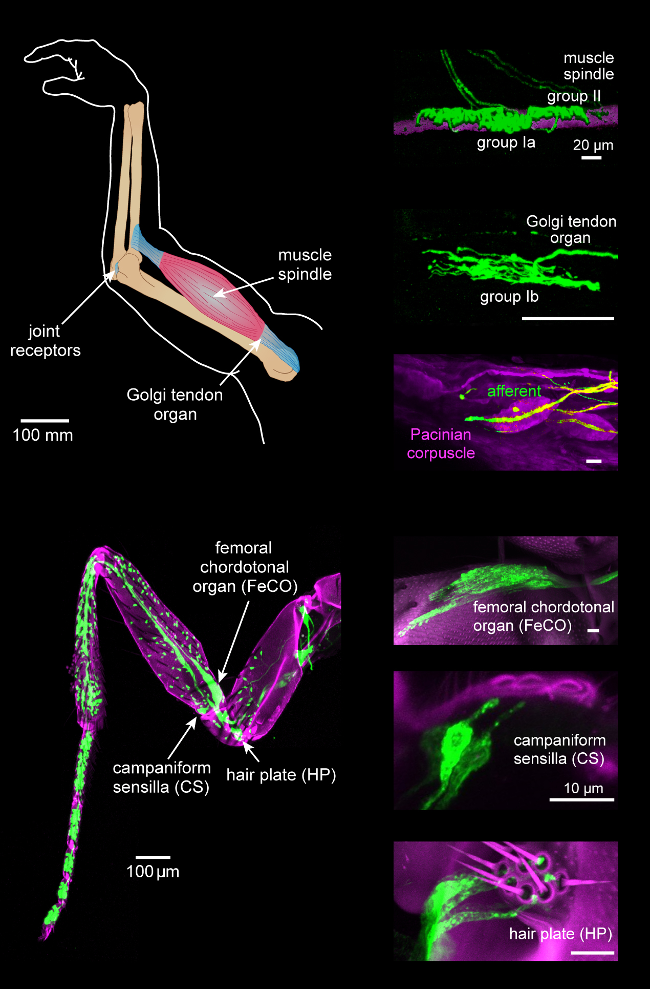
Proprioception ( ) is the sense of self-movement, force, and body position. Proprioception is mediated by proprioceptors, a type of sensory receptor, located within muscles, tendons, and joints. Most animals possess multiple subtypes of proprioceptors, which detect distinct kinesthetic parameters, such as joint position, movement, and load. Although all mobile animals possess proprioceptors, the structure of the sensory organs can vary across species. Proprioceptive signals are transmitted to the central nervous system, where they are integrated with information from other Sensory nervous system, sensory systems, such as Visual perception, the visual system and the vestibular system, to create an overall representation of body position, movement, and acceleration. In many animals, sensory feedback from proprioceptors is essential for stabilizing body posture and coordinating body movement. System overview In vertebrates, limb movement and velocity (muscle length and the rate ... [...More Info...] [...Related Items...] OR: [Wikipedia] [Google] [Baidu] |
Muscle Spindle
Muscle spindles are stretch receptors within the body of a skeletal muscle that primarily detect changes in the length of the muscle. They convey length information to the central nervous system via afferent nerve fibers. This information can be processed by the brain as proprioception. The responses of muscle spindles to changes in length also play an important role in regulating the contraction of muscles, for example, by activating motor neurons via the stretch reflex to resist muscle stretch. The muscle spindle has both sensory and motor components. * Sensory information conveyed by primary type Ia sensory fibers which spiral around muscle fibres within the spindle, and secondary type II sensory fibers * Activation of muscle fibres within the spindle by up to a dozen gamma motor neurons and to a lesser extent by one or two beta motor neurons ''.'' Structure Muscle spindles are found within the belly of a skeletal muscle. Muscle spindles are fusiform (spindle-shaped), a ... [...More Info...] [...Related Items...] OR: [Wikipedia] [Google] [Baidu] |
Nuclear Chain Fiber
A nuclear chain fiber is a specialized sensory organ contained within a muscle. Nuclear chain fibers are intrafusal fibers that, along with nuclear bag fibers, make up the muscle spindle responsible for the detection of changes in muscle length. There are 3–9 nuclear chain fibers per muscle spindle that are half the size of the nuclear bag fibers. Their nuclei are aligned in a chain and they excite the secondary nerve. They are static, whereas the nuclear bag fibers are dynamic in comparison. The name "nuclear chain" refers to the structure of the central region of the fiber, where the sensory axons wrap around the intrafusal fibers. The secondary nerve association involves an efferent and afferent pathway that measure the stress and strain placed on the muscle (usually the extrafusal fibers connected from the muscle portion to a bone). The afferent pathway resembles a spring wrapping around the nuclear chain fiber and connecting to one of its ends away from the bone. ... [...More Info...] [...Related Items...] OR: [Wikipedia] [Google] [Baidu] |
Type Ia Fiber
A type Ia sensory fiber, or a primary afferent fiber, is a type of afferent nerve fiber. It is the sensory fiber of a stretch receptor called the muscle spindle found in muscles, which constantly monitors the rate at which a muscle stretch changes. The information carried by type Ia fibers contributes to the sense of proprioception. Function of muscle spindles For the body to keep moving properly and with finesse, the nervous system has to have a constant input of sensory data coming from areas such as the muscles and joints. In order to receive a continuous stream of sensory data, the body has developed special sensory receptors called proprioceptors. Muscle spindles are a type of proprioceptor, and they are found inside the muscle itself. They lie parallel with the contractile fibers. This gives them the ability to monitor muscle length with precision. Types of sensory fibers This change in length of the spindle is transduced (transformed into electric membrane potentials) ... [...More Info...] [...Related Items...] OR: [Wikipedia] [Google] [Baidu] |


