|
Auditory Cortex
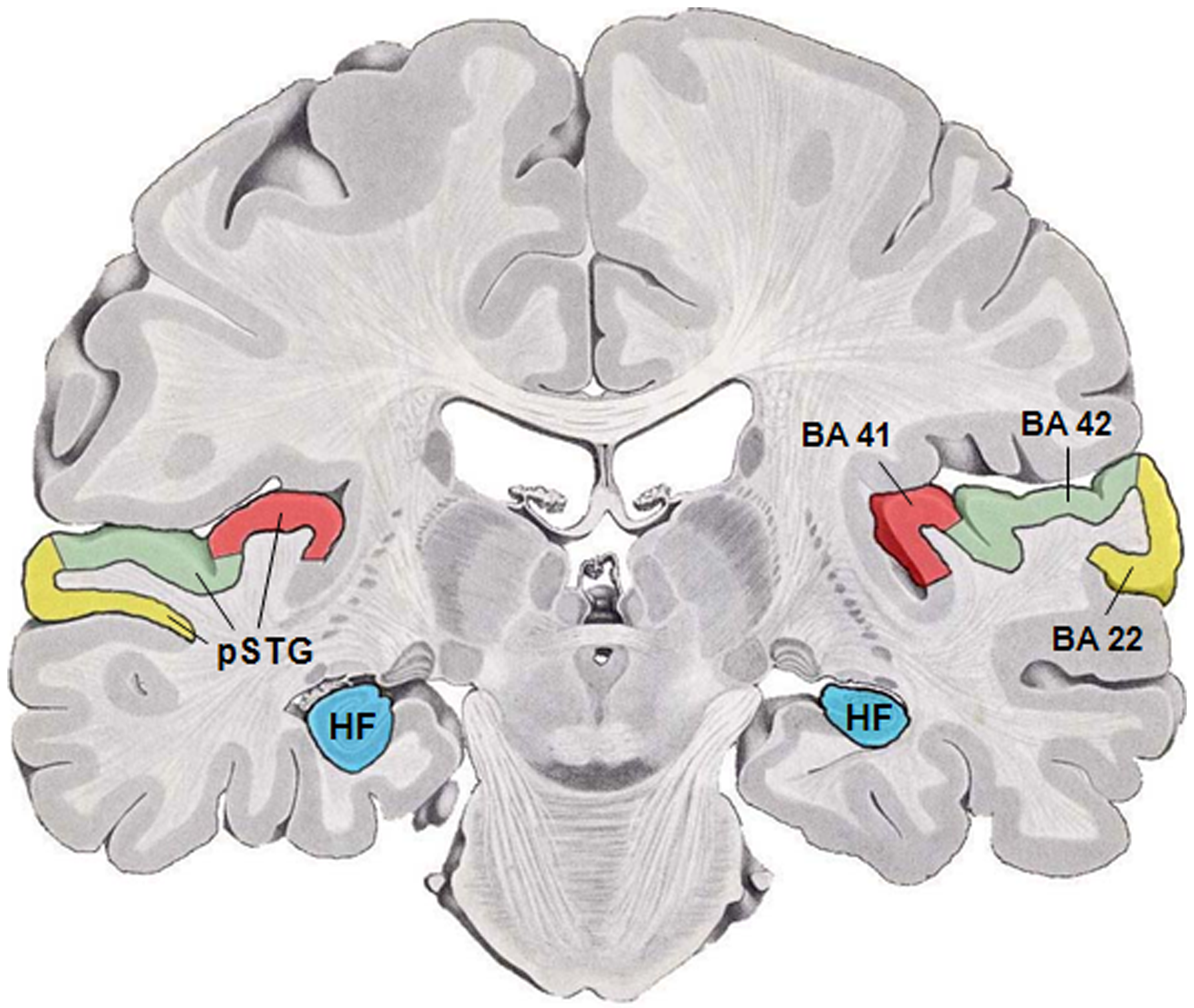
The auditory cortex is the part of the temporal lobe that processes auditory information in humans and many other vertebrates. It is a part of the auditory system, performing basic and higher functions in hearing, such as possible relations to language switching.Cf. Pickles, James O. (2012). ''An Introduction to the Physiology of Hearing'' (4th ed.). Bingley, UK: Emerald Group Publishing Limited, p. 238. It is located bilaterally, roughly at the upper sides of the temporal lobes – in humans, curving down and onto the medial surface, on the superior temporal plane, within the lateral sulcus and comprising parts of the transverse temporal gyri, and the superior temporal gyrus, including the planum polare and planum temporale (roughly Brodmann areas 41 and 42, and partially 22). The auditory cortex takes part in the spectrotemporal, meaning involving time and frequency, analysis of the inputs passed on from the ear. The cortex then filters and passes on the information to ... [...More Info...] [...Related Items...] OR: [Wikipedia] [Google] [Baidu] |
Human Brain
The human brain is the central organ (anatomy), organ of the nervous system, and with the spinal cord, comprises the central nervous system. It consists of the cerebrum, the brainstem and the cerebellum. The brain controls most of the activities of the human body, body, processing, integrating, and coordinating the information it receives from the sensory nervous system. The brain integrates sensory information and coordinates instructions sent to the rest of the body. The cerebrum, the largest part of the human brain, consists of two cerebral hemispheres. Each hemisphere has an inner core composed of white matter, and an outer surface – the cerebral cortex – composed of grey matter. The cortex has an outer layer, the neocortex, and an inner allocortex. The neocortex is made up of six Cerebral cortex#Layers of neocortex, neuronal layers, while the allocortex has three or four. Each hemisphere is divided into four lobes of the brain, lobes – the frontal lobe, frontal, pa ... [...More Info...] [...Related Items...] OR: [Wikipedia] [Google] [Baidu] |
Functional Magnetic Resonance Imaging
Functional magnetic resonance imaging or functional MRI (fMRI) measures brain activity by detecting changes associated with blood flow. This technique relies on the fact that cerebral blood flow and neuronal activation are coupled. When an area of the brain is in use, blood flow to that region also increases. The primary form of fMRI uses the blood-oxygen-level dependent (BOLD) contrast, discovered by Seiji Ogawa in 1990. This is a type of specialized brain and body scan used to map neuron, neural activity in the brain or spinal cord of humans or other animals by imaging the change in blood flow (hemodynamic response) related to energy use by brain cells. Since the early 1990s, fMRI has come to dominate brain mapping research because it does not involve the use of injections, surgery, the ingestion of substances, or exposure to ionizing radiation. This measure is frequently corrupted by noise from various sources; hence, statistical procedures are used to extract the underlying si ... [...More Info...] [...Related Items...] OR: [Wikipedia] [Google] [Baidu] |
Brain Scan
Neuroimaging is the use of quantitative (computational) techniques to study the structure and function of the central nervous system, developed as an objective way of scientifically studying the healthy human brain in a non-invasive manner. Increasingly it is also being used for quantitative research studies of brain disease and psychiatric illness. Neuroimaging is highly multidisciplinary involving neuroscience, computer science, psychology and statistics, and is not a medical specialty. Neuroimaging is sometimes confused with neuroradiology. Neuroradiology is a medical specialty that uses non-statistical brain imaging in a clinical setting, practiced by radiologists who are medical practitioners. Neuroradiology primarily focuses on recognizing brain lesions, such as vascular diseases, strokes, tumors, and inflammatory diseases. In contrast to neuroimaging, neuroradiology is qualitative (based on subjective impressions and extensive clinical training) but sometimes uses basic ... [...More Info...] [...Related Items...] OR: [Wikipedia] [Google] [Baidu] |
Visual Cortex
The visual cortex of the brain is the area of the cerebral cortex that processes visual information. It is located in the occipital lobe. Sensory input originating from the eyes travels through the lateral geniculate nucleus in the thalamus and then reaches the visual cortex. The area of the visual cortex that receives the sensory input from the lateral geniculate nucleus is the primary visual cortex, also known as visual area 1 ( V1), Brodmann area 17, or the striate cortex. The extrastriate areas consist of visual areas 2, 3, 4, and 5 (also known as V2, V3, V4, and V5, or Brodmann area 18 and all Brodmann area 19). Both hemispheres of the brain include a visual cortex; the visual cortex in the left hemisphere receives signals from the right visual field, and the visual cortex in the right hemisphere receives signals from the left visual field. Introduction The primary visual cortex (V1) is located in and around the calcarine fissure in the occipital lobe. Each h ... [...More Info...] [...Related Items...] OR: [Wikipedia] [Google] [Baidu] |
Neuron
A neuron (American English), neurone (British English), or nerve cell, is an membrane potential#Cell excitability, excitable cell (biology), cell that fires electric signals called action potentials across a neural network (biology), neural network in the nervous system. They are located in the nervous system and help to receive and conduct impulses. Neurons communicate with other cells via synapses, which are specialized connections that commonly use minute amounts of chemical neurotransmitters to pass the electric signal from the presynaptic neuron to the target cell through the synaptic gap. Neurons are the main components of nervous tissue in all Animalia, animals except sponges and placozoans. Plants and fungi do not have nerve cells. Molecular evidence suggests that the ability to generate electric signals first appeared in evolution some 700 to 800 million years ago, during the Tonian period. Predecessors of neurons were the peptidergic secretory cells. They eventually ga ... [...More Info...] [...Related Items...] OR: [Wikipedia] [Google] [Baidu] |
Midbrain
The midbrain or mesencephalon is the uppermost portion of the brainstem connecting the diencephalon and cerebrum with the pons. It consists of the cerebral peduncles, tegmentum, and tectum. It is functionally associated with vision, hearing, motor control, sleep and wakefulness, arousal (alertness), and temperature regulation.Breedlove, Watson, & Rosenzweig. Biological Psychology, 6th Edition, 2010, pp. 45-46 The name ''mesencephalon'' comes from the Greek ''mesos'', "middle", and ''enkephalos'', "brain". Structure The midbrain is the shortest segment of the brainstem, measuring less than 2cm in length. It is situated mostly in the posterior cranial fossa, with its superior part extending above the tentorial notch. The principal regions of the midbrain are the tectum, the cerebral aqueduct, tegmentum, and the cerebral peduncles. Rostral and caudal, Rostrally the midbrain adjoins the diencephalon (thalamus, hypothalamus, etc.), while Rostral and caudal, cau ... [...More Info...] [...Related Items...] OR: [Wikipedia] [Google] [Baidu] |
Auditory Pathway
The auditory system is the sensory system for the sense of hearing. It includes both the sensory organs (the ears) and the auditory parts of the sensory system. System overview The outer ear funnels sound vibrations to the eardrum, increasing the sound pressure in the middle frequency range. The middle-ear ossicles further amplify the vibration pressure roughly 20 times. The base of the stapes couples vibrations into the cochlea via the oval window, which vibrates the perilymph liquid (present throughout the inner ear) and causes the round window to bulb out as the oval window bulges in. Vestibular and tympanic ducts are filled with perilymph, and the smaller cochlear duct between them is filled with endolymph, a fluid with a very different ion concentration and voltage. Vestibular duct perilymph vibrations bend organ of Corti outer cells (4 lines) causing prestin to be released in cell tips. This causes the cells to be chemically elongated and shrunk ( somatic m ... [...More Info...] [...Related Items...] OR: [Wikipedia] [Google] [Baidu] |
Awareness
In philosophy and psychology, awareness is the perception or knowledge of something. The concept is often synonymous with consciousness. However, one can be aware of something without being explicitly conscious of it, such as in the case of blindsight. The states of awareness are also associated with the states of experience so that the structure represented in awareness is mirrored in the structure of experience. Concept Awareness is a relative concept. It may refer to an internal state, such as a visceral feeling, or on external events by way of sensory perception. It is analogous to sensing something, a process distinguished from observing and perceiving (which involves a basic process of acquainting with the items we perceive). Awareness can be described as something that occurs when the brain is activated in certain ways, such as when the color red is seen once the retina is stimulated by light waves. This conceptualization is posited due to the difficulty in developin ... [...More Info...] [...Related Items...] OR: [Wikipedia] [Google] [Baidu] |
Stroke
Stroke is a medical condition in which poor cerebral circulation, blood flow to a part of the brain causes cell death. There are two main types of stroke: brain ischemia, ischemic, due to lack of blood flow, and intracranial hemorrhage, hemorrhagic, due to bleeding. Both cause parts of the brain to stop functioning properly. Signs and symptoms of stroke may include an hemiplegia, inability to move or feel on one side of the body, receptive aphasia, problems understanding or expressive aphasia, speaking, dizziness, or homonymous hemianopsia, loss of vision to one side. Signs and symptoms often appear soon after the stroke has occurred. If symptoms last less than 24 hours, the stroke is a transient ischemic attack (TIA), also called a mini-stroke. subarachnoid hemorrhage, Hemorrhagic stroke may also be associated with a thunderclap headache, severe headache. The symptoms of stroke can be permanent. Long-term complications may include pneumonia and Urinary incontinence, loss of b ... [...More Info...] [...Related Items...] OR: [Wikipedia] [Google] [Baidu] |
Tumor
A neoplasm () is a type of abnormal and excessive growth of tissue. The process that occurs to form or produce a neoplasm is called neoplasia. The growth of a neoplasm is uncoordinated with that of the normal surrounding tissue, and persists in growing abnormally, even if the original trigger is removed. This abnormal growth usually forms a mass, which may be called a tumour or tumor.'' ICD-10 classifies neoplasms into four main groups: benign neoplasms, in situ neoplasms, malignant neoplasms, and neoplasms of uncertain or unknown behavior. Malignant neoplasms are also simply known as cancers and are the focus of oncology. Prior to the abnormal growth of tissue, such as neoplasia, cells often undergo an abnormal pattern of growth, such as metaplasia or dysplasia. However, metaplasia or dysplasia does not always progress to neoplasia and can occur in other conditions as well. The word neoplasm is from Ancient Greek 'new' and 'formation, creation'. Types A neopla ... [...More Info...] [...Related Items...] OR: [Wikipedia] [Google] [Baidu] |
Lesion
A lesion is any damage or abnormal change in the tissue of an organism, usually caused by injury or diseases. The term ''Lesion'' is derived from the Latin meaning "injury". Lesions may occur in both plants and animals. Types There is no designated classification or naming convention for lesions. Because lesions can occur anywhere in the body and their definition is so broad, the varieties of lesions are virtually endless. Generally, lesions may be classified by their patterns, sizes, locations, or causes. They can also be named after the person who discovered them. For example, Ghon lesions, which are found in the lungs of those with tuberculosis, are named after the lesion's discoverer, Anton Ghon. The characteristic skin lesions of a varicella zoster virus infection are called '' chickenpox''. Lesions of the teeth are usually called dental caries, or "cavities". Location Lesions are often classified by their tissue types or locations. For example, "skin lesions" or ... [...More Info...] [...Related Items...] OR: [Wikipedia] [Google] [Baidu] |
Cerebral Cortex
The cerebral cortex, also known as the cerebral mantle, is the outer layer of neural tissue of the cerebrum of the brain in humans and other mammals. It is the largest site of Neuron, neural integration in the central nervous system, and plays a key role in attention, perception, awareness, thought, memory, language, and consciousness. The six-layered neocortex makes up approximately 90% of the Cortex (anatomy), cortex, with the allocortex making up the remainder. The cortex is divided into left and right parts by the longitudinal fissure, which separates the two cerebral hemispheres that are joined beneath the cortex by the corpus callosum and other commissural fibers. In most mammals, apart from small mammals that have small brains, the cerebral cortex is folded, providing a greater surface area in the confined volume of the neurocranium, cranium. Apart from minimising brain and cranial volume, gyrification, cortical folding is crucial for the Neural circuit, brain circuitry ... [...More Info...] [...Related Items...] OR: [Wikipedia] [Google] [Baidu] |


